Article Text

Statistics from Altmetric.com
Obesity is associated with an increased risk of adverse cardiovascular outcomes; a global public health problem given the high and ever increasing prevalence of obesity in most developed countries. In addition to the increased prevalence of conventional cardiovascular risk factors in obese patients, obesity results in both structural and electrical alterations in cardiac function that might be associated lead to an increased risk of sudden cardiac death. In the large multicenter population based Atherosclerosis Risk in Communities (ARIC) study, Dr. Adabag and colleagues (see page 215) found that sudden cardiac death was associated positively with body mass index, waist circumference and waist-hip ratio in non-smokers, although this association was not seen in smokers. After adjustment for other cardiac risk factors and known heart disease, the risk of sudden cardiac death was doubled for nonsmokers with a waist-hip ratio greater than 0.95 for women and 1.01 in men Figure 1.
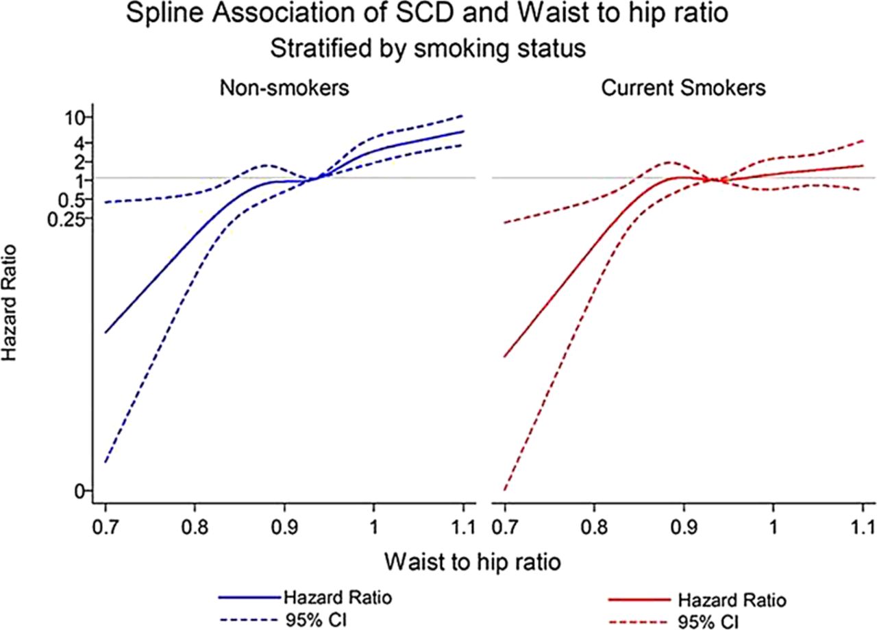
Cubic spline model for risk of sudden cardiac death (SCD) in relation to waist hip ratio, stratified by smoking status and adjusted for age, sex and race, field centre and education level.
In the accompanying editorial, Professors Reinier and Chugh (see page 165) comment that the association between waist-hip ratio and sudden cardiac death, even after adjustment for other risk factors, suggests the possibility other mechanisms, such pro-inflammatory effects of visceral adipose tissue, might be involved in the link between obesity and adverse outcomes. Regardless of mechanism, from the clinical point of view both overall weight and waist-hip ratio are potentially modifiable cardiovascular risk factors, providing an opportunity to improve cardiovascular health by increased efforts to reduce visceral adiposity both at the individual and population levels.
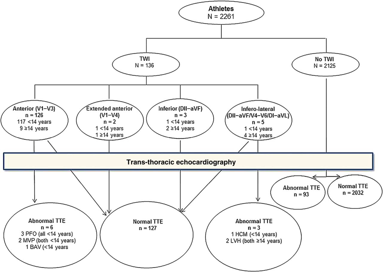
Efforts to reduce the risk of sudden cardiac death in young athletes have focused on pre-participation screening tests, typically including an electrocardiogram (ECG) as well as history and physical examination, to detect underlying structural heart disease. In a study of 2261 consecutive male soccer players, with a mean age of 12 years, Professor Calò and colleagues (see page 193) observed T-wave inversion on ECG in 6% of subjects, most often in the anterior leads. Echocardiography revealed structural heart disease in only 6 of 126 subjects (4.8%) with anterior T-wave inversion compared to 4.4% of those with normal T-waves. In contrast, inferolateral T-wave inversion was rare but was associated with structural heart disease in 3 of 5 (60%) patients–one with hypertrophic cardiomyopathy and the other two with nonspecific ventricular hypertrophy Figure 2.

Relationship between ECG and transthoracic echocardiogram (TTE) findings in athletes with or without T wave inversion (TWI) at ECG. Arrows indicate subgroups. PFO, MVP and BAV indicate patent foramen ovale, mitral valve prolapse and bicuspid aortic valve, respectively.
Drs Wasfy and Baggish (see page 167) put these findings into context. “Contemporary athlete ECG criteria rely on the principle that ECG patterns among athletes can be divided into ‘common and training-related ECG changes' (ie, benign adaptive findings) and ‘uncommon and training-unrelated ECG changes’, with this latter category touted as being highly suggestive of underlying disease”. The current study is helpful in identifying a high rate of underlying pathology in young male athletes with inferolateral T-wave inversion. In contrast, T-wave inversion in the anterior leads is likely to be benign. However, caution still is needed as mild early structural heart disease may have been missed and subsequent development of heart disease cannot be excluded in this cross-sectional study. They suggest: “Future efforts should follow the lead of Professor Calò and colleagues by pairing ECG data with non-invasive imaging and should ideally be conducted in a longitudinal fashion with ample numbers to assess clinical outcomes”.
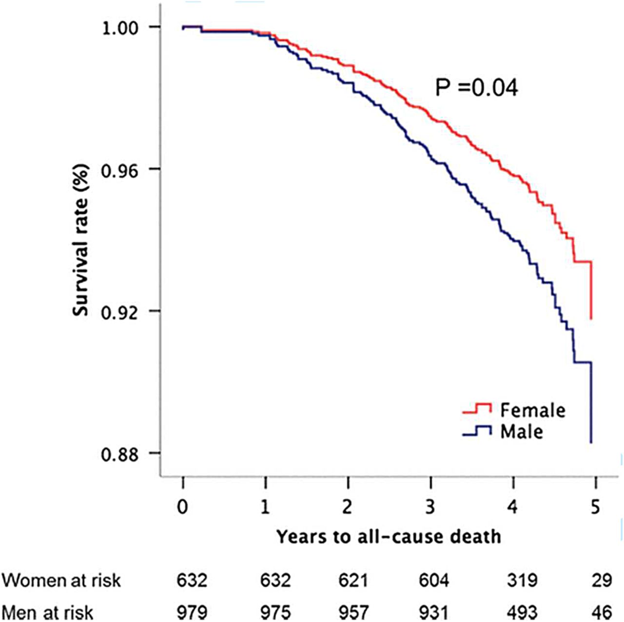
Management of elderly adults with calcific aortic stenosis is of increasing importance with the aging of our population and with the availability of non-surgical approaches to relief of valve obstruction. Bicuspid aortic valve disease is more common in men than women, so that men typically present with severe symptomatic aortic stenosis at a younger age than women. In addition, the ventricular changes associated with valvular aortic stenosis differ with women typically having a smaller, more hypertrophied ventricle with predominant diastolic dysfunction, and a low stroke volume index. However, there is little data on possible sex differences in rates of hemodynamic progression or clinical outcomes in adults with aortic stenosis. In the current volume of Heart, Dr. Cramariuc and colleagues (see page 209) used the large cohort of patients in the Simvastatin Ezetimibe in Aortic Stenosis (SEAS) study data to examine this issue. Interestingly, there were no sex related differences in the rate of hemodynamic progression, although women had less reduction in ejection fraction over time, congruent with previous research publications and clinical experience. Somewhat surprisingly, women had a lower rate of coronary ischemic events (40% lower) and coronary bypass grafting (50% lower), risk of stroke (50% lower), and all-cause mortality (31% lower) compared to men even though there was no difference in aortic stenosis event rates Figure 3.

Overall survival in women and men during progression of aortic valve stenosis with adjustment for covariates (the means of age, hypertension, active study treatment, energy loss index, low EF and midwall shortening, and abnormal LV geometry) and p value of significance based on Cox proportional hazard analysis.
Differences between women and men were independent of active study treatment, age, hypertension, stenosis severity, or ventricular function. These findings contrast with the prevalent erroneous clinical concern that women might be at higher risk than men due to frailty or other factors. This data also underscores the importance of providing guideline based care to both men and women with aortic stenosis.
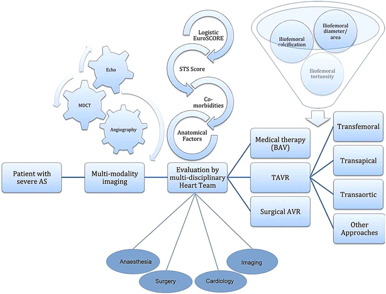
For readers interested in updating their knowledge of transcatheter aortic valve replacement, Dr. Kapadia and colleagues (see page 169) provide a contemporary review with sections on the workflow process for patient evaluation and management, details of pre-procedural imaging in patient selection and valve sizing, risk assessment, procedural considerations, and valve and access selection, as well as an update on complications, mortality and long term outcomes. In addition, a video abstract provides a panel discussion by the authors on more controversial aspects of transcatheter aortic valve implantation Figure 4. http://heart.bmj.com/content/early/2015/01/02/heartjnl-2014-306254.full

Workflow process involved in the evaluation and management of a patient presenting with severe symptomatic AS. The figure illustrates the importance of multimodality imaging and the central role of the multidisciplinary heart team in the evaluation of these patients. AS, aortic stenosis; AVR, aortic valve replacement; BAV, balloon aortic valvuloplasty; MDCT, multidetector CT; STS, Society of Thoracic Surgeons; TAVR, transcatheter aortic valve replacement.
The Education in Heart article (see page 230) in this issue focuses on the role of cardiac synchronization therapy in patients who do not fit the specific criteria in randomized controlled clinical trials. These patients include those with a wide QRS but without a left bundle branch morphology, atrial fibrillation, renal dysfunction, older age, diabetes, or congenital heart disease. Further clinical trials are needed in each of these patient groups.
Be sure to see if you can identify the unusual cardiac anomaly shown on echocardiography and CT-imaging in this week's Image Challenge (see page 184).
Linked Articles
- Education in Heart
- Editorial
- Review
- Special populations
- Valvular heart disease
- Editorial
- Image challenge
- Cardiac risk factors and prevention