Article Text

Abstract
Background and purpose Arrhythmogenic right ventricular cardiomyopathy (ARVC) is an inherited disorder that causes ventricular arrhythmias and sudden death. The disease manifests predominantly in the right heart and is more common in endurance athletes. ARVC patients also show increased atrial arrhythmias. Loss-of-function mutations in desmosomal proteins such as plakoglobin are associated with ARVC, and endurance training exacerbates ventricular arrhythmias and right ventricular enlargement in mice with reduced expression of plakoglobin (Plako+/–). Chronic intensive endurance training has recently been observed to increase susceptibility to atrial arrhythmias in rats. We therefore tested whether standardised endurance training affects atrial electrophysiology or increases in mice with heterozygous plakoglobin deficiency (Plako+/–).

S2-induced atrial arrhythmias >1 second in trained wildtype and plakoglobin-deficient hearts *p < 0.05 Fisher’s exact test
Methods Plako+/– and WT mice group swim-trained for 5 d/week over 8 weeks, gradually increased from 2 to 90 min/d (average swim times 50h). Atrial size and function was assessed by echocardiography. Left atrial (LA) monophasic action potentials (MAPs) were recorded from Langendorff-perfused hearts. Action potential durations (APDs) were measured during RA pacing at 100ms paced cycle lengths (CL). Atrial arrhythmias were provoked by programmed stimulation (S1-S2). Atrial arrhythmias were defined as fast irregular atrial activity >1s). Transmembrane action potentials (TAPs) were recorded at 100ms CL from isolated, superfused LA using floating glass microelectrodes. All continuous parameters are expressed as mean±SEM.

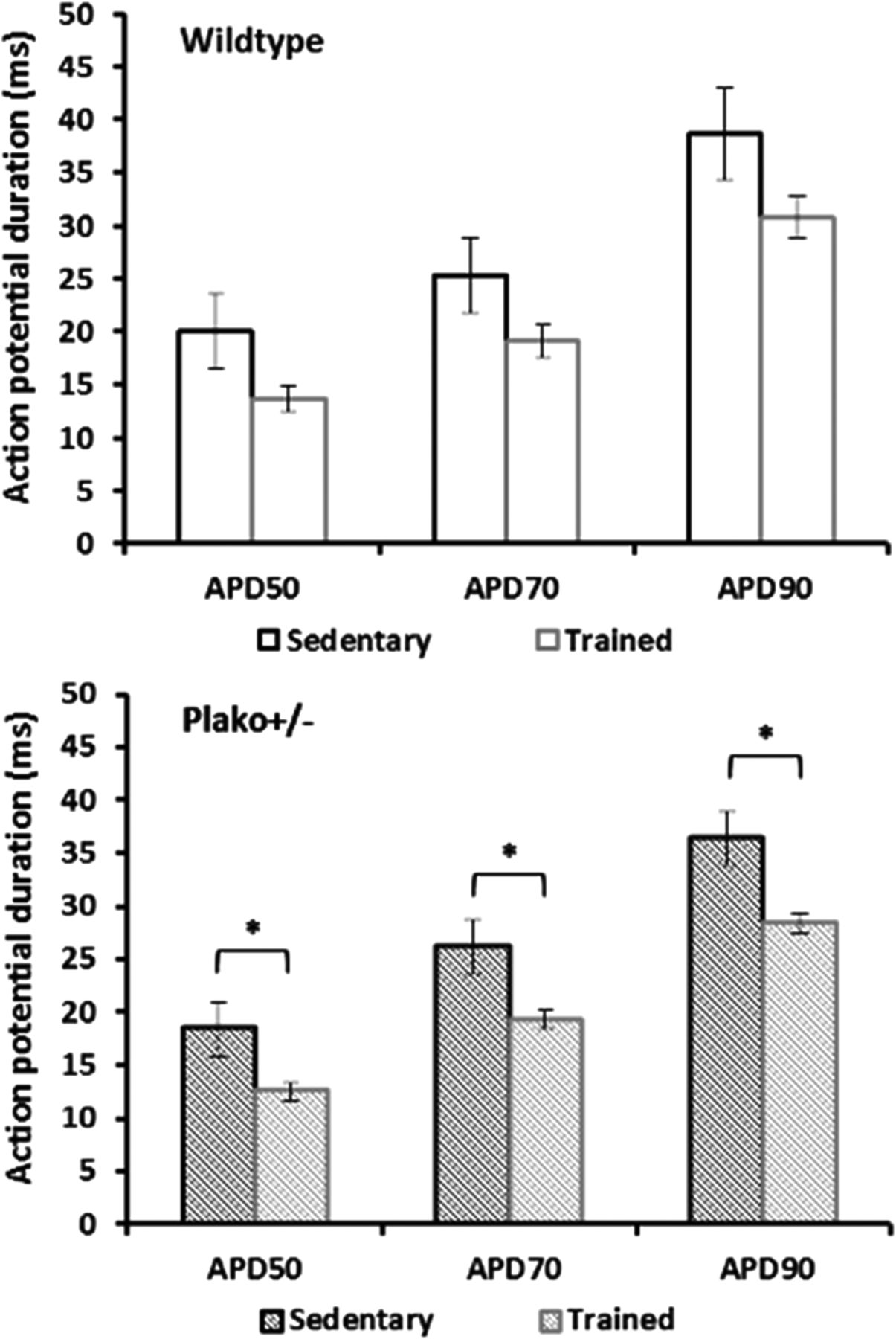
Action potential durations at 100 ms paced cycle length in sedentary and trained hearts *p < 0.05 ANOVA
Results Swim-training increased atrial arrhythmia susceptibility in Plako+/– but not WT mice (Plako+/– sedentary: 1/9 vs trained: 9/17 hearts),(WT sedentary: 3/11 hearts vs trained: 1/11 hearts). Training induced mild leftventricular hypertrophy in both genotypes (LV mass increase 6–14%, p < 0.05). As shown previously, training also increased right ventricular size in Plako+/– vs. trained WT, e.g. Parasternal RV diameter Plako+/– (1.79 ± 0.03 mm) vs. WT (1.66 ± 0.02 mm) after training, p < 0.05. Training increased LA size as assessed in echocardiography in both genotypes versus sedentary (LA size WT: 3.11 ± 0.14 sedentary vs 3.79 ± 0.17 mm2 trained, p < 0.05; Plako+/–: 3.23 ± 0.1 mm2 sedentary vs 4.14 ± 0.23 mm2 trained, p < 0.05). LA APD from MAPs tended to be shorter in trained WT and were significantly shorter in trained Plako+/- mice (Figure 2). RA APD90s measured from TAPs were also shorter after training (WT: 22 ± 1 sedentary vs 19 ± 1 ms trained; Plako+/- 24 ± 1 sedentary vs 18 ± 1 ms trained).
Conclusion Our observations suggest that endurance training shortens atrial action potential duration and increases left atrial size in both WT and plakoglobin deficient atria. Endurance training increased atrial arrhythmia susceptibility in mice with a deficiency in the mechanical cell-cell contact protein plakoglobin.
- Desmosome
- ARVC
- Endurance exercise