Article Text

Abstract
Introduction Optical coherence tomography (OCT) is a relatively new catheter-based imaging modality utilising frequencies near-infrared light (as opposed to ultrasound) to visualise intracoronary dimensions. This allows spatial resolution of 20 nm compared with 200 nm observed in intravascular ultrasound. Several studies, including both the ODESSA trial and a subanalysis of the HORIZONS-AMI trial both describe the ability of OCT to assess stent deployment characteristics such as stent strut coverage. There are however no previously reported case series evaluating the use of intracoronary OCT in clinical practice.
Aim The aim of this study was to evaluate the use of intracoronary OCT in routine clinical practice in an Irish Tertiary referral centre, and assess for stent thrombosis/ restenosis at follow-up.
Methods A Retrospective cohort analysis was performed on all patients undergoing coronary optical coherence tomography (OCT) over an 18-month period from 01/08/2012 to 31/12/2014. Paper and electronic medical records were sourced for demographic and follow-up data.
Results A total of 99 patients underwent OCT during the study period (see Table 1). The average age was 63 (range 29–78), with a male: female ratio of 4:1. The use of intracoronary OCT was used in a variety of presenting complaints, from stable angina (n = 59 [59.6%]), to unstable angina (n = 7 [7.1%]), NSTEMI (n = 16 [16.2%]) and STEMI (n = 12 [12.1%]). OCT was used most commonly in the left anterior descending (LAD) artery (n = 65 [65.7%]); proximal LAD (n = 39 [82.3%]), mid LAD (n = 6 [12.7%]) and distal LAD (n = 2 [4.2%]). Other vessels assessed by OCT included the left circumflex artery (n = 27 [27.3%]), the left main artery (n = 18 [18.2%]) and the right coronary artery (n = 16 [16.2%]). OCT was used to guide stent deployment in 59 patients (59.6%), to evaluate intraluminal dimensions in 37 patients (37.4%) (Figure 1) and used to assess for intimal lesions (such as plaque rupture or dissection) in 15 cases (15.2%) (Figure 2). In 12 instances (12.1%), OCT was used to both assess the primary lesion and then to guide stent deployment after PCI. At an average follow-up of 314 days, of the 59 patients who underwent OCT-guided stent deployment and post-dilatation, 14 underwent repeat angiography. There were no incidences of stent thrombosis in this group and 2 incidences of instent restenosis requiring angioplasty.
Demographics of cohort

Instent restenosis of proximal RCA

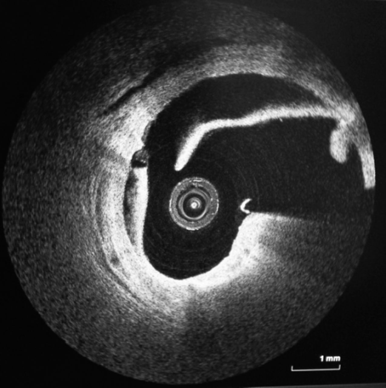
OCT image of spontaneous proximal LAD dissection
Conclusion Intracoronary OCT has been shown to provide valuable information for integrated assessment of coronary lesions and the direction of stent deployment in routine clinical practice.