Article Text

Abstract
Objective A proportion of patients with suspected ST-elevation myocardial infarction (STEMI) presenting for primary percutaneous coronary intervention (PPCI) do not have obstructive coronary disease and other conditions may be responsible for their symptoms and ECG changes. In this study, we set out to determine the prevalence and aetiology of alternative diagnoses in a large PPCI cohort as determined with multimodality imaging and their outcome.
Methods From 2009 to 2012, 5238 patients with suspected STEMI were referred for consideration of PPCI. Patients who underwent angiography but had no culprit artery for revascularisation and no previous history of coronary artery disease were included in the study. Troponin values, imaging findings and all-cause mortality were obtained from hospital and national databases.
Results A total of 575 (13.0%) patients with a mean age of 58±15 years (69% men) fulfilled the inclusion criteria. A specific diagnosis based on imaging was made in 237 patients (41.2%) including cardiomyopathies (n=104, 18%), myopericarditis (n=48, 8.4%), myocardial infarction/other coronary abnormality (n=27, 4.9%) and severe valve disease (n=23, 4%). Pulmonary embolism and type A aortic dissection were identified in seven (1.2%) and four (0.7%) cases respectively. A total of 40 (7.0%) patients died over a mean follow-up of 42.6 months.
Conclusions A variety of cardiac and non-cardiac conditions are prevalent in patients presenting with suspected STEMI but culprit-free angiogram, some of which may have adverse outcomes. Further imaging of such patients could thus be useful to help in appropriate management and follow-up.
This is an Open Access article distributed in accordance with the Creative Commons Attribution Non Commercial (CC BY-NC 4.0) license, which permits others to distribute, remix, adapt, build upon this work non-commercially, and license their derivative works on different terms, provided the original work is properly cited and the use is non-commercial. See: http://creativecommons.org/licenses/by-nc/4.0/
Statistics from Altmetric.com
Introduction
The diagnosis of ST-elevation myocardial infarction (STEMI) is made from a history of acute-onset chest pain, ST-elevation on ECG and subsequent release of cardiac biomarkers suggesting myocardial necrosis.1 Guidelines recommend urgent revascularisation, ideally with primary percutaneous coronary intervention (PPCI) of the culprit artery.1 The levels of biomarkers such as troponin are generally not available at the time of revascularisation.
A subset of patients (2.6%–14%) with suspected STEMI do not have a culprit lesion or obstructive coronary artery disease (CAD) on the initial diagnostic coronary angiogram (DCA).2–5 Some of these patients may have other non-atheromatous cardiac or non-cardiac causes for their symptoms and ECG abnormalities as well as biomarker release.6 ,7 In the last few years, there has been an increasing interest in alternative causes of acute chest pain associated with troponin rise particularly with non-invasive diagnosis of myocarditis on cardiac magnetic resonance (CMR) or demonstration of myocardial infarction (MI) in the presence of recanalised arteries.8–10 However, such studies have mainly focused on small series of patients presenting within a broader spectrum of acute coronary syndrome (ACS) with raised troponin with the aim of evaluating the usefulness of CMR. The prevalence of non-coronary causes of acute chest pain and their prognosis, particularly those presenting with suspected STEMI, is less well described.
The aim of this study was to determine the prevalence and outcome of different cardiac and non-cardiac conditions as identified on multimodality imaging in a large cohort of patients presenting with suspected STEMI, who had no culprit lesion on the angiogram.
Methods
Patient recruitment
Our institution is a tertiary cardiothoracic centre with an established 24-hour PPCI service with a catchment population of about 2 million people. Patients with suspected STEMI on the basis of clinical signs and symptoms, and 12-lead ECG are transferred directly by the ambulance service (diagnosis made autonomously in the field without the use of teletransmission) to our centre for consideration of PPCI.11 Patients on arrival are reviewed by the on-call cardiology team on site to confirm the diagnosis and proceed to PPCI, if appropriate. Some patients are also transferred from nearby general hospitals, if they are found to have suffered a STEMI.
Consecutive patients who were admitted to the PPCI programme between 1 January 2009 and 31 December 2012 were identified from the programme registry (n=5238). Patients were excluded as per the criteria shown in figure 1. All patients without a culprit lesion on angiogram and no known CAD were included in the study. Absence of a culprit lesion was defined as absence of a significant stenosis (>70% luminal narrowing) in the vessel supplying the area of acute ECG change. Known CAD was defined as history of previous ACS, PCI or coronary artery bypass surgery. The National Health Service ethics approval was obtained from the hospital's research department (Project ID: 199060).
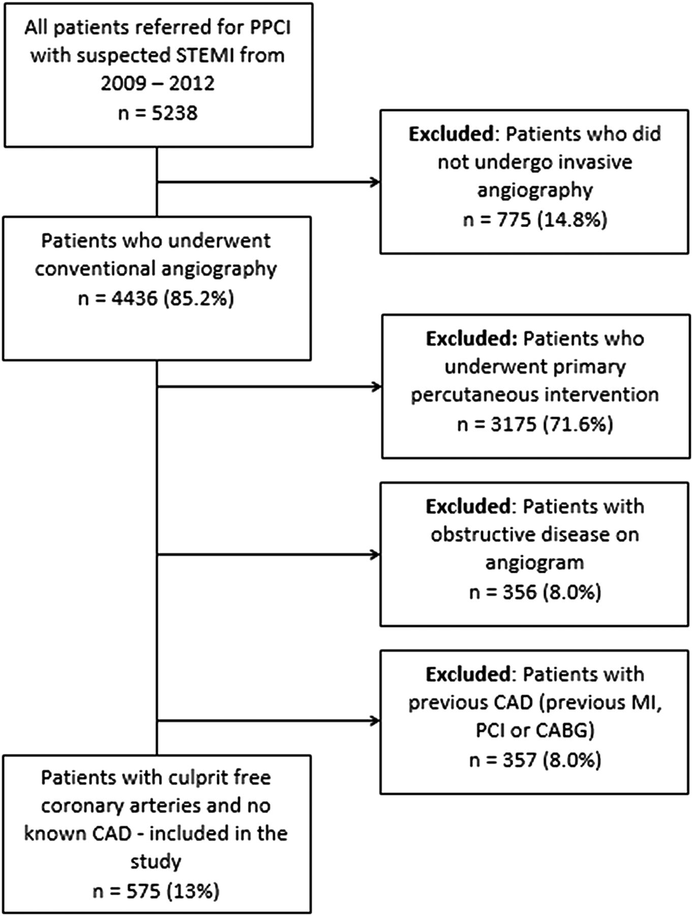
Flow chart showing exclusion criteria and final patient population included in the study. CABG, coronary artery bypass surgery; CAD, coronary artery disease; MI, myocardial infarction; PCI, percutaneous coronary intervention; PPCI, primary percutaneous coronary intervention; STEMI, ST-elevation myocardial infarction.
Data collection and imaging indications
Patient demographics, traditional risk factors for CAD and presenting ECG as recorded in the database were identified. For all included patients, the highest initial troponin result during the admission was recorded (Access AccuTnI assay, Beckman Coulter, Brea, CA, USA) from laboratory results in the electronic patient records (EPR). The different non-invasive imaging procedures performed as a result of the primary admission episode, their findings and subsequent management were recorded from the hospital Picture Archiving and Communication System and the EPR. Images were not reviewed in all cases unless there was uncertainty about a diagnosis or clarification was required. Our hospital has a protocol for routine performance of plain chest X-rays (CXR) and echocardiograms for all PPCI admissions once the patient is stable. CMR is performed in those with non-obstructed coronary arteries on DCA and unexplained rise in troponin or if required from echocardiogram findings. A chest or ECG-gated cardiac CT is performed if clinically indicated in patients with suspected pulmonary embolism (PE), acute aortic dissection, pericarditis or other coronary abnormality not identified on initial DCA. All imaging was performed within the acute admission with follow-up scans as required.
Imaging equipment and protocol
Echocardiography was performed on different commercially available ultrasound machines (Vivid-7 or 9, GE Healthcare, Milwaukee, WI, USA and IE33, Phillips Healthcare, Andover, MA, USA). CT scans of the chest or ECG-gated scans of the heart were carried out on a 64-slice scanner (Aquilion 64, Toshiba Medical Systems, Japan). CMR scans of the heart were performed on a cardiac optimised 1.5T system (Avanto, Siemens Medical Systems, Germany). Standard protocols were used for all imaging as recommended and described by various societies,12–14 with echocardiograms being performed by British Society of Echocardiography certified sonographers and cardiologists. CT and CMR interpretation was carried out by level 3 certified cardiothoracic radiologists (TKM, BA) and/or imaging cardiologists (JW, SR-H).
Imaging diagnostic criteria and outcome
All imaging diagnoses (called primary diagnosis) were made according to standard criteria using one or more imaging modalities and classified under a broader diagnostic category. In some patients, additional imaging findings were recorded if they were considered important and not being a result of the primary diagnosis.
Myocarditis was diagnosed on CMR as per the Lake Louise consensus criteria based on T2-weighted oedema and late gadolinium enhancement (LGE) sequences.15 A diagnosis of pericarditis was made in the presence of a pericardial effusion or thickening on any imaging modality in the absence of other findings. MI was diagnosed if there was a typical LGE pattern on CMR involving the subendocardium.16 Dilated cardiomyopathy (DCM) was defined as left ventricular (LV) dilatation with generalised hypokinesia and ejection fraction (EF) of <50% in the absence of CAD or valvular disease.17 Hypertrophic cardiomyopathy (HCM) was diagnosed in the presence of unexplained myocardial thickness of ≥15 mm on echocardiography and CMR in the absence of another cardiac or systemic disorder that could explain the hypertrophy.18 A diagnosis of Takotsubo cardiomyopathy (TTCM) was made when there was dilatation and dysfunction of the distal half of the LV on ventriculogram, which normalised on subsequent imaging scan in the absence of MI or other significant pathologies.19 Hypertensive heart disease was defined as concentric myocardial thickening >12 mm associated with systolic and/or diastolic dysfunction in patients with a known long-standing history of hypertension or newly diagnosed persistent hypertension for the purpose of this study.20 Different valvular diseases were included if they were found to be severe in degree as per guidelines.21 Pulmonary infection was defined as air space shadowing on the CXR or CT, associated with raised inflammatory markers. Filling defects in the pulmonary arteries on CT pulmonary angiogram were diagnostic of PE. Aortic pathology included patients with imaging findings of acute aortic dissection (type A or B), intramural haematoma or unknown dilatation of the aortic root and/or thoracic aorta of ≥5 cm in maximum diameter.
Finally, patient outcome was determined as all-cause mortality from UK's Health & Social Care Information Centre, which tracks deaths of people residing within the country and registered with a general practitioner. Cause of death was identified from hospital records and general practitioners.
Statistical methods
Continuous variables that were normally distributed are expressed as mean±SD. Other continuous variables such as troponin levels that were not normally distributed are described as median with interquartile range and their groups were analysed using Mann-Whitney test. Categorical variables are expressed as percentages and the χ2 test was performed for analysis. A two-tailed p value of <0.05 was considered statistically significant.
Cox regression with univariable and multivariable analyses was performed to examine factors associated with survival times. All variables were entered into the multivariable analysis regardless of statistical significance. Data were collected from all patients for all variables except for EF, which was only available for 429 patients from echocardiography and CMR. Kaplan-Meier survival graphs were drawn with survival times measured from the time of acute presentation to the time of death. Patients who did not die were censored at the time of last follow-up. Assumptions of proportional hazards were checked by graphing −In{−In(survival)} against In(analysis time).
All statistical analysis was performed using Stata V.13.1 (StataCorp LP, College Station, TX, USA).
Results
During the 4-year study period, 575 patients (13.0%) fulfilled the inclusion criteria. The mean age of the included patients was 57.7 years (±15.2) with 397 (69%) being men. Two hundred twenty-nine patients (39.8%) had a raised troponin. Other baseline characteristics are given in table 1.
Patient characteristics and imaging performed
A total of 499 patients (86.8%) had one or more imaging procedure performed with a specific imaging-based diagnosis made in 237 patients (41.2%) (table 2). Besides, there were 10 additional important findings including 3 cases of aortic aneurysm, 5 cases of pulmonary infection and 2 cases of additional pulmonary cancers that were present apart from the primary diagnosis (table 2).
Primary imaging diagnosis
The different imaging modalities used for the various diagnostic categories are given in table 3. Multiple imaging techniques were used in most cases as thought to be appropriate to arrive at a diagnosis.
Imaging modalities used for different diagnostic categories
Out of the patients who had one or more imaging test performed, 229 (46%) were troponin positive (TP). Among these, an imaging diagnosis was made in 171 patients (74.7%). One hundred twenty-two (53%) TP patients underwent CMR scan as the rest either had diagnosis made from other imaging techniques or were considered unsuitable (due to low clinical risk or contraindications). The TP patients who did not undergo a CMR scan and had no diagnosis made on other imaging modalities had a much lower troponin level (median 0.095±0.48 vs 1.58±11.55 in those with diagnosis, p<0.0001). Of the 270 (54%) troponin-negative patients, an imaging diagnosis was made in 66 patients (24.4%).
A total of 40 deaths (7.0%) from all causes occurred during a mean follow-up of 42.6±17 months with mortality varying widely between different conditions (table 2). Twelve of these deaths occurred within 30 days giving a 30-day all-cause mortality rate of 2.1%. Different causes of death and mean time to death is given in table 4. Cox regression revealed that on multivariate analysis, only age, out-of-hospital cardiac arrest (OHCA) and EF<50% were significantly associated with survival times (table 5). None of the patients with LGE on CMR died. Kaplan-Meier survival curves demonstrating the effect of significant variables on survival are given in figure 2.
Different causes of death and average time to death
Predictors of all-cause mortality
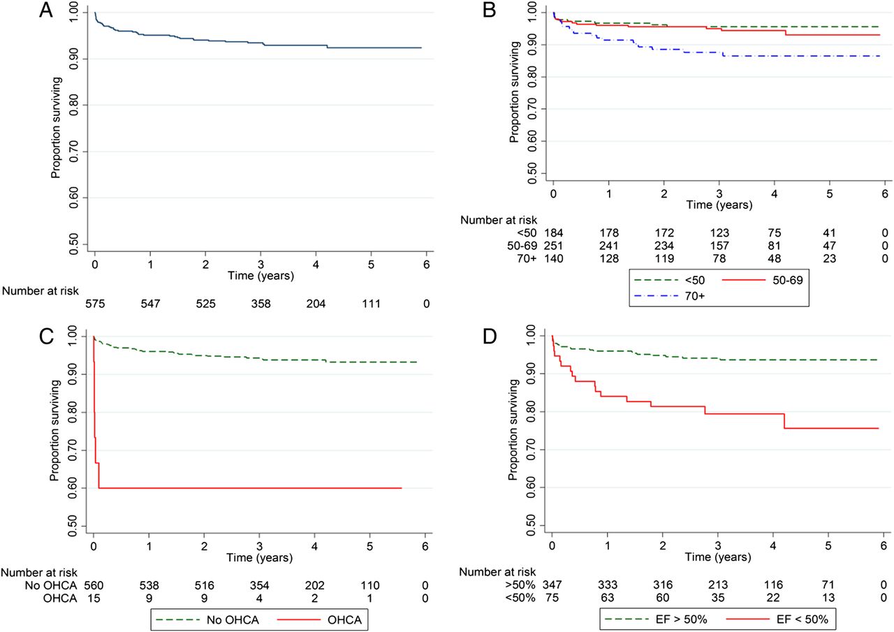
Kaplan-Meier survival curves (A) for the whole study group, (B) by age groups, (C) by out-of-hospital cardiac arrest (OHCA) and (D) and by ejection fraction (EF).
Discussion
Our study demonstrates the prevalence of alternative diagnoses in patients admitted with suspected STEMI who had culprit-free coronary arteries on angiography and no known CAD. This is the largest study of patients presenting to a large PPCI programme with further clinically directed but comprehensive multimodality non-invasive imaging. It demonstrates that just under half the patients are discharged with an alternative diagnosis, some of which are potentially associated with an adverse outcome. However, not all the diagnoses may be responsible for patients' presentation and some could be purely coincidental.
The prevalence of culprit-free coronary arteries in our patient cohort (13.0%) is similar to that described in some studies,4 ,5 while other studies have reported a lower rate of up to 5%.2 ,3 This variation can be attributed to the local arrangements for PPCI referral including the use of teletransmission in some centres and differences in patient population. Like in our hospital's protocol, where the patients are brought straight on the basis of initial diagnosis of STEMI made by the trained ambulance crew, the risk of false primary activation has to be weighed against that of missing a true STEMI. This would explain the presence of substantial number of troponin-negative patients in our cohort.
Cardiomyopathies formed the most prevalent group of alternative diagnosis in our study population (18.1%). This was a heterogeneous group, with HCM being the most common, followed by DCM, TTCM and hypertensive heart disease. HCM is well known to be associated with myocardial ischaemia due to morphological abnormalities of the intramural coronary arterioles22 ,23 and is the most common cause for sudden cardiac death in the young.18 However, presence of ischaemia in HCM is under-investigated in clinical practice and prevalence of HCM in patients presenting with suspected STEMI has been unknown.
Existence of TTCM has increasingly been recognised in patients presenting with ACS. TTCM was equally prevalent as HCM in our study cohort. Eitel et al24 reported 38 patients with confirmed diagnosis of TTCM with CMR in 6100 patients presenting with ACS, but did not specify the number of patients with non-culprit angiogram to make a comparison. In a systemic review of previous studies, Gianni et al25 reported an average prevalence of 2% in both Caucasian and Japanese populations presenting with ACS.
DCM was the third most common cardiomyopathy in our study. Existence of DCM has been described in patients with ACS in other studies,2 ,9 ,26 but in much smaller numbers. The diagnosis of DCM in this patient group may be coincidental, but would be relevant in view of adverse prognosis.
The second most common group of diagnosis was myopericarditis, but myocarditis itself was the most common condition (6.3% in all patients and 15.2% in TP patients). Studies that have mainly focused on the usefulness of CMR in patients with normal angiograms, without reference to the total number of patients with culprit-free angiograms, usually report a much higher prevalence of myocarditis, varying between 50% and 80%,8 ,9 ,26 although one study reports a prevalence of 16%.10 A study by Larson et al,4 which is more similar to ours demonstrated myocarditis in 19% of patients. However, the prevalence of clinical myocarditis is likely to be higher as CMR criteria have a sensitivity of up to 70%.15 ,27
The diagnosis of MI in the absence of culprit disease on the angiogram has been made possible with LGE on CMR. The prevalence of MI demonstrated in studies4 ,8–10 ,26 comprising TP ACS patients varies from 5% to 23% (mean=13.5%), which is similar to our patient cohort (13.6%) if only patients who underwent CMR are included. These patients had a low mortality rate in the follow-up period (table 2).
Among the valve diseases, severe aortic valve stenosis (AVS) is well known to present with symptoms of angina. The prevalence of AVS in patients presenting as ACS for PPCI is not well known with two studies describing a single case each.3 ,4 A prevalence of 2.8% (n=16) is possibly the highest reported with 75% having raised troponin.
Pulmonary abnormalities including embolism as the primary diagnosis formed the fourth largest group. Pulmonary infection was the most common followed by PE, both of which are accepted causes of raised troponin.6 The cases of lung tumours picked up on the CXR are likely to be coincidental, but would have a definite bearing on patient management and prognosis.
Acute aortic syndromes are well-known causes of acute chest pain with an incidence of about 6 per 100 000 persons per year in a recent study.28 While the majority are identified with the appropriate clinical features and imaging, about 10% cause coronary obstruction and thus present as STEMI.29 The patients included in our study did not have coronary obstruction as patients with latter have been excluded. Lastly, a number of acute abdominal conditions including cholecystitis, pancreatitis and rupture of abdominal aortic aneurysms are known to mimic STEMI. The prevalence of these conditions is low, but early identification with imaging and management is important.
A substantial proportion of patients (45.6%) in our study cohort did not have any other diagnosis made on clinically guided imaging, although all of them had abnormal ECGs and clinical history sufficient enough to warrant primary activation and undergo DCA. Fifty-eight of these patients also had mild rise in troponin level raising the possibility of microemboli causing myocardial necrosis, but with partial or complete recanalisation of the coronary arteries, or due to coronary embolism or spasm.9 If CMR with LGE was performed in all these patients, it is possible that some may have demonstrated areas of MI.
The study cohort contains a group of patients with heterogeneous diagnoses and hence a variable prognosis. The all-cause mortality was worse in patients with severe AVS, aortic dissection and pulmonary neoplasia, while benign in those with myocarditis. The patient with myocarditis who died was diagnosed to have giant-cell myocarditis on endocardial biopsy, which is known to have a poor outcome. However, irrespective of the diagnosis, increasing age, presentation with OHCA and EF of <50% were associated with worse outcomes. All deaths in patients with OHCA occurred within 35 days of presentation, resulting in an excellent late survival (60%) in those who survived (figure 2C).
Limitations
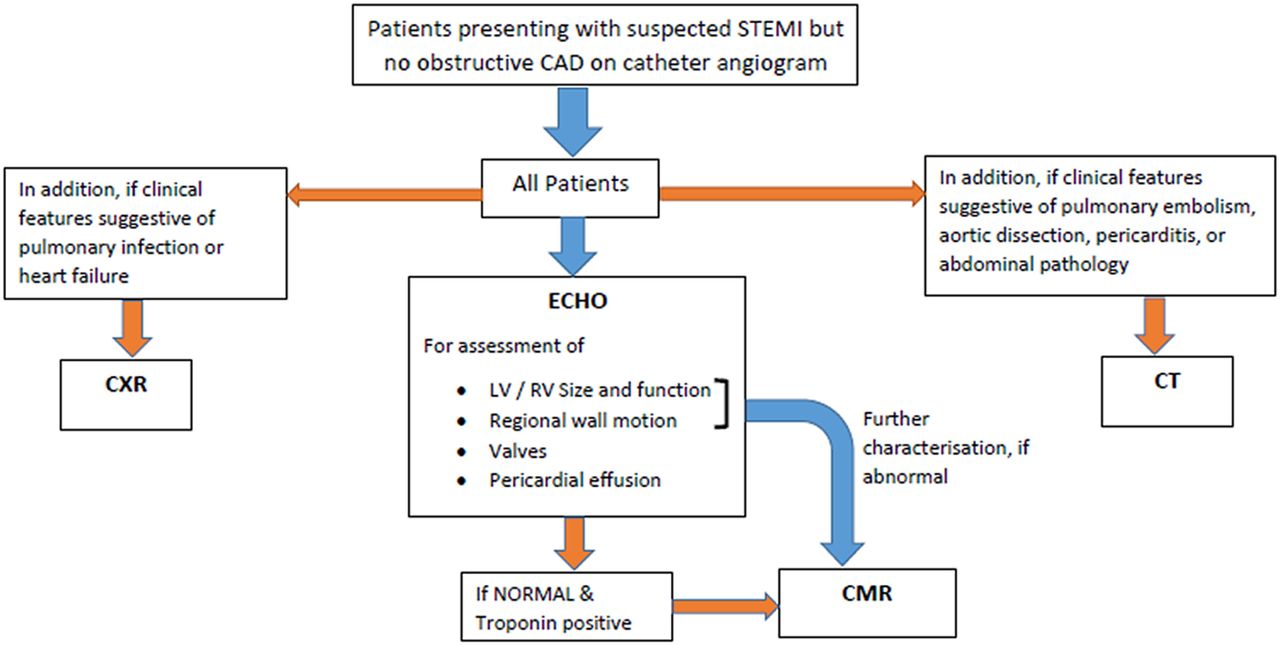
The limitations of our study pertain to its retrospective observational nature. Not all patients had every type of non-invasive imaging, which would be impractical in a clinical set up. This may have led to underestimation of prevalence for certain conditions such as myocarditis and infarction; non-invasive diagnosis of which is particularly dependent upon CMR. However, we think that this underestimation would be low as the troponin levels were much lower in patients who did not undergo CMR imaging. Also even though not all patients underwent an echocardiogram, a ventriculogram was performed in every patient during DCA. This allowed identification of LV dysfunction from TTCM and other causes. Therefore, our findings are representative of a real-life scenario rather than a unified controlled approach throughout a study group. Based on our experience, we suggest a pathway for non-invasive imaging in this patient cohort (figure 3).
Flow chart of suggested non-invasive imaging pathway in patients with suspected STEMI but no obstructive CAD. CAD, coronary artery disease; CXR, chest X-ray; CMR, cardiac magnetic resonance; ECHO, echocardiogram; STEMI, ST-elevation myocardial infarction.
In conclusion, just under half of patients with suspected STEMI with a culprit-free coronary angiogram and no history of previous CAD have an alternate diagnosis made with multimodality imaging. These diagnoses are heterogeneous involving both cardiac and non-cardiac abnormalities with a variable prognosis. The use of non-invasive imaging techniques in this group of patients is, therefore, helpful as otherwise important findings could be missed and go untreated.
Key messages
What is already known on this subject?
A proportion of patients with suspected ST-elevation myocardial infarction (STEMI) with no obstructive coronary artery disease have other diagnosis, which could be identified with one or more imaging techniques.
There is a high prevalence of myocarditis in this patient cohort as identified on cardiac MRI studies.
What might this study add?
About 13% of patients presenting with suspected STEMI may not have obstructive coronary artery disease. The most common alternative diagnosis in these patients as identified on multimodality imaging include cardiomyopathies (18%), myopericarditis (8.4%), myocardial infarction without coronary artery obstruction (4.9%), severe valve disease (4%), pulmonary embolism (1.2%) and type A aortic dissection (0.7%).
Some of the conditions have an adverse prognosis.
How might this impact on clinical practice?
Diagnosis of alternative diagnosis with imaging in patients with suspected STEMI can be important to implement appropriate management strategies.
Acknowledgments
The authors would like to gratefully acknowledge the statistical assistance provided by Paul Bassett and Winston Banya for this study.
References
Footnotes
Contributors TKM initiated and proposed the study. PPGR and LR collected the data. TKM, AG, SR-H and JW checked the data for their respective imaging modalities. TKM and LR performed the statistical analysis and initial draft of the manuscript. BA, TKM, LR, AG, SR-H, TK, JW and MD revised the manuscript. All authors read and approved the final manuscript.
Competing interests None declared.
Ethics approval Royal Brompton & Harefield NHS Foundation Trust.
Provenance and peer review Not commissioned; externally peer reviewed.