Article Text

Abstract
Introduction Nicotinic acid adenine dinucleotide phosphate (NAADP) is a calcium-mobilising messenger that acts via two-pore calcium channels (TPCs). NAADP releases calcium from a lysosome-related acidic compartment distinct from the endo-sarcoplasmic reticulum and has been identified as an important mediator of acute and chronic beta-adrenergic signalling in the heart. Genetic or pharmacological manipulation of NAADP signalling appears to be protective in pre-clinical models of cardiovascular diseases, such as ischaemia-reperfusion injury, arrhythmia and cardiac hypertrophy. Recent evidence has indicated that TPC2, rather than TPC1, mediates the effects of beta-adrenergic-evoked NAADP signalling, although whether this reflects differing subcellular distributions of TPC1 and TPC2 remains to be established.
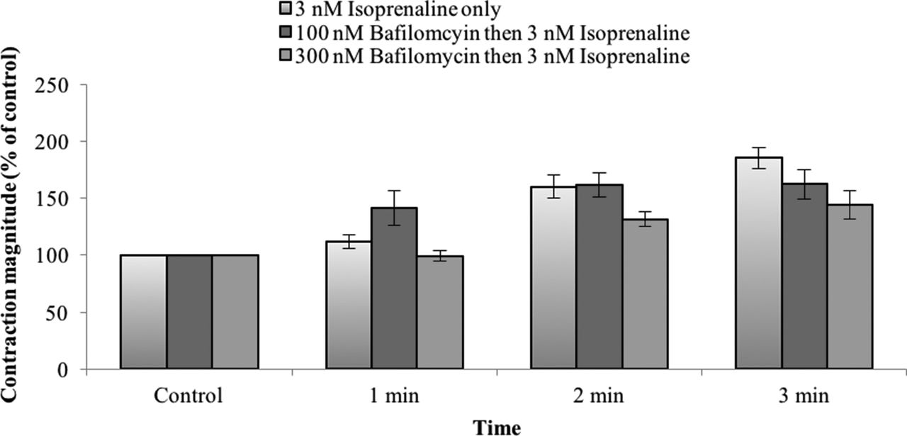
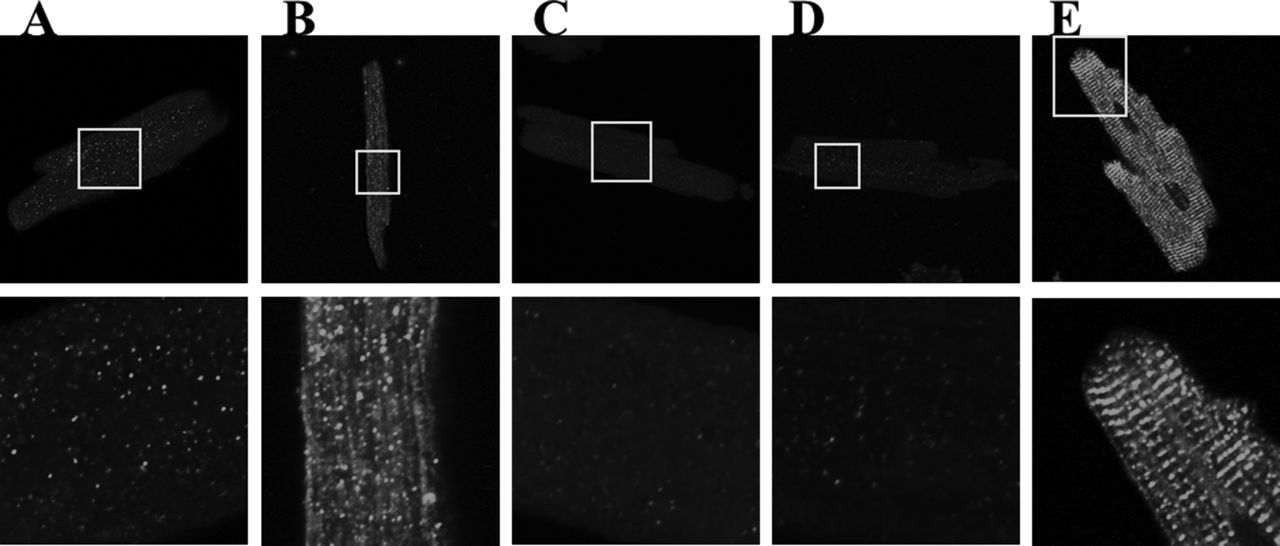
Methods Single ventricular cells were isolated enzymatically from guinea pig hearts and superfused with physiological saline solution (36°C). Cells were field-stimulated (3 ms pulse duration) to fire action potentials. Cell shortening was ?measured with an edge-detection system. To confirm the involvement of a lysosome-related acidic compartment in beta-adrenergic signalling, the effect of isoprenaline (a beta-adrenoceptor agonist) on cell contraction was investigated in the presence and absence of bafilomycin (a vacuolar H+-ATPase inhibitor that depletes acidic calcium stores). Data are ?presented as mean±SEM; comparisons were made using one-way ANOVA. In additional experiments, standard immunocytochemical techniques were employed to investigate the cellular distributions of TPC1, TPC2, transient receptor potential channel mucolipin-1 (TRP-ML1), lysosome-associated membrane protein-2 (LAMP2) and ryanodine receptor-2 (RyR2). Immunofluorescence was visualised using a Zeiss LSM 510 confocal microscope.
Results Isoprenaline (3 nM, 3 min) increased myocyte contraction (relative to control) by 86±9% (n=8) in the absence of bafilomycin, 62±13% (n=6) in the presence of 100 nM bafilomycin, and 44±12% (n=8) in the presence of 300 nM bafilomycin (Figure 1). The effect of bafilomycin tended closely toward statistical significance (p<0.06, one-way ANOVA). TPC1 and TPC2 displayed qualitatively similar patterns of punctate intracellular labelling. This punctate pattern resembled that for the lysosomal markers TRP-ML1 and LAMP2. In contrast, RyR2 labelling showed a striated appearance, consistent with the known organisation of the sarcoplasmic reticulum in cardiac myocytes (Figure 2).
Conclusions/implications This is the first direct immunocytochemical evidence in cardiac cells describing the localisation of TPC1 and TPC2. The subcellular distribution of TPC1 and TPC2 does not appear to explain their differing contributions to beta-adrenergic signalling. Our data are also consistent with a role for lysosomal calcium stores in the inotropic effect of beta-adrenoceptor agonists.

Effect of bafilomycin on the contractile response of cardiac ventricular myocytes to isoprenaline.

Representative images (immunocytochemistry) showing the subcellular distributions of: (A) TRP-ML1; (B) LAMP2; (C) TPC1; (D) TPC2; (E) RyR2.
- Beta Adrenergic Signalling
- Pharmacology
- Two Pore Channels