Article Text

Abstract
Introduction Ventricular thrombus is a serious complication in a subgroup of left ventricular dysfunction (LVD) patients because of the risk of embolisation. Thrombus occurs as a consequence of stasis, hypercoagulability and endothelial injury. There are no reliable predictors for which patients will develop thrombus. 4D flow CMR may allow insights into thrombus formation by intra-cardiac blood flow visualisation. We hypothesise that in patients with LV dysfunction and thrombus, compared to those without thrombus, the residual volume would constitute a similar proportion of the LV end diastolic volume (EDV) but possess less kinetic energy, thereby predisposing the blood to stasis and therefore thrombus formation.
Methods 100 participants (47 LV dysfunction but no thrombus (LVD) patients, 17 LV dysfunction and thrombus patients and 36 controls underwent CMR (Table 1)). LV flow was analysed as 4 components; direct flow, retained inflow, delayed ejection flow and residual volume. Each components volume was calculated in proportion to the EDV. The kinetic energy of the blood per millilitre was summed throughout the cardiac cycle and divided by the cycle length to calculate the average kinetic power. 25 controls, 14 LVD and 14 thrombus patients returned for an interval scan to assess the stability of flow parameters.
Results Both patient groups had significantly increased residual volume (LVD 50±10%, thrombus 51±12% vs 30±4% controls, p 0.001) and decreased direct flow (LVD 11±7%, thrombus 16±10% vs 38±4% controls, p 0.001). There was no difference between the 2 patient groups (Fig 1A). The average kinetic power of the residual volume was significantly higher in the LVD group (0.55±0.30 microJ/ml) compared to the thrombus group (0.38±0.16 microJ/ml, p 0.02) (Fig 1B). No difference between patient groups was seen for the direct flow average kinetic power (Fig 1C). 4D flow parameters were similar between visits with no significant change on paired t-tests (Table 2). The average kinetic power of the residual volume was higher in the LV dysfunction than thrombus group at visit 1 and 2, but failed to reach statistical significance with the smaller cohorts.
Discussion The residual volume blood of thrombus patients possessed less kinetic power than that of LV dysfunction patients with a well matched LV size, impairment and proportion of residual volume. Residual volume blood resides within the ventricle for at least two cardiac cycles; reduced movement of this blood component may be a contributing factor to stasis and hence thrombus formation. Similar results at interval studies propose that the residual volume average kinetic power is a temporally stable parameter.
This study suggests that the average kinetic power of the residual volume is a novel imaging biomarker which may allow identification, monitoring and potentially aid anticoagulation decisions in patients with LV dysfunction at higher risk of thrombus formation.
Clinical characteristics of controls, LV dysfunction and thrombus patients.
Results for conventional and 4D flow cardic remodelling parameters for participants who attended two study visits. There were no significant differences between visit 1 and visit 2 when paired t-tests were performed for each participant for the parameters shown above.

A) The volume of the four flow components by mean percentage ± SD in relation to the end diastolic volume. There is increased residual volume and decreased direct flow in the LV dysfunction and thrombus groups compared to controls. B) The residual volume summed kinetic energy per millilitre, normalised to duration of heart cycle for each group. The LV thrombus group have significantly lower residual volume summed kinetic energy, compared to the LV dysfunction group. C) Direct flow summed kinetic energy; no difference is seen between the LV dysfunction and thrombus groups.
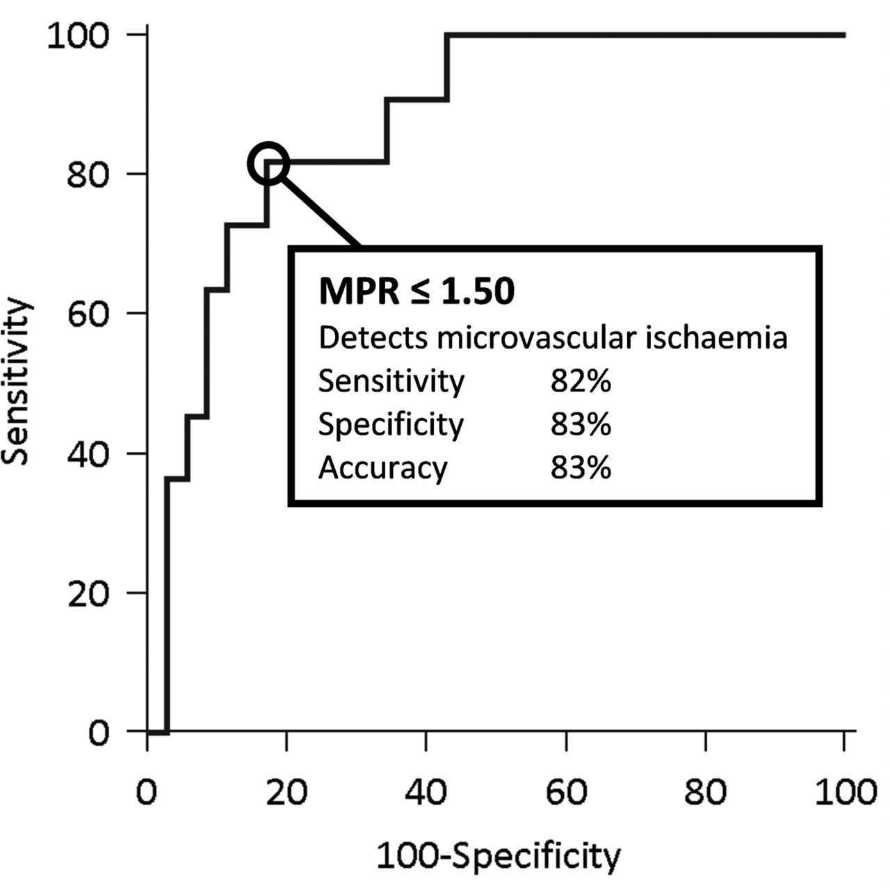
ROC analysis of MPR assessed using CMR for detecting microvascular ischaemia as defined by high IMR (> 40) in the absence of significant epicardial stenosis (FFR >0.8). True positives: high IMR>40; true negatives: normal IMR<20. Area under the curve 0.87,±0.06.
- 4D Flow
- Kinetic Energy
- Ventricular Thrombus