Article Text

Statistics from Altmetric.com
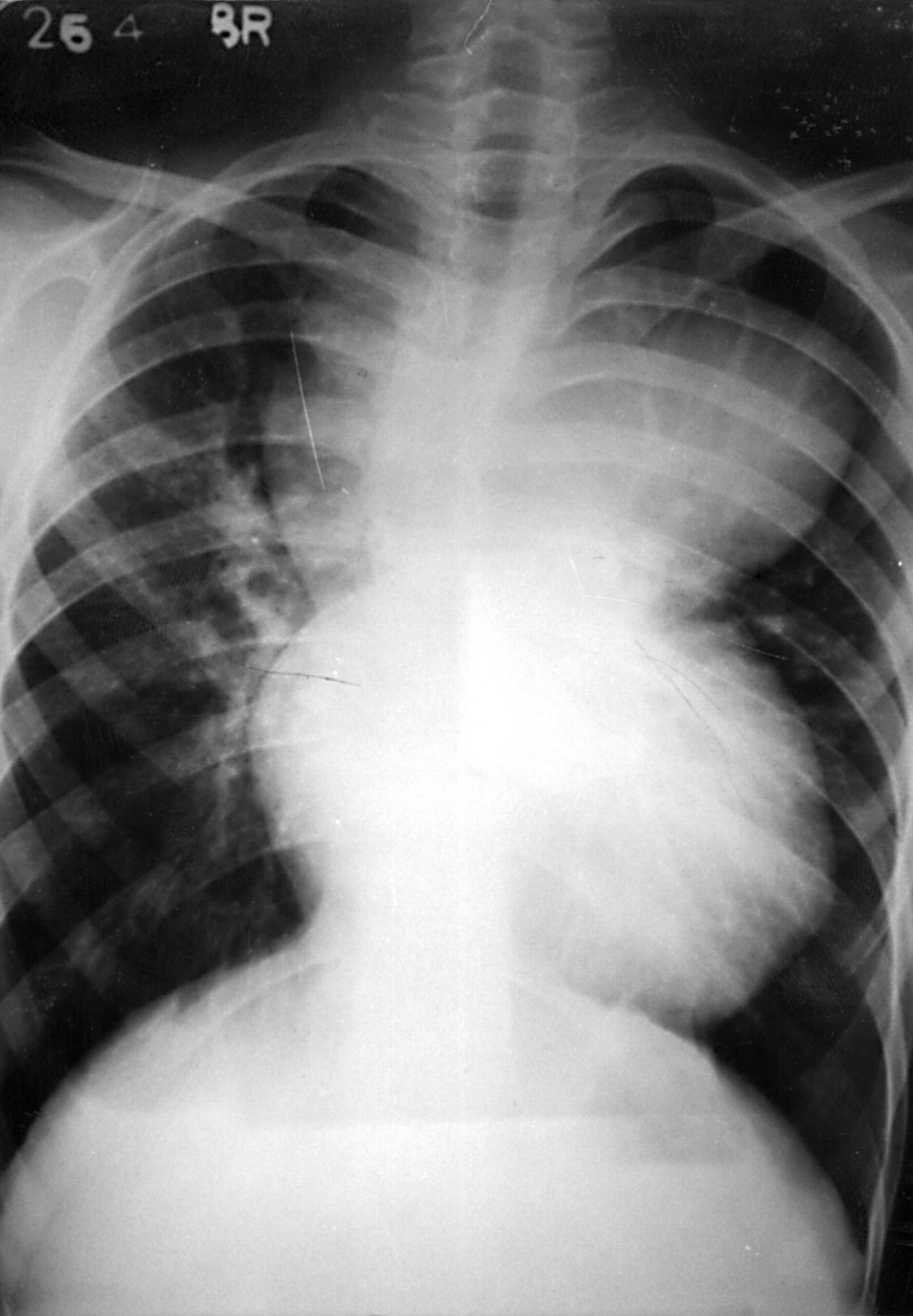
A 22 year old man had total anomalous pulmonary venous connection to the left vertical vein, which drained into the left brachiocephalic vein. He had cyanosis and clubbing. There was no cardiomegaly. Tricuspid flow murmur was absent. Chest radiography in the postero-anterior view showed a “figure of eight” or “snowman's” appearance. The upper half of the figure of eight was formed by the dilated superior vena cava on the right side, left brachiocephalic vein in the top, and the dilated vertical vein on the left side. The lower portion of the figure of eight was formed by the dilated right atrium and ventricle. He had dilated proximal pulmonary arteries with peripheral pruning of pulmonary vasculature, suggesting pulmonary vascular obstructive disease. No surgery was offered.