Article Text
Correction
Correction for vol. 84, p. 442

Statistics from Altmetric.com
Non-invasive coronary artery imaging with electron beam computed tomography and magnetic resonance imaging. PJ de Feyter, K Nieman, P van Ooijen, M Oudkerk.Heart2000;84 :442–8.
In this article fig 2 was incorrect, as certain elements were missing. The correct fig 2 is reproduced here.

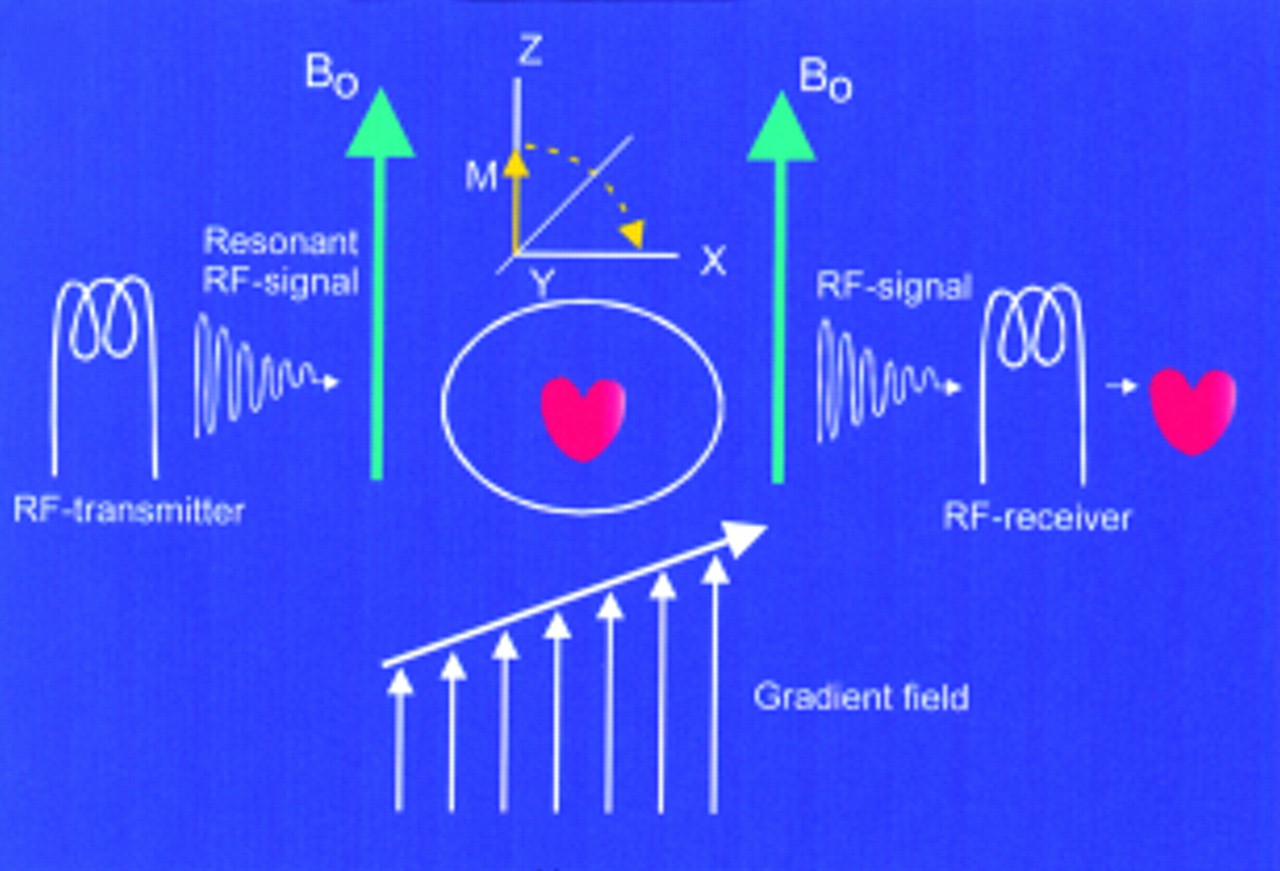
Figure 2
The patient (heart) is placed within a strong external magnetic field (Bo). The RF transmitter rotates the net tissue magnetisation in the transverse plane, and after termination, relaxation occurs which emits a signal detected by the RF receiver. The gradient coils produce a supplemental magnetic field gradient to allow precise location of the excited protons. The received signals have certain signal intensity (brightness) and location, both of which are processed to form the desired image.