
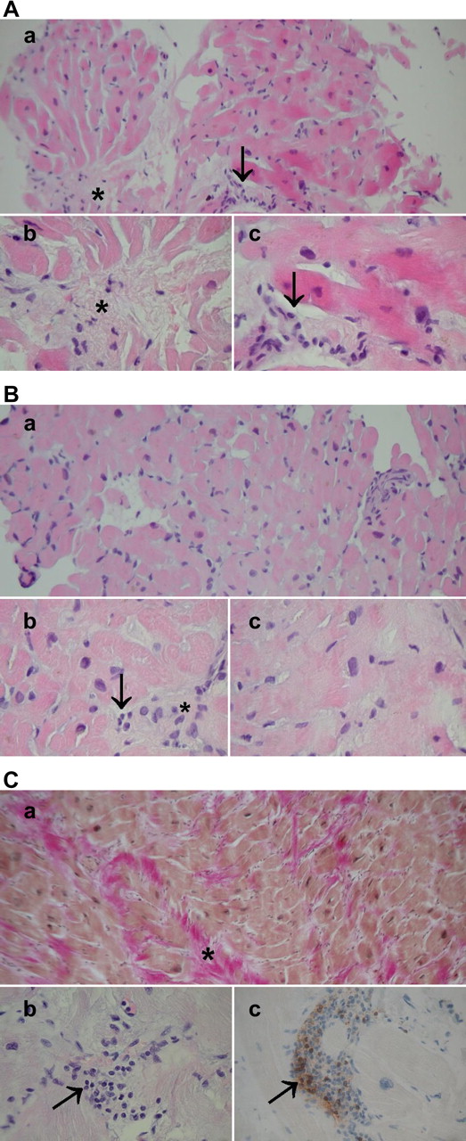
(A) Typical histopathology in an endomyocardial biopsy of a patient with hypertrophic cardiomyopathy (HCM). (a) A general view, haematoxylin and eosin (H&E) ×200; (b, c) moderate fibre disarray, interstitial fibrosis (asterisk), moderate myocyte size heterogeneity and hypertrophy and scattered mononuclear inflammatory cells (arrows), H&E ×400, ×630. (B) Mild histopathological findings in a patient with HCM. (a) A general view, H&E ×200; (b, c) mild fibre disarray, fibrosis (asterisk), myocyte hypertrophy and occasional mononuclear inflammatory cells (arrow), H&E ×400, ×630. (C) Severe HCM. (a) A general view: Weigert van Gieson staining highlights marked fibrosis (red, shown by asterisk), ×200; (b) multiple mononuclear inflammatory cells (arrow); (C) showing CD3 positivity in immunohistochemistry (staining in brown, arrow). Original magnification ×400. The patient had severe symptoms, marked left ventricular hypertrophy and inducible ventricular arrhythmia in ventricular stimulation. An intracardiac defibrillator was subsequently implanted.


Share this article
Click the icon of the social media platform on which you would like to share this article.


