Article Text

Abstract
Aims Clinical studies failed to prove convincingly efficiency of intravenous infusion of neseritide during heart failure and evidence suggested a pro-adrenergic action of B-type natriuretic peptide (BNP). However, subcutaneous BNP therapy was recently proposed in heart failure, thus raising new perspectives over what was considered as a promising treatment. We tested the efficiency of a combination of oral β1-adrenergic receptor blocker metoprolol and subcutaneous BNP infusion in decompensated heart failure.
Methods and results The effects of metoprolol or/and BNP were studied on cardiac remodelling, excitation–contraction coupling and arrhythmias in an experimental mouse model of ischaemic heart failure following postmyocardial infarction. We determined the cellular and molecular mechanisms involved in anti-remodelling and antiarrhythmic actions. As major findings, the combination was more effective than metoprolol alone in reversing cardiac remodelling and preventing ventricular arrhythmia. The association of the two molecules improved cardiac function, reduced hypertrophy and fibrosis, and corrected the heart rate, sympatho-vagal balance (low frequencies/high frequencies) and ECG parameters (P to R wave interval (PR), QRS duration, QTc intervals). It also improved altered Ca2+ cycling by normalising Ca2+-handling protein levels (S100A1, SERCA2a, RyR2), and prevented pro-arrhythmogenic Ca2+ waves derived from abnormal Ca2+ sparks in ventricular cardiomyocytes. Altogether these effects accounted for decreased occurrence of ventricular arrhythmias.
Conclusions Association of subcutaneous BNP and oral metoprolol appeared to be more effective than metoprolol alone. Breaking the deleterious loop linking BNP and sympathetic overdrive in heart failure could unmask the efficiency of BNP against deleterious damages in heart failure and bring a new potential approach against lethal arrhythmia during heart failure.
- ARRHYTHMIAS
- HEART FAILURE
- PHARMACOLOGY
- AUTONOMIC NERVOUS SYSTEM
Statistics from Altmetric.com
Introduction
A major source of preventable cardiac death in heart failure is the ventricular arrhythmia (VA).1 VA involves ventricular remodelling and alterations of Ca2+ homeostasis following chronic adrenergic overactivation.2 We showed that B-type natriuretic peptide (BNP) promotes Ca2+-dependent VA via a similar mechanism.3 The clinical advantages of the use of recombinant intravenous BNP nesiritide are also subject to debate despite its favourable haemodynamic effects.4 One explanation could be that intravenous BNP further compromises autonomic regulation in heart failure5 ,6 via a pro-adrenergic action unmasking its beneficial effects.3 ,7 ,8
Subcutaneous BNP administration has recently yielded promising results in systolic heart failure.9 We thus aimed to determine the effects of a combination of the selective β1-adrenergic blocker (BB) metoprolol associated with subcutaneous BNP infusion in a mouse model of decompensated heart failure. Until now, no study in human or animal had specifically tested this combination and investigated cellular and molecular mechanisms. This combination could associate the efficiency of the main antiarrhythmic in use, particularly in postmyocardial ischaemic heart failure with a reduced LV EF,10 and unmasks the beneficial antifibrotic, antiapoptotic and antihypertrophic properties of BNP by abolishing the BNP-associated adrenergic effects.
We showed that the combination was more effective than metoprolol or BNP alone in preventing cardiac remodelling and VA, with better benefits on cardiac morphology, function and Ca2+ homeostasis.
Materials and methods
Please refer online supplement for methods which is available online.
Study design
Procedures conformed to European Parliament Directive 2010/63/EU and Council on the protection of animals were approved by our institutional animal research committee (CE-LR-0714). Seven-week-old male C57Bl/6 mice (Janvier, France) were randomly assigned to the following groups: (1) postmyocardial infarction (PMI); (2) PMI treated with BNP (BNP-PMI); (3) PMI treated with metoprolol (BB-PMI); (4) PMI treated with metoprolol and BNP (BB-BNP-PMI); and (5) sham-operated mice (Shams). For PMI, the coronary artery was ligated 1–2 mm beyond its emergence from the left atrium, under anaesthesia and cardiac monitoring (2% isoflurane/O2, Aerrane, Baxter). Buprenorphine (0.3 mg/mL) was injected for postoperative analgesia.3 Metoprolol (Sigma-Aldrich, 100 mg/kg/day) was administered in the drinking water. The mouse BNP (14-5-30A, American Peptide, USA) was subcutaneously administered at 0.03 µg/kg/min for 14 days (Alzet-1002 osmotic pumps).3 Following in vivo investigations, heart was explanted after cervical dislocation for single-cell experiments. The time sequence of the protocol is shown in figure 1.
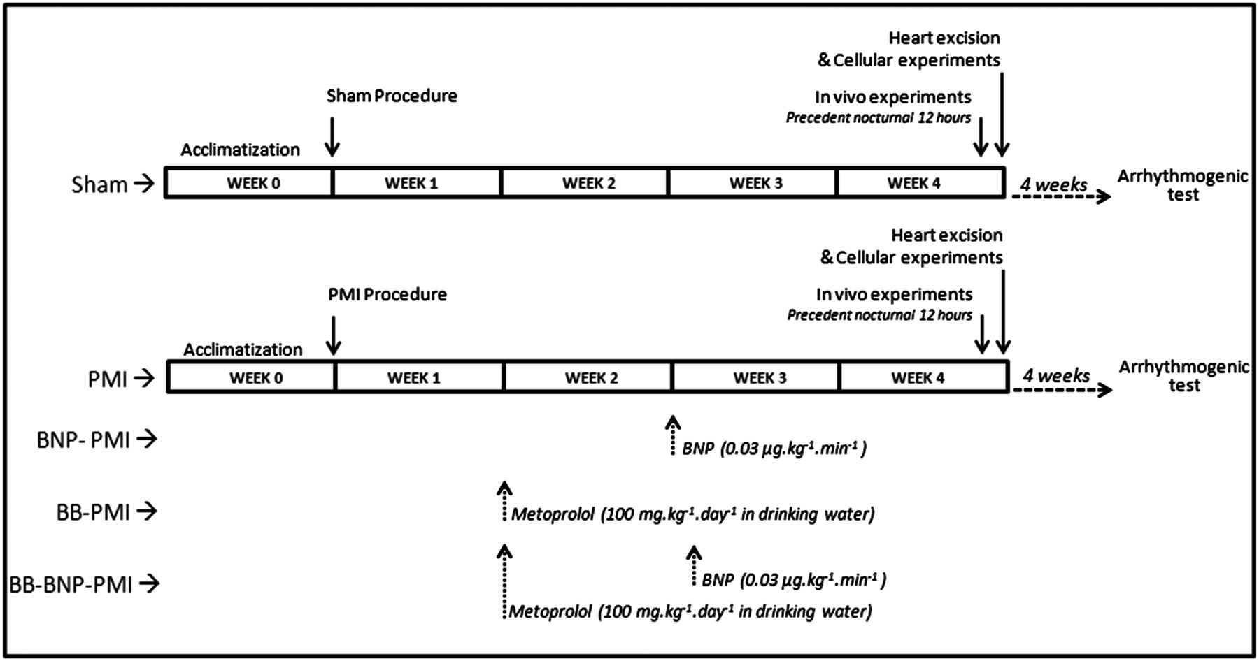
Time sequence of experimental procedure.
In vivo analysis
Telemetric ECGs were recorded (DSI, USA) and analysed in respect of the Lambeth conventions. Heart rate variability, PR, QRS, corrected QT (QTc) intervals, short term variability of QT (QTSTV) and spontaneous arrhythmias were analysed (EMKA, France). To test the contribution of long term anti-remodelling effect of treatments on arrhythmogenic susceptibility, the β-adrenergic catecholamine isoproterenol (2.5 mg/kg intraperitoneal) was injected during and 4 weeks after the treatment. The triggering of sustained ventricular tachycardia (SVT) was monitored. At the same time-points, systolic, diastolic and mean arterial blood pressure were measured with a tail-cuff and pulse transducer (ML125/M NIBP System, ADInstruments, UK) in triplicate in conscious mice.
LV mass, LV shortening fraction, end-diastolic and end-systolic LV dimensions were measured by echocardiography (Vivid7Pro, GE Medical Systems, USA).3 Survival throughout experimental protocol was followed (see online supplementary table S1 and S4).
Autopsy and heart excision
Autopsies were performed to verify pleural effusion and lungs congestion. The heart and lungs were excised and weighed, and the heart weight index determined (heart weight/body weight). Interstitial fibrosis was measured in 10 μm thick transverse sections of hearts in the peri-infarcted area (H&E and Sirius red staining). Results indicated the area of Sirius red-stained tissue (percentage of total area of myocardial tissue).
RNA extraction and RT-qPCR
Total RNA was extracted from LV tissue using TRIzol, and treated with DNase I at 37°C for 30 min. cDNA was synthesised using superscript II reverse transcriptase (Invitrogen, France). RT-qPCR was performed for myocardin-related transcription factor A (MRTF-A), serum response factor (SRF), Na+-Ca2+ exchanger (NCX1), sarcoplasmic reticulum (SR) Ca2+-ATPase (SERCA2a) and Ca2+-binding protein S100a1 in duplicate (LightCycler, Roche, France) and normalised to GAPDH (eight mice/group).
Ca2+ handling and patch-clamp
Experiments were performed on freshly isolated LV myocytes.3 Cardiomyocytes were loaded with Indo-1AM (10 µM, Invitrogen, France) and field-stimulated at 1.0 Hz with 1 ms current pulses (IonOptix system, USA).3 Indo-1 fluorescence emitted at 405 (F405) and 480 nm (F480) were recorded to estimate intracellular Ca2+ level (F405 to F480 ratio) during a 30 s pacing period, followed by a 30 s rest period. Diastolic Ca2+ level, Ca2+ transient decay time (tau) and percentage of cells developing spontaneous Ca2+ waves were quantified during the rest period. Ca2+ sparks (frequency, amplitude and spatiotemporal characteristics) were recorded by following variations of fluorescence at 505 nm (ΔF) divided by initial fluorescence at 505 nm (F0) (ΔF/F0, Fluo-4AM, 5 µM, 1.5 ms/line; LSM510 Zeiss confocal microscope, 63X water-immersion objective, NA: 1.2). Cell volume was estimated using Z-stack (x-y projection, front view) image acquisition.3 Electrophysiological profiles of cardiomyocytes were investigated by current-clamp (action potential (AP)) and voltage-clamp approaches (ICa,L, IK) using the patch-clamp technique.
Ca2+-handling proteins
LV were homogenised into lysis buffer (0.3% CHAPS, 1 µg/mL leupeptin, 1 µg/mL pepstatin and, in mM; HEPES 20, KCl 40, DTT 1, PMSF 1, EDTA 1, pH7.4) and centrifugated (6000×g, 5 min). After protein quantification (DC Protein Assay, Bio-Rad), total proteins (50 mg) were loaded on SDS-PAGE and transferred on nitrocellulose membrane (GE Healthcare). The membranes were blocked (Thermoscientific) and incubated with primary antibodies at 4°C overnight: SERCA2a (1:5000) (A010-20, Badrilla, UK), NCX1 (1:1000) (R3F1, Swant), ryanodine receptor RyR2 (1:1000) (Covalab, France) and PhosphoSer2808-RyR2 (1:1000) (A010-30, Badrilla), phospholamban (1:20 000) (A010-14, Badrilla) and PhosphoSer16-PLB (1:5000) (A010-12, Badrilla), and S100A1 (1:2500) (SP5355P, Acris antibodies, Germany). After incubation with secondary antibody 800 nm (1:30 000): antirabbit (SERCA2a, PhosphoPLB, RyR2, PhosphoRyR2, S100A1) or antimouse (NCX1, PLB, GAPDH), membranes were washed and scanned (Odyssey, LI-COR Biosciences). Results were expressed relative to GAPDH (1:60 000) (ab8245, Abcam).
Statistical analysis
All data are reported as means±SD (mean±SE for patch-clamp experiments). Statistical analyses were performed using GraphPad Prism and Origin Softwares. One-way ANOVA for multiple comparisons was used, followed by a parametric t test with Bonferroni’s correction. Percentage data were analysed by a χ2 test. A p value of 0.05 or less indicates a statistically significant difference.
Results
BNP reduced fibrosis and metoprolol improved cardiac remodelling
MI mice exhibited heart failure with cardiac hypertrophy, increased LV end-diastolic dimensions, decreased LV shortening fraction and interstitial fibrosis (table 1; see online supplementary figure S1). The systolic, diastolic and mean arterial blood pressures were decreased (see online supplementary table S1). BNP did not improve morpho-functional remodelling after MI but reduced interstitial fibrosis (table 1, see online supplementary figure S1). In contrast, metoprolol reversed the increases of the heart weight index and LV end-diastolic dimensions in PMI, while pleural effusion and lung congestion worsened (table 1). Consistently, metoprolol reduced cell hypertrophy as confirmed by the quantification of MRTF-A and SRF mRNAs (see online supplementary table S2).
Morphological and histological parameters
BB+BNP normalised morpho-functional parameters
The BB+BNP combination was far more effective than metoprolol on cardiac hypertrophy, pleural effusion, lung congestion and LV end-diastolic dimensions (table 1). The combination reduced fibrosis and hypertrophy more efficiently than BB (table 1, see online supplementary figure S1). BB and BB+BNP had no detrimental effect on the systolic blood pressure during treatment and conferred a persistent beneficial effect over time (see online supplementary table S1).
BB+BNP corrected rhythm disturbances better than monotherapy
PMI mice presented higher heart rate, and prolonged QRS and QTc intervals (table 2) when compared with Shams. The typical collapsed low frequencies to high frequencies ratio indicated that the sympathetic system was overactivated, as observed in heart failure.3 Moreover, the ventricular repolarisation instability expressed as the QTSTV was increased (table 2, see online supplementary figure S2). All these parameters are well-recognised prognostic markers for VA. PMI mice displayed a higher incidence of VA than Shams. BB and BB+BNP both normalised the heart rate and improved heart rate variability in PMI (table 2). The QTSTV and the number of VA were also diminished (table 2, see online supplementary figure S2). Similar results have been reported in heart failure patients under β-blockers.11 Overall, BB+BNP was more effective than BB in correcting the heart rate variability and QTSTV and in reducing VA (table 2).
ECG analysis
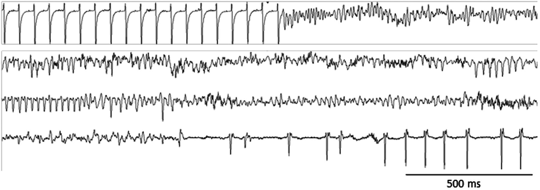
Since catecholamines are a potent trigger of VA, mice were challenged with isoproterenol at two time-points. (1) During treatment, 33% PMI and 66% BNP-PMI mice developed SVT whereas Sham did not (table 2). BB and BB+BNP prevented SVT. (2) When all treatments were stopped, 58% of PMI developed SVT, and 33% triggered ventricular fibrillation (figure 2, table 2). In the BNP-PMI group, 60% of mice developed SVT, and 33% developed ventricular fibrillation followed by cardiac death (4/12, p<0.05, χ2 test vs PMI). The BB+BNP therapy remained highly beneficial in successfully preventing SVT (8%) (χ2 test, p<0.05, table 2).

Arrhythmic events. Typical sustained ventricular tachycardia and ventricular fibrillation in postmyocardial infarction mice during isoproterenol challenge (2.5 mg/kg intraperitoneal).
BB+BNP normalised intracellular Ca2+ homeostasis
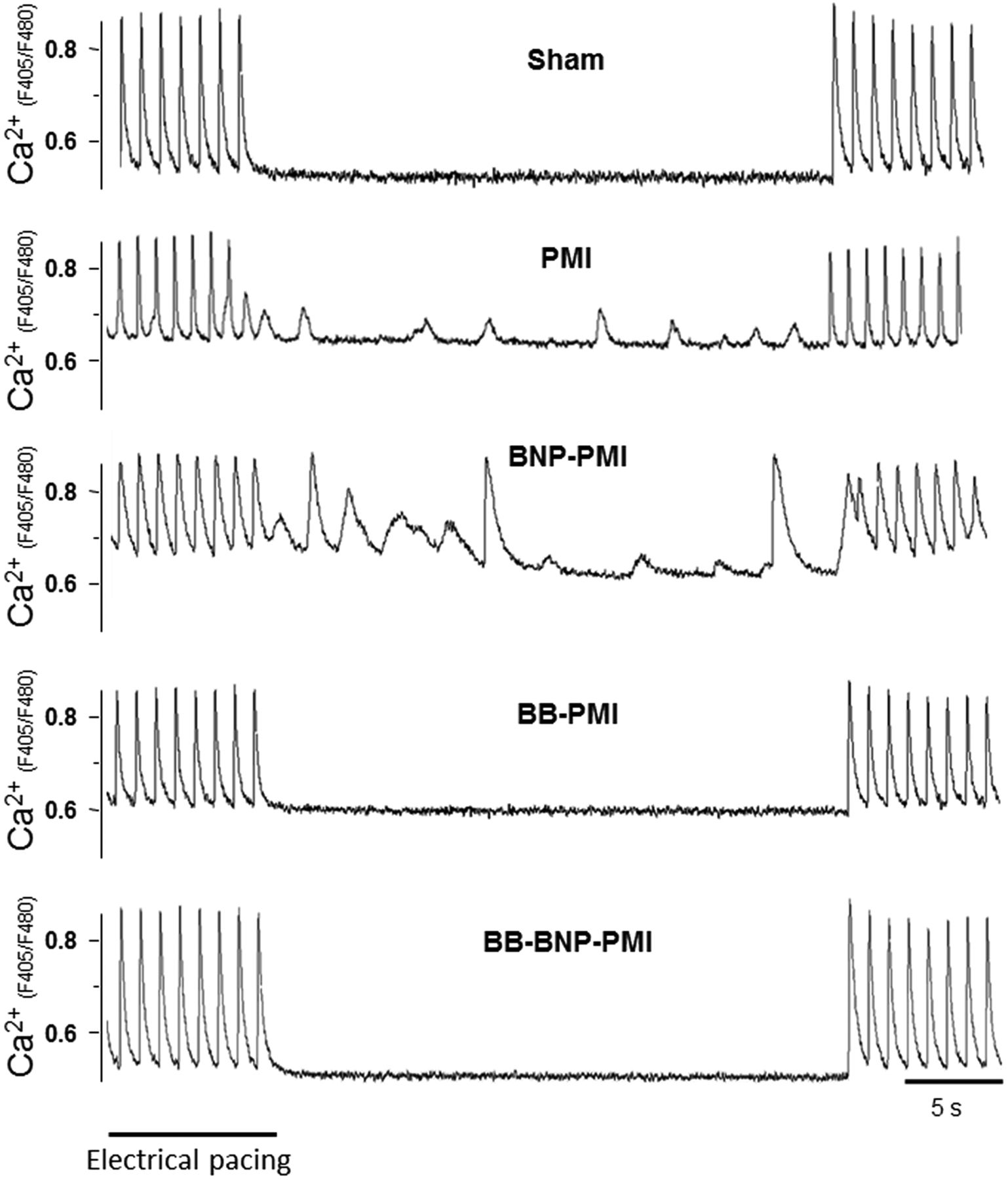
VA could originate from AP lengthening and/or disturbed Ca2+ handling. Whereas AP shape was altered in PMI and BNP-PMI, AP duration and underlying currents (Ca2+ and K+) were comparable between these two groups (figure 3, see online supplementary table S3). So we looked for differences in intracellular Ca2+ level that could trigger afterdepolarisations (figure 3B). In PMI, Ca2+ transient was altered with smaller amplitude and slower decay kinetics together with reduced SR-Ca2+ content and higher diastolic Ca2+ levels than in Shams (figure 4, table 3). These changes accounted for, respectively, the decrease in sarcomeres shortening and the triggering of spontaneous irregular Ca2+ waves (figure 4, table 3). Chronic BNP worsened all these alterations and irregular Ca2+ waves persisted (figure 4, table 3). In contrast, both BB and BB+BNP improved Ca2+ homeostasis and reduced the number of pro-arrhythmogenic waves (table 3). Nevertheless, BB+BNP was more effective than BB alone to reduce Ca2+ sparks frequency and occurrence of Ca2+ waves (figure 4 and figure 5; table 3). Thus, the beneficial effects of BB+BNP on Ca2+ cycling could explain its antiarrhythmic advantage.
Intracellular Ca2+ signalling in LV cardiomyocytes, Ca2+ transients and cell shortening

Cellular electrophysiological profiles. Current clamp studies: (A) Representative action potentials were recorded from LV cardiomyocytes isolated from Sham, postmyocardial infarction (PMI) and B-type natriuretic peptide (BNP)-PMI mice. (B) Typical early afterdepolarisations obtained in BNP-PMI mice. Voltage clamp studies: Ionic currents in Sham (open square), PMI (filled square) and BNP-PMI mice (filled circle). (C) Mean±SE current/voltage relationships of the total voltage-gated K+ currents (IKpeak), (D) transient outward K+ current (Ito,F), (E) IKslow, (F) IK1 and (G) ICa,L (n=13–22 cells). **p<0.01 versus Sham.

Ca2+ transients. BB+B-type natriuretic peptide (BNP) decreased cellular susceptibility to arrhythmia by normalising Ca2+ homeostasis. Indo1-AM fluorescence ratio at 405 and 480 nm (F405 to F480) reflected intracellular Ca2+ level variations during and after electrical pacing in LV cardiomyocytes. Postmyocardial infarction (PMI) and BNP-PMI developed abnormal spontaneous activities during non-stimulated period. BB, β1-adrenergic blocker.

Ca2+ sparks. BB+B-type natriuretic peptide (BNP) prevented Ca2+ leakage from RyR2. Representative variations of fluorescence at 505 nm (F505) during line scan acquisition in Fluo-4 AM-loaded cardiomyocytes from Sham, postmyocardial infarction (PMI), BNP-PMI, BB-PMI and BB-BNP-PMI animals. Each sporadic elevation of fluorescence (indicated by white arrows) represents a Ca2+ spark due to spontaneous activation of ryanodine receptors. Whereas Sham cells presented few Ca2+ sparks, PMI presented an increased sparks frequency, reflecting a severe Ca2+ leakage from the reticulum sarcoplasmic. BB and, to a larger extent, BB-BNP reduced Ca2+ sparks frequency. BB, β1-adrenergic blocker.
BB+BNP normalised changes in Ca2+-handling proteins
The alterations of Ca2+ homeostasis in heart failure resulted from modifications of Ca2+-handling proteins as we observed for SERCA2a, S100A1 and NCX in PMI (table 4, see online supplementary figure S3). In addition, PLB phosphorylation (Ser16) was decreased while RyR2 phosphorylation (Ser2808) was increased (table 4). BNP had no major effect on SERCA2a, NCX, or the phosphorylation of PLB and RyR2, but it further reduced S100A1 expression (table 4, see online supplementary figure S3). This additional reduction of S100A1 accounted for the higher Ca2+ sparks frequency in BNP-PMI.12 ,13 In contrast, BB+BNP normalised SERCA2a, NCX and S100A1 expression, and the phosphorylation of PLB and RyR2 (table 4, see online supplementary figure S3). The benefits of BB were therefore enhanced by BNP, with an additional decrease in NCX1 expression and the P-RyR2 to RyR2 ratio, and normalisation of PLB phosphorylation. These results accounted for the improved control of Ca2+ leakage and inotropy, and prevention of irregular Ca2+ waves.
Ca2+-handling proteins
Discussion
β-Blockers are commonly used as first-line treatment after MI and heart failure, with unquestionable benefits on mortality.14 Here, we conclude that a combination of the selective BB metoprolol with subcutaneous BNP is more effective to prevent cardiac remodelling than metoprolol alone in decompensated heart failure. We also provide novel insights into how the combination prevents Ca2+-handling alterations and subsequent morbid arrhythmias (figure 6).

Beneficial effect of combination therapy in the concept of Coumel’s triangle of arrhythmogenesis.30 In addition to its beneficial effects on the ‘substrate’ (cardiac remodelling) and the ‘trigger’ (Ca2+ cycling), β1-adrenergic receptor antagonism also abolishes the adverse consequences of B-type natriuretic peptide (BNP) on the ‘modulator’ (sympathetic system overactivation).
Higher benefits of BB+BNP on cardiac remodelling
Favourable effects of BNP have been reported in hypertension-induced heart failure,15 but they are still debated in postischaemic heart failure.16–18 We showed that subcutaneous BNP was beneficial when combined with metoprolol in ischaemic heart failure. Furthermore, the overall benefit was independent of heart rate and haemodynamic changes. In particular, no severe hypotension was observed.4 The combination clearly associated the antifibrotic effect of BNP and the anti-remodelling action of metoprolol that account for efficient antiarrhythmic properties. Even if in the ASCEND-HF trial almost 60% of patients received β-blocker therapy and/or ACE inhibitors with intravenous BNP infusion,4 it is the first time that a study specifically addressed the utility to use subcutaneous BNP associated with oral metoprolol in heart failure and reveals beneficial effects. In the ASCEND-HF trial no benefit was observed. The discrepancy may reflect differences of dosage, administration route (intravenous 0.01 μg/kg/min for 24 h or more for up to 7 days in acute decompensated heart failure vs subcutaneous 0.03 μg/kg/min for 15 days in established chronic heart failure) and of model.
The normalisation of the sympatho-vagal balance by the combination was a key mechanism contributing to its overall therapeutic effect. Metoprolol not only provided its well-known benefits, but it broke the deleterious loop linking BNP and sympathetic overdrive.3 Indeed, BNP promotes adrenergic signalling through two distinct pathways. BNP induces norepinephrine release from sympathetic cardiac neurons via protein kinase G-induced inhibition of PDE3-mediated cAMP hydrolysis,7 which is likely to offset its desirable effects.7 ,8 ,19 BNP also inhibits PDE3 through the activation of NPR-B,8 ,20 which is the predominant natriuretic peptide receptor in failing hearts.21 Moreover, whereas low doses of nesiritide have beneficial effect on autonomic nervous system, high doses of intravenous BNP could induce prolonged hypotension and activate the sympathetic system.5 ,22 Altogether, these effects could account for the increased propensity of BNP-PMI mice to develop catecholamines-induced VT, fibrillation and death. Such pro-arrhythmic effect was observed in patients in whom high doses of intravenous nesiritide induced a minor increase of VA (ventricular tachcardia (VT), couplets and triplets).23 However, in this study, the antiarrhythmic treatment of patients may have prevented the effect of parenteral vasoactive therapy on the occurrence of VAs.23 In addition, non-sustained VT was also reported during study drug infusion of nesiritide in three patients receiving a high dose of nesiritide (0.03 µg/kg/min) in clinical trial.22 We therefore propose that the combination retains the beneficial effects of subcutaneous BNP but attenuates the deleterious consequences mediated by β1-adrenergic pathway. In line, the combination reduced fibrosis and corrected the QRS lengthening and QT dispersion, which both correlate with a lowered risk of developing VA.11 ,24 Importantly, this antiarrhythmic benefit persisted over time, and lasted longer than that of metoprolol alone, which is in line with a protective long-term anti-remodelling effect.
Mechanisms of higher benefits of BB+BNP combination therapy
Alterations in Ca2+ homeostasis are responsible for excitation–contraction coupling defects and VA.2 Aberrant ryanodine receptor (RyR) opening in diastole, observed functionally as the abnormal occurrence of Ca2+ sparks, generates spontaneous irregular Ca2+ waves involved in the triggering of VA/SVT.25 ,26 Increase in Ca2+ sparks frequency could result from increased cytosolic Ca2+ level due to a blunted SERCA2a activity, associated with a modulation of the intrinsic properties of the RyR2 complex (see online supplement). Sympathetic overdrive, leading to SR-Ca2+ leakage, may be involved since high diastolic Ca2+ levels could also participate in the triggering of afterdepolarisations. The combination effectively reduced SR-Ca2+ leakage, and improved SR-Ca2+ load and transient amplitude by normalising proteins alterations which accounted for the maintained cell contraction and the antiarrhythmic properties. Interestingly, the combination restored baseline levels of S100A1 and increased SR-Ca2+ content. Restoration of S100A1 expression, as a Ca2+-dependent molecular inotrope regulating cardiac SR-Ca2+ cycling, was suggested to treat heart failure.27
Clinical implication
The most important finding of this study is that association of oral metoprolol with subcutaneous BNP infusion is more effective than monotherapy with the β-blocker in reducing ventricular remodelling and VA following MI. The interest of combinations, elevating circulating plasma concentration of natriuretic peptides associated with classical drugs, has been recently highlighted.28 ,29 For example, neutral endopeptidase inhibitors have per se limited clinical benefice. However, their association with ACE or angiotensin receptor antagonists is promising despite limitations such as incidence of multilevel potentially life-threatening angioedema.28 Our results may challenge this innovative concept because β1-adrenergic receptor antagonisation abolished the main adverse effects of BNP (figure 6). The therapeutic benefits resulted from an improved balance of the BNP and adrenergic systems, and from mechanisms intrinsic to the two pathways at the cardiomyocytes level. These promising results obtained in an experimental model of ischaemic heart failure warrant further evaluation and optimisation (dose) in humans.
Key messages
-
What is already known on this subject
-
Chronic B-type natriuretic peptide (BNP) administration alters excitation–contraction coupling (Ca2+ signalling) in mouse ventricular cardiomyocytes, which triggers ventricular arrhythmia through activation of the sympathetic system. However, chronic subcutaneous BNP improves cardiac function and avoids the severe hypotensive effect of BNP.
-
What this study adds
-
We determined the effects of a combination of the selective β1-adrenergic blocker metoprolol associated with subcutaneous BNP infusion in a mouse model of decompensated heart failure. Until now, no study in humans or animals had specifically tested this combination and investigated both cellular and molecular mechanisms. We showed that metoprolol unmasks beneficial effects of BNP. The combination of the two molecules reduced the occurrence of both spontaneous and catecholamines-induced ventricular tachycardia in postischaemic heart failure.
References
Supplementary materials
Supplementary Data
This web only file has been produced by the BMJ Publishing Group from an electronic file supplied by the author(s) and has not been edited for content.
Files in this Data Supplement:
- Data supplement 1 - Online supplement
- Data supplement 2 - Online figures
- Data supplement 3 - Online tableS1
- Data supplement 4 - Online tableS2
- Data supplement 5 - Online tableS3
- Data supplement 6 - Online tableS4
Footnotes
-
SK and SR contributed equally to this study.
-
Contributors (1) Conception and design or analysis and interpretation of data, or both, in addition to experimental work: JT, SK, SR, JR, CC, AG, JF, FA. (2) Drafting of the manuscript or revising it critically for important intellectual content: JT DB, J-Y LG, AL, SR. (3) Final approval of the manuscript submitted: JT, SR.
-
Funding This work was supported by Fondation de France (Project PepNaRhythm, N° 2068001722) and INSERM. JT, SR, JF and AL hold CNRS positions.
-
Competing interests None.
-
Provenance and peer review Not commissioned; externally peer reviewed.