Article Text

Abstract
Objectives To evaluate the diagnostic and prognostic benefits of CT coronary angiography (CTCA) using the 2016 National Institute for Health and Care Excellence (NICE) guidelines for the assessment of suspected stable angina.
Methods Post hoc analysis of the Scottish COmputed Tomography of the HEART (SCOT-HEART) trial of 4146 participants with suspected angina randomised to CTCA. Patients were dichotomised into NICE guideline-defined possible angina and non-anginal presentations. Primary (diagnostic) endpoint was diagnostic certainty of angina at 6 weeks and prognostic endpoint comprised fatal and non-fatal myocardial infarction (MI).
Results In 3770 eligible participants, CTCA increased diagnostic certainty more in those with possible angina (relative risk (RR) 2.22 (95% CI 1.91 to 2.60), p<0.001) than those with non-anginal symptoms (RR 1.30 (1.11 to 1.53), p=0.002; pinteraction <0.001). In the possible angina cohort, CTCA did not change rates of invasive angiography (p=0.481) but markedly reduced rates of normal coronary angiography (HR 0.32 (0.19 to 0.52), p<0.001). In the non-anginal cohort, rates of invasive angiography increased (HR 1.82 (1.13 to 2.92), p=0.014) without reducing rates of normal coronary angiography (HR 0.78 (0.30 to 2.05), p=0.622). At 3.2 years of follow-up, fatal or non-fatal MI was reduced in patients with possible angina (3.2% to 1.9%%; HR 0.58 (0.34 to 0.99), p=0.045) but not in those with non-anginal symptoms (HR 0.65 (0.25 to 1.69), p=0.379).
Conclusions NICE-guided patient selection maximises the benefits of CTCA on diagnostic certainty, use of invasive coronary angiography and reductions in fatal and non-fatal myocardial infarction. Patients with non-anginal chest pain derive minimal benefit from CTCA and increase the rates of invasive investigation.
Trial registration number ClinicalTrials.gov: NCT01149590;post results.
- Cardiac computer tomographic (CT) imaging
- Coronary artery disease
This is an Open Access article distributed in accordance with the Creative Commons Attribution Non Commercial (CC BY-NC 4.0) license, which permits others to distribute, remix, adapt, build upon this work non-commercially, and license their derivative works on different terms, provided the original work is properly cited and the use is non-commercial. See: http://creativecommons.org/licenses/by-nc/4.0/
Statistics from Altmetric.com
Introduction
Chest pain is a common symptom within the community and is responsible for at least 1% of all presentations to general practitioners.1 2 It is frequently a cause of concern for patients and clinicians alike, with both eager to identify or exclude potentially serious underlying conditions. Although stable coronary heart disease (CHD) is responsible for only 10% of such presentations,3 the resources required to exclude this diagnosis have important public health implications. Clearly, it is in the interests of all parties to develop an efficient and effective strategy for the assessment and management of these symptoms.
In response to this clinical need, the National Institute of Health and Care Excellence (NICE) first published an innovative guideline (CG95) on the assessment of chest pain of recent onset in 2010.4 This publication encouraged a systematic approach to determining the pre-test probability of CHD using routinely available clinical features. Furthermore, it established explicit thresholds of risk, below which additional investigation for coronary disease was regarded as unnecessary and unhelpful. These changes were met with initial scepticism related to the potential for increased costs,5 underestimation of disease prevalence6 and frequent pathway non-adherence arising from the unacceptability of discharging low risk patients without further investigation.7 Fortunately, the ensuing years have allayed many of these concerns with more recent studies demonstrating an association between increasing guideline compliance, reduced diagnostic testing and lower overall expenditure.8–10
In November 2016, the NICE guideline was updated with two important changes made to the recommendations.11 First, the abolition of the explicit approach to the estimation of pretest probability with patients now selected for further testing based simply on the description of chest pain or the presence of an abnormal resting ECG. Second, driven by technological developments and cost reductions, non-invasive testing for myocardial ischaemia has been replaced with broad indications for CT coronary angiography (CTCA). However, concerns have already been raised that this new strategy has not been adequately assessed and should be tested in a clinical trial.12 We therefore aimed to determine the diagnostic and prognostic implications of these changes to the assessment of patients presenting with stable chest pain of recent onset using the Scottish COmputed Tomography of the HEART (SCOT-HEART) trial dataset.
Methods
Study design and population
The SCOT-HEART study is a prospective, multicentre, randomised controlled trial investigating the role of CTCA in patients referred to a specialist clinic with suspected angina due to CHD. The study design13 and principal findings14 have previously been reported. The study population comprised individuals without a documented history of prior CHD referred for assessment of suspected stable angina of recent onset who were randomised 1:1 to CTCA plus standard care or standard care alone (see online supplementary material). Chest pain symptoms were defined as typical angina, atypical angina or non-anginal according to established criteria (see online supplementary table 1).11 An abnormal resting ECG was determined by the presence of any of the following: pathological Q waves, left bundle branch block or either ST segment or T wave abnormalities. As per the 2016 NICE guideline recommendations, participants were categorised into two groups: those with non-anginal chest pain and a normal ECG (non-anginal cohort) and those with either typical or atypical chest pain, or non-anginal chest pain and an abnormal ECG (possible angina cohort).
Outcomes
The primary (diagnostic) endpoint was clinician certainty (yes/no vs unlikely/probable) in the diagnosis of angina secondary to CHD at 6 weeks. The prognostic endpoint for this study was a composite of fatal and non-fatal myocardial infarction. Additional secondary endpoints included the requirement for invasive coronary angiography, changes in clinician prescribing of cardiovascular pharmacotherapy, coronary revascularisation, all-cause death and non-fatal stroke.
Outcome data were updated on 29 June 2016 and were identified via record linkage from regional and national registries provided by the Information and Statistics Division of the National Health Service (NHS) Scotland and when appropriate, confirmed by review of patient health records. Within Scotland, this has previously been demonstrated as a robust approach to clinical trial endpoint identification.15 Categorisation for analysis was performed while masked to randomised allocation.
Statistical analysis
Statistical analysis was performed using R V.3.3.0 (R Foundation for Statistical Computing, Vienna, Austria). All analyses were post hoc and were performed stratified by study cohort and according to intention-to-treat, irrespective of compliance with scanning. Diagnostic endpoints were analysed using log-binomial regression16 17 or log-Poisson regression employing robust variance estimates for analysis of those secondary endpoints where log-binomial regression failed to converge.18 For ease of interpretability, results are reported as the relative risk (RR) with 95% CIs and p value.19 20 The diagnosis of CHD and angina due to CHD (diagnostic endpoints) was assessed for certainty (yes/no vs unlikely/probable) and change within these four categories. Clinical outcome endpoints were analysed with Cox regression and reported as HR with cumulative incidence plots constructed. In addition to these stratum specific analyses, we modelled interaction terms for allocation and study cohort to provide hypothesis testing for interaction on the relative scale. All primary and secondary endpoints are reported after adjustment for the minimisation variables of age, sex, body mass index, diabetes mellitus and atrial fibrillation. Data are presented as mean ± SD or mean differences with 95% CIs. Statistical significance was taken as two-sided p<0.05.
Results
Data collection and study population
The study population of the SCOT-HEART trial has previously been described.14 Between 18 November 2010 and 24 September 2014, 4146 participants were recruited of whom four patients had incomplete description of their chest pain symptoms recorded and were excluded from the analysis. As recommended in the updated NICE guidelines, a further 372 participants were excluded from the primary analysis due to a documented history of prior CHD. The median duration of follow-up was 3.2 years (IQR 2.5 to 4.1). In total 1884 were randomly assigned to standard care and 1886 to standard care plus CTCA. Of these, three participants allocated to standard care and 1616 within the standard care plus CTCA arms underwent CTCA at one of three sites (figure 1).
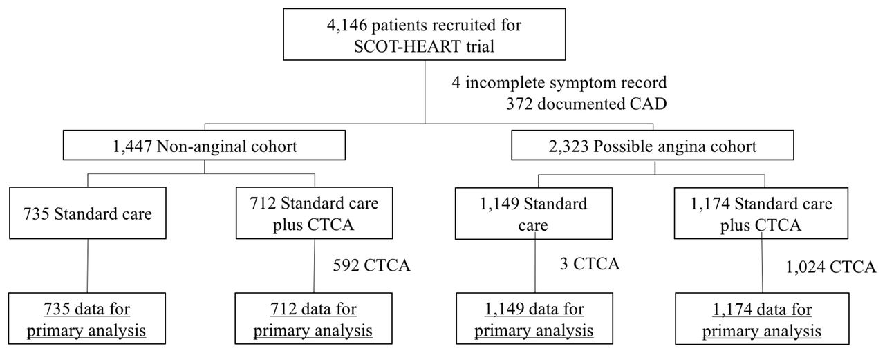
Consort diagram. CTCA, CT coronary angiography.
The mean age of the participants was 56.6±9.7 years and 1721 (45.6%) were women. Overall, 1447 (38.3%) of participants had non-anginal symptoms and a normal ECG while 2323 (61.6%) participants had symptoms or ECG changes consistent with possible angina. The non-anginal cohort were typically younger and had fewer cardiovascular risk factors than those with possible angina (table 1).
Baseline characteristics
CT coronary angiography
Compared with the non-anginal cohort, participants with possible angina were more likely to have obstructive coronary disease identified on CTCA (29.7% vs9.5%; RR 2.81, 95% CI 2.15 to 3.68, p<0.001) and less likely to have normal coronary arteries (33.1% vs 50.1%; RR 0.74, 95% CI 0.66 to 0.83, p<0.001) (table 2). The pretest probability assessment recommended in the 2010 NICE guidelines substantially overestimated the risk of obstructive coronary disease (see online supplementary table 3), while the revised diagnostic cohorts demonstrated persistent heterogeneity in risk by age and sex (see online supplementary table 3). The average rate of the primary diagnostic and prognostic endpoints for both men and women is presented in online supplementary table 4, and additional, non-coronary findings made on CTCA are reported in online supplementary table 5.
Findings of CTCA
Diagnostic certainty and additional investigations
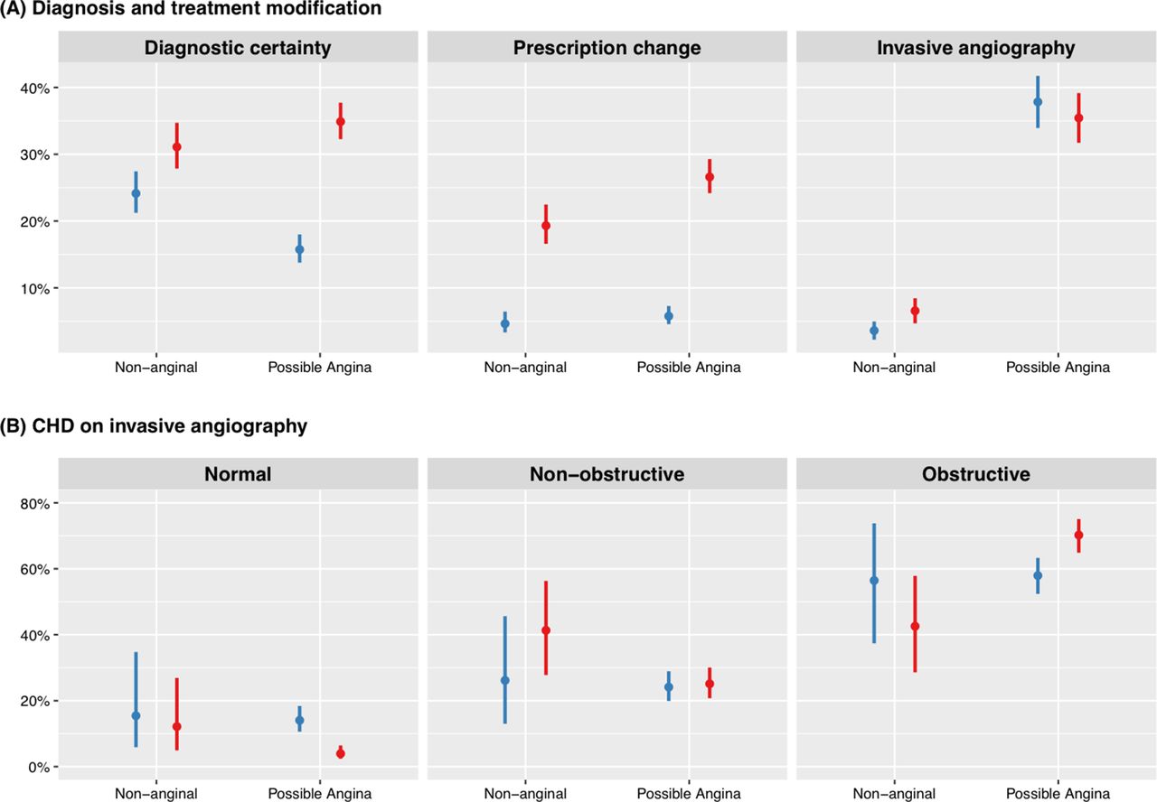
The use of CTCA increased the certainty with which a diagnosis of angina was made (figure 2a). This benefit was greatest (cohort interaction: p<0.001) in those with possible angina where the proportion of participants with a certain diagnosis of angina at 6 weeks was 34.9% with CTCA and 15.7% with standard care (RR 2.22, 95% CI 1.91 to 2.60, p<0.001). The improvement in diagnostic certainty remained, although attenuated, in the non-anginal cohort (CTCA 32%, standard care 25.2%; RR 1.30, 95% CI 1.11 to 1.53, p=0.002). The use of CTCA was associated with a change in diagnosis in 341 (29.0%) participants with possible angina and 120 (16.9%) participants with non-anginal symptoms. These improvements were associated with treatment changes in 26.8% of those with possible angina and 19.4% in those with non-anginal chest pain.

Diagnostic certainty, pharmacotherapeutic changes and effect on invasive angiography with standard care (blue) or standard care plus CTCA (red) according to diagnostic cohort. CHD, coronary heart disease; CTCA, CT coronary angiography.
The use of CTCA was associated with an increase in new requests for invasive coronary angiography at 6 weeks in both the possible angina (71 (6.0%) vs 7 (0.6%)) and the non-anginal (12 (1.7%) vs 0 (0.0%)) groups. Overall, CTCA only increased the total number of angiograms performed during the complete follow-up period in the non-anginal cohort (6.6% vs 3.7%; HR 1.82, 95% CI 1.13 to 2.92, p=0.014), with no change in the possible angina cohort (30.2% vs 32.1%; HR 0.95, 95% CI 0.82 to 1.10, p=0.481) (figure 2a). In participants with possible angina, CTCA was associated with a reduced likelihood of the invasive angiogram revealing normal coronary arteries (RR 0.32; 95% CI 0.19 to 0.52, p<0.001) and an increased likelihood of identifying obstructive disease (RR 1.18; 95% CI 1.07 to 1.32, p=0.002). In contrast, invasive angiography performed in the non-anginal cohort demonstrated similar rates of normal arteries (RR 0.78; 95% CI 0.30 to 2.05, p=0.622) and obstructive coronary disease (RR 0.82; 95% CI 0.50 to 1.34, p=0.422) in both treatment arms (figure 2b).
Clinical outcomes
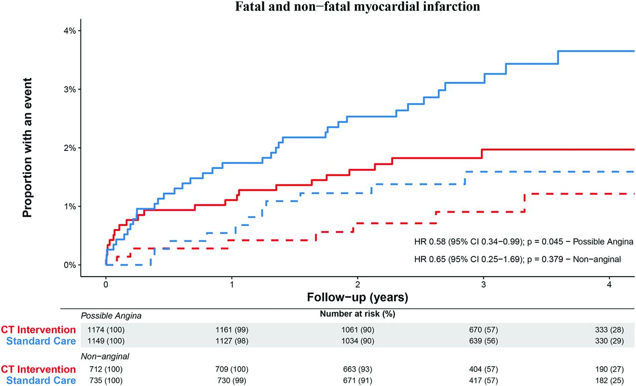
During follow-up, 18 (1.2%) and 59 (2.5%) of participants experienced a fatal or non-fatal myocardial infarction in the non-anginal and possible angina groups, respectively (table 3). Allocation to standard care with CTCA reduced the likelihood of this endpoint in the cohort with possible angina from 3.2% to 1.9% (HR 0.58; 95% CI 0.34 to 0.99; p=0.045, figure 3a). This was predominantly related to a reduction in non-fatal myocardial infarction from 3.0% to 1.6% (HR 0.55, 95% 0.31 to 0.96; p=0.034). Although a similar effect size was seen in those allocated to CTCA in the non-anginal cohort, the CI was wide, reflecting the lower event rate, and this did not achieve statistical significance (HR 0.65; 95% CI 0.25 to 1.69; p=0.379, figure 3b). The treatment-group interaction p value was 0.836.

Cumulative event curves for fatal and non-fatal myocardial infarction in the possible angina (solid lines) and non-anginal (dashed lines) cohorts in patients assigned to standard care (blue) and standard care plus CTCA (red). CTCA, CT coronary angiography.
Clinical endpoints according to diagnostic cohort
The use of CTCA was not associated with an increase in coronary revascularisation in either the possible angina (18.7% vs 16.5%; HR 1.16, 95% CI 0.95 to 1.41, p=0.140) or non-anginal cohorts (2.2% vs 1.9%; HR 1.20, 95% CI 0.59 to 2.46, p=0.619).
Discussion
We have applied the updated 2016 NICE guideline criteria to a prior large multicentre randomised controlled trial population. We have demonstrated that the selective investigation of patients with possible angina produced the greatest absolute benefits in terms of diagnostic certainty, use of invasive angiography, targeting of therapies and ultimately improving clinical outcome. In contrast, CTCA was not associated with a significant improvement in outcomes in patients with non-anginal symptoms and a normal resting ECG despite nearly doubling rates of invasive coronary angiography. These findings provide robust evidence to support the diagnostic strategy recommended within the new NICE guidelines.
This study has five notable strengths. First, the study participants were recruited from an unselected patient population referred to 12 chest pain clinics across Scotland and thereby accurately reflect the target cohort of the new guidelines. Second, as participants were allocated to CTCA in a randomised manner regardless of the typicality of chest pain symptoms, we minimised the potential for case ascertainment bias. Third, by not dictating the use of additional investigations in the standard care arm, we have focused on the effect of CTCA on clinically significant outcomes rather than comparing the diagnostic accuracy of different imaging modalities. Fourth, all scans were performed on CT scanners meeting or exceeding the guideline technological requirements and were reported in accordance with the recommended definitions for obstructive CHD. Finally, the prospective nature of the SCOT-HEART trial enabled detailed and accurate phenotypic characterisation of patients at baseline and comprehensive clinical follow-up.
The use of CTCA in the assessment of patients with possible angina results in a 1.3% absolute risk reduction in the prognostic endpoint of fatal or non-fatal myocardial infarction over 3.2 years. This corresponds to 74 CTCA referrals (65 completed scans) to prevent a myocardial infarct. It should be noted that both cohorts had numerically similar HRs and failed to demonstrate a statistically different treatment effect on formal interaction testing. Consequently, the non-significant risk reduction in the non-anginal cohort likely relates to the very low event rate observed within this group. While this study was underpowered to reliably exclude a benefit in the non-anginal cohort, the results do suggest that the clinical significance of any benefits are likely to be small with an estimated number of 195 CTCA referrals to prevent a myocardial infarct.
An important innovation of the 2010 NICE guidelines was the recommendation to avoid further testing in patients with a low likelihood (<10%) of CHD. This has a sound theoretical basis in probability theory, reduces unnecessary investigations and is similar to the approach adopted by the European Society of Cardiology.21 Despite this, the explicit calculation of risk has been removed from the updated recommendations due to the questionable applicability of the established scoring system—developed in 1979 within a US population—to the modern UK context, resulting in potential overestimation of disease prevalence. Fortuitously, in this study, the cohort with non-anginal symptoms and a normal ECG had a prevalence of obstructive CHD of 9.5%, suggesting that the updated approach continues to provide an implicit method of pretest probability estimation. It is important to note, however, that within the non-anginal group, there is some underlying heterogeneity in risk by age and sex—the predicted risk of obstructive CHD does vary from 3.4% in women aged less than 60 years of age to 20.2% in men aged over 60 years—and it is likely that including routinely recorded clinical variables such as these could further optimise the assessment process.22 Nonetheless, our study suggests that deferring the use of additional cardiac testing in these patients is safe, with an incidence of fatal or non-fatal myocardial infarction during the follow-up period of 1.5%, which was not reduced with the use of CTCA.
Within this study, CTCA increased the identification of both obstructive and non-obstructive coronary atherosclerosis that led to an increase in new requests for invasive coronary angiography in both patient cohorts. This increase in referrals has been raised as a potential drawback of adopting an anatomical approach to coronary assessment given the associated costs of unnecessary downstream testing.12 Such concern is justified if CTCA is applied in an indiscriminate manner. Indeed, this study found no decrease in the likelihood of finding normal coronary arteries in those patients with non-anginal symptoms who underwent invasive evaluation, implying that CTCA did not improve appropriate test selection in this group. In contrast, when restricted to use in patients with possible angina, there was a reduction in the likelihood of normal coronary arteries and an increase in the rate of obstructive disease found on angiography suggesting that candidates for further testing had been appropriately selected. Furthermore, although both groups demonstrated higher rates of invasive angiography at 6 weeks, this increase only persisted in the non-anginal cohort by the conclusion of study follow-up. Interestingly, despite the increased detection of coronary obstruction on angiography, there was no increase in coronary revascularisation in patients with possible angina. This suggests the adoption of a more nuanced approach to coronary intervention in the modern era. Our findings therefore refute previous commentators’ criticisms of the 2016 NICE guidance and their assumptions regarding CTCA-guided use of both angiography and revascularisation.12
Limitations
Although this was a post hoc analysis of the SCOT-HEART trial, it took place during the prespecified period of follow-up of clinical events with systematic and robust collection of outcome data. Furthermore, the original trial was pragmatically designed in order to recruit patients with suspected stable angina of recent onset in a non-selective manner, and the population enrolled is reflective of the heterogenous group seen in chest pain clinics with an even spread of chest pain symptom typicality. In addition, participants had a broad range of estimated pretest probability of CHD, thereby ensuring direct applicability of the study outcomes to the proposed setting for implementation of the updated NICE guidelines.
It should be noted that, within this study, clinicians made use of additional ischaemia tests, particularly exercise ECG, that are no longer recommended by current guidelines. This does not necessarily detract from the overall findings. Indeed, it could be claimed that the high use of exercise ECG in both treatment arms would likely reduce the incremental benefit of CTCA compared with the recommended avoidance of this investigation.
Finally, it is uncommon for trials of diagnostic investigations to demonstrate improvements in clinical outcomes, and this study cannot answer the question of how this reduction in event rates was achieved. It seems plausible that the identification of CHD initiated a series of management changes including more personalised patient education, greater adherence to healthy lifestyle recommendations and more appropriate use of risk-modifying medications.23 Uncertainty persists concerning how to manage patients with no evidence of atherosclerosis on CTCA, specifically whether this warrants the cessation of preventative medications even in the presence of other cardiovascular risk factors. Furthermore, we have previously demonstrated a gradient of risk between the categories of normal, non-obstructive and obstructive coronary artery disease23 and the ability to robustly quantify plaque burden is an important strength of CTCA. How this information is best used to inform treatment decisions, however, remains an important unanswered question, particularly in light of recent effective but costly pharmacological interventions.24
Conclusions
The clinical characterisation of symptoms is central to the 2016 updated NICE guidelines for the assessment of chest pain. When applied to a modern chest pain cohort, this revised approach appropriately selects patients requiring further investigation for CHD and minimises unnecessary testing in low-risk individuals. Once patients with possible angina are identified, the use of CTCA is associated with greater diagnostic certainty, more appropriate use of invasive angiography and a reduced risk of fatal and non-fatal myocardial infarction.
Key questions
What is already known on this subject?
CT coronary angiography (CTCA) enhances the assessment of patients with suspected stable angina by increasing diagnostic certainty when applied to a broad population referred to a specialist chest pain clinic. The recently updated 2016 National Institute for Health and Care Excellence (NICE) guidelines advocate use of CTCA for a subset of this population only.
What might this study add?
The present study investigated the diagnostic and prognostic impact of CTCA when used in accordance with the new NICE guidance. Selective use of CTCA in patients with possible angina maximises the benefits of CTCA including greater diagnostic certainty, avoidance of normal invasive coronary angiography and reductions in fatal and non-fatal myocardial infarction. It also demonstrates that patients with non-anginal symptoms are at low risk and do not derive major diagnostic or prognostic benefit from CTCA, but its use is associated with greater rates of invasive angiography.
How might this impact on clinical practice?
These findings offer clinicians robust evidence of the safety and efficacy of the revised NICE guidelines and provide healthcare services some reassurance that such an approach does not increase downstream use of invasive coronary angiography. In keeping with these updated NICE guidelines, CTCA should only be used where there is diagnostic uncertainty in patients with possible angina.
Acknowledgments
DEN (CH/09/002), MCW (FS/11/014) and NLM (FS/16/14/32023) are supported by the British Heart Foundation. DEN is the recipient of a Wellcome Trust Senior Investigator Award (WT103782AIA). AT is supported by Barts Cardiovascular Biomedical Research Unit, funded by the National Institute for Health Research. EJRvB is supported by the Scottish Imaging Network: A Platform of Scientific Excellence (SINAPSE). The Royal Bank of Scotland supported the provision of 320-multidetector CT for NHS Lothian and the University of Edinburgh. The Edinburgh Imaging Facility QMRI (Edinburgh) is supported by the National Health Service Research Scotland (NRS) through National Health Service Lothian Health Board. The Clinical Research Facility Glasgow and Clinical Research Facility Tayside are supported by National Health Service Research Scotland (NRS).
References
Footnotes
PDA and AH contributed equally.
Contributors PDA, AH, MCW, MRD, DAM and DEN contributed to the conception and design of this work. PDA, AH, MCW, ASVS, TAP, MRD, CB, NAB, MF, JF, SM, GR, EJRvB, ADT and DEN contributed to the acquisition of study data. PDA, AH, MCW, MRD, ASVS, DAM and DEN contributed to the analysis and interpretation of data and drafting of the manuscript. PDA, AH, MCW, ASVS, DAM, TAP, MRD, NLM, CB, NAB, EC, MF, JF, SM, GR, EJRvB, ADT and DEN contributed to the revision of the manuscript. PDA and DEN are responsible for the overall content of this work. The SCOT-HEART Investigators contributed to the conception or design of the work, or the acquisition, analysis or interpretation of data for the work. They were involved in drafting the manuscript and revising it and have given final approval of the version to be published. The SCOT-HEART investigators are accountable for the work.
Competing interests DEN, EJRvB and GR have received honoraria and consultancy from Toshiba Medical Systems. GR has received honoraria from companies (Bracco, Bayer-Schering, GE Healthcare and Guerbet) producing contrast media.
Patient consent Obtained.
Ethics approval South East Scotland Research Ethics Committee.
Provenance and peer review Not commissioned; externally peer reviewed.