Article Text

Statistics from Altmetric.com
Clinical introduction
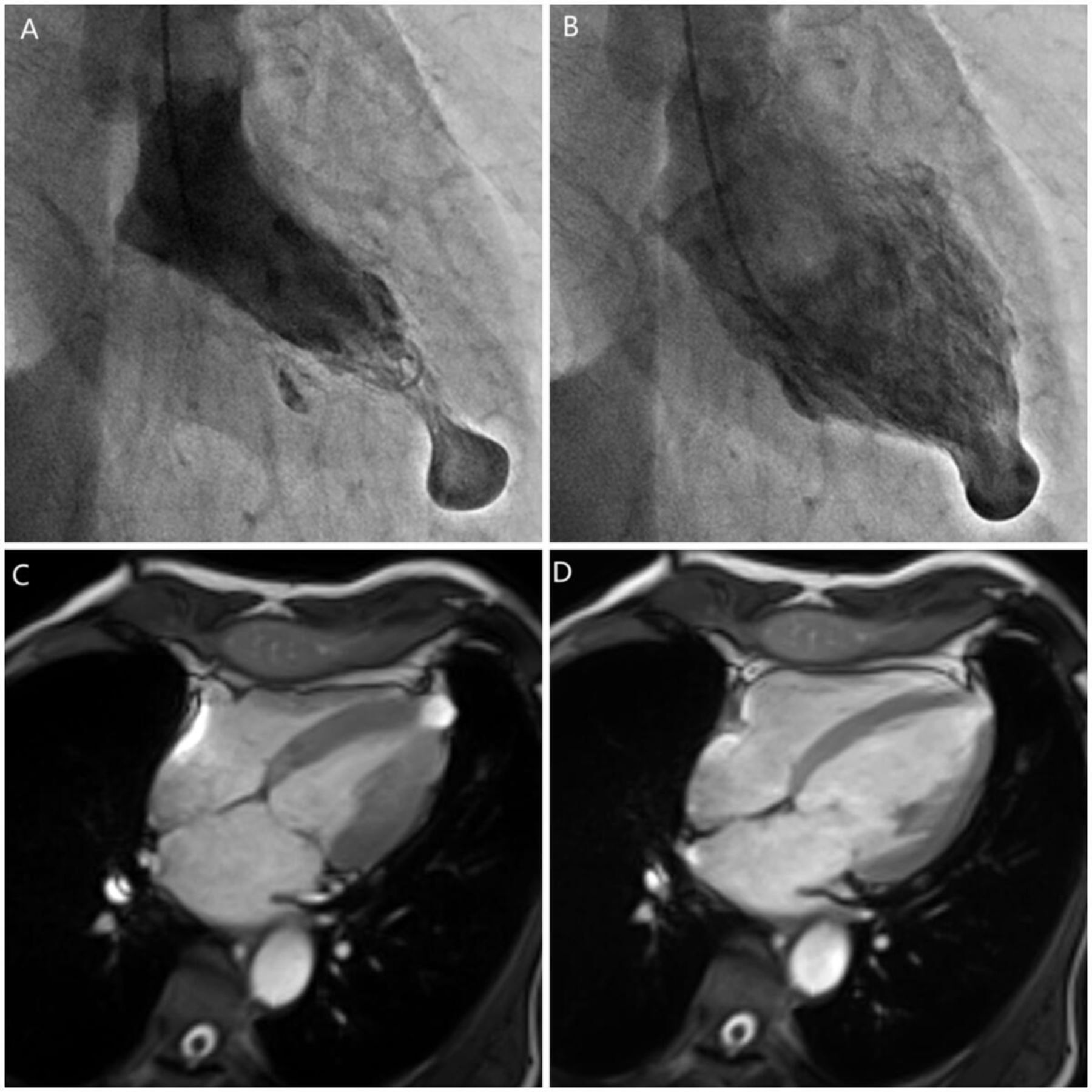
A man in his 60s with hypertension and tobacco use presented to the outpatient clinic with 3-month progressive exertional dyspnoea. ECG showed sinus rhythm with poor R-wave progression in the precordial leads. Transthoracic echocardiography (TTE) revealed normal left ventricular (LV) size and function. Coronary angiography showed no significant stenosis. However, left ventriculography found a narrow neck, thin-walled outpouching at the apex with synchronous contraction (figure 1A,B and video 1). Subsequently, MRI confirmed a small LV outpouching on the apex measuring 17×12 mm, which contained myocardium (figure 1C,D and video 2). On late gadolinium enhancement images, the entire LV myocardium was nulled without evidence of scar. During the period of follow-up, the size and location of his LV outpouching remained unchanged over time.
Ventriculogram (right anterior oblique projection) showing the contrast-filled outpouching at the apex during systole (A) and diastole (B). MRI (long axial plane of the left ventricular view) showing a small outpouching on the apex during systole (C) and diastole (D).
Question
What is the most probable diagnosis of this patient?
LV pseudoaneurysm.
LV aneurysm.
LV diverticulum.
Apical ballooning syndrome.
Answer: C
LV diverticulum is defined by a narrow-necked saccular outpouching that extends beyond the confines of the myocardium and contracts with the ventricle, as is seen in this case. Aneurysms are nearly always associated with total occlusion of the coronary artery, usually the left anterior descending. Pseudoaneurysm is a false aneurysm, resulting from a contained rupture of the LV wall, a rare complication following myocardial infarction (MI). Apical ballooning syndrome is a usually reversible cardiomyopathy that may be precipitated by stress or critical illness and can mimic MI. In our case, coronary angiography demonstrated normal coronary arteries and MRI found no signs of MI, making aneurysm and pseudoaneurysm unlikely.
Two types of diverticulum have been reported: muscular and fibrous types.1 2 The fibrous type is located at the ventricular apex or a subvalvular position, without distinct contraction. It is not associated with other congenital cardiac malformations, and thus may be diagnosed only in adulthood. The diverticulum in our patient was consistent with the fibrous type of congenital LV diverticulum. In adult presentation, diverticula are discovered incidentally during cross-sectional diagnostic imaging procedures, such as echocardiography, cardiac MRI or cardiac catheterisation.3 Diverticula can be difficult to diagnosis by TTE, but contrast echocardiography can be useful. Management of ventricular diverticula remains uncertain and treatment options include either observation and surgery.4 If the diverticulum is small and asymptomatic, as in our case, a conservative treatment with follow-up is often recommended.
Abstract translation
This web only file has been produced by the BMJ Publishing Group from an electronic file supplied by the author(s) and has not been edited for content.Ethics statements
Ethics approval
The case report was approved by Beijing Friendship Hospital’s committee on human research (approval number 2020-P2-138-01).
Footnotes
Contributors All authors gave the final approval of the version published. LZ wrote the first draft of the manuscript. LZ and HC were involved in the care of the patient. HL made revisions to the manuscript.
Funding LZ is funded by the Beijing Municipal Administration of Hospitals Incubating Program (code PX2020006).
Competing interests None declared.
Patient and public involvement Patients and/or the public were not involved in the design, or conduct, or reporting, or dissemination plans of this research.
Provenance and peer review Not commissioned; internally peer reviewed.