Article Text

Statistics from Altmetric.com
Image challenge
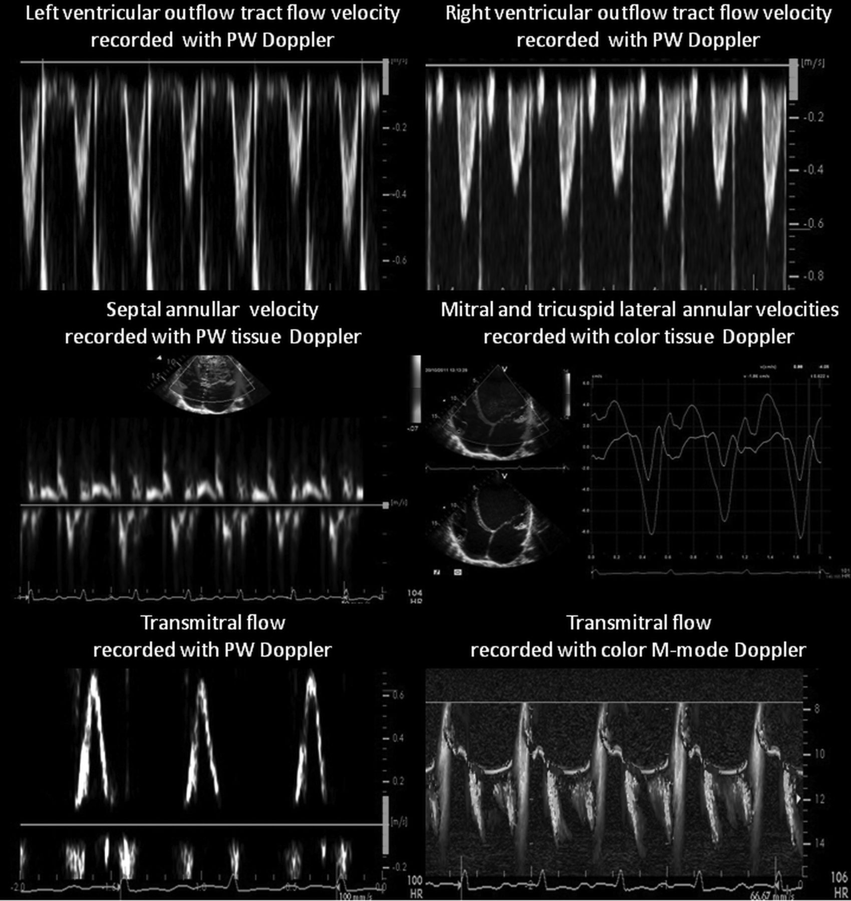
A young patient with dilated cardiomyopathy and a history of pulmonary embolism presented with severe heart failure. An echocardiographic study showed both left and right ventricular dilatation and severe systolic dysfunction of both ventricles (see online supplementary video). The Doppler recordings of left and right ventricular outflow tract velocities, pulse wave Doppler and colour M-mode Doppler recordings of transmitral flow as well as tissue Doppler recordings of mitral and tricuspid annulus velocities are presented in Figure 1.
Doppler recordings of left and right ventricular outflow tract velocities (top panels), mitral and tricuspid annular velocities (mid-panels) and transmitral flow (bottom panels). Access the article online to view this figure in colour.
Question
The presented Doppler tracings are typical for:
-
Constrictive pericarditis
-
Restrictive cardiomyopathy
-
Left bundle branch block
-
Biventricular pulsus alternans
For answers see page 90
Answer
From question on page 83
The correct answer is: biventricular, systolic and diastolic pulsus alternans. Beat-to-beat alterations of left and right ventricular ejection (top panels) as well as beat-to-beat systolic alterations of myocardial tissue velocities of both mitral and tricuspid annuli are apparent (mid-panels). There is no easily visible beat-to beat change of transmitral flow recorded with pulse wave Doppler; however, close inspection of colour M-mode Doppler recording reveals alternating transmitral flow (alternating diastolic aliasing area) (bottom panels). Diastolic beat-to-beat alterations are also apparent on Doppler myocardial velocities of mitral and tricuspid annulus (mid-panels). Constriction is associated with respiratory rather than beat-to-beat cardiac output changes. Neither restrictive cardiomyopathy nor left bundle branch block is associated with significant beat-to-beat cardiac output changes, unless associated with severe systolic dysfunction of the left ventricle.
Pulsus alternans characterised by beat-to-beat variations in stroke volume with alternate cardiac cycles is usually associated with a severe ventricular dysfunction.1 The two principal mechanisms proposed to account for its development are based on the Frank–Starling relationship and intrinsic beat-to-beat alternation in myocardial contractility due to alternation of the Ca2+ release from the sarcoplasmic reticulum.2
The first medical description of pulsus alternans was provided by Traube in 1872 in a patient with advanced alcoholic cardiomyopathy. In 1913, based on observations in patients with advanced cardiomyopathy, Windle concluded that alternans ‘always adds to the gravity of the prognosis’.3 At this time, D H Lawrence provided its literary description in a classic novel Sons and Lovers—‘He felt her pulse. There was a strong stroke and a weak one, like a sound and its echo. That was supposed to betoken the end’. Recently, right sided mechanical alternans was identified as an unfavourable prognostic sign in pulmonary arterial hypertension.4
Supplementary materials
Supplementary Data
This web only file has been produced by the BMJ Publishing Group from an electronic file supplied by the author(s) and has not been edited for content.
Files in this Data Supplement:
- Data supplement 1 - Online video
Footnotes
-
Contributors Conception and design: PS. Acquisition of data: PS and ML. Analysis and interpretation of data: PS, ML, AK and PH. Drafting the article: PS. Revising it critically for important intellectual content: PS, ML, AK and PH. Final approval of the version published: PS, ML, AK and PH.
-
Competing interests None.
-
Provenance and peer review Not commissioned; internally peer reviewed.