Article Text

Abstract
Objective Perivascular adipose tissue (PVAT) exerts an anticontractile effect that is lost in obesity. A recent study reported that bariatric surgery can reverse the damaging effects of obesity on PVAT function with accompanying reduction in systolic blood pressure. However, PVAT function has not previously been characterised following weight loss induced by caloric restriction, which is often the first line treatment for obesity. This study investigated the role of PVAT in control of vascular function in rat models of diet-induced obesity and weight loss.
Design and Methods Male Sprague Dawley rats were fed high-fat diet (45% fat) ad libitum for 16 weeks to induce obesity, they were then split into 2 groups; obese rats maintained on the diet and weight loss rats subjected to 50% caloric restriction for a further 4 weeks. A control group was also provided with a 10% fat diet during the 20 week period. Blood pressure was recorded in conscious animals at 8 week intervals using a CODA tail cuff blood pressure monitoring system. The effect of PVAT on the contractility of isolated rat mesenteric arteries (250–300 µm internal diameter) in response to noradrenaline (1 × 10⁻⁵ – 3 × 10⁻⁹ mol.l-1) was investigated using wire myography. Data were expressed as mean ± SEM and analysed using two-way ANOVA.
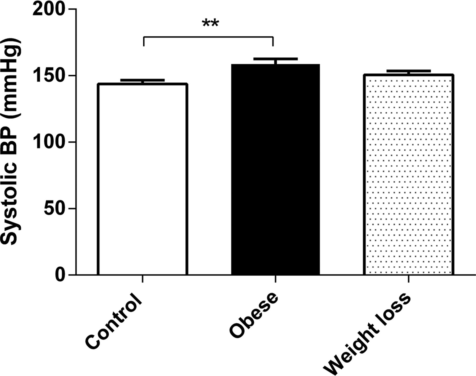
Results and Conclusions In control animals, the vasoconstrictor response to noradrenaline was reduced in the presence of PVAT through an endothelium-dependent mechanism (weight = 591 g, endo: PVAT vs no PVAT P < 0.05, no endo: PVAT vs no PVAT P = 0.998, n = 5). The PVAT anticontractile effect was abolished in obese animals (weight = 818 g, P = 0.4248, n = 5) with an accompanying increase in systolic blood pressure (Figure A). The PVAT anticontractile effect was partially restored following diet-induced weight loss (weight = 620 g, n = 6, Figure B) and this was accompanied by a reduction in obesity-induced hypertension (Figure A, systolic BP: obese = 159 ± 3.8 mmHg, weight loss = 148 ± 2.9 mmHg). The vasoconstrictor response was unaltered between the groups in the absence of PVAT suggesting that changes in body weight do not affect vascular smooth muscle or endothelial cell function.
Overall, our data indicate that obesity-induced hypertension may be a consequence of PVAT damage and this can be partially reversed following diet-induced weight loss.

Systolic blood pressure
Constriction in the presence of PVAT
- perivascular adipose tissue
- obesity
- weight loss