Article Text

Abstract
Introduction Left ventricular noncompaction (LVNC) is characterised by prominent ventricular myocardial trabeculations, composed of sheets of cardiomyocytes. They form early during cardiogenesis but their development is complex. Measuring trabecular complexity in animal models of cardiac disease is important as abnormal trabecular patterns are increasingly been recognised to coexist with several other cardiac conditions, not just LVNC. We describe an innovative approach that utilises fractal algorithms and high-resolution episcopic microscopy (HREM) to study the developmental timing of myocardial trabeculation in mouse and we validate it using a recently described LVNC mouse model (NOTCH pathway regulator Mib1 mutant).
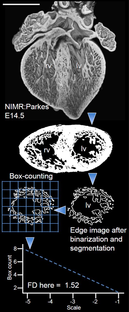
Methods HREM (2–3 μm resolution) analysis was performed prospectively on 123 embryonic mouse hearts consisting of wild-type (WT) NIMR:Parkes, WT C57BL/6 and Mib1 flox; cTnT-cre mutant and WT littermates. HREM permits the 2D/3D imaging of tissue samples as they are physically sectioned. Datasets underwent fractal analysis using a box-counting approach (Figure 1).

Schematic representation of the fractal procedure applied to a single HREM slice for calculating trabecular complexity in the LV of the embryonic mouse heart. Each HREM dataset comprised of approximately 1,200 slices, is imported into OsiriX as a double-oblique short-axis stack. Binarization, inversion, backgroundfilling, region of interest selection and edge-detection are carried out to generate a stack of endocardial contours spanning the length of the LV from base to apex. These contours undergo fractal analysis using the box-counting method.

LV trabeculae during cardiac development in wild-type mouse. The complete murine atlas of embryonic LV trabecular development in WT NIMR:Parkes. Heart sections represent the embryonic stages: E14.5, 5; E15.5, 4; E16.5, 3, E17.5, 2, E18.5, n = 14. Solid lines, mean heart FD; coloured ribbons, 95% CI. Scale bars, 0.5 mm. * .05, ** .001 determined by Student’s t test. Absent ‘*’ indicate no statistically significant difference in FD.


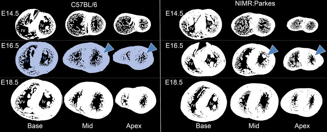
Trabecular development in WT C57BL/6 and comparison with NIMR:Parkes. (a) Trabecular development in WT C57BL/6 (E14.5, n = 3; E16.5, E18.5, n = 0) differs from that observed in NIMR:Parkes (.001 for NIMR:Parkes vs. C57BL/6 comparisons at stages E14.5, E16.5 and E18.5). (b) The inter-strain difference is most marked at E16.5 (blue arrow heads) where C57BL/6 embryos (left) retain substantially higher trabecular complexity across the mid and basal LV. This reduces by E18.5 approaching that seen in NIMR:Parkes.
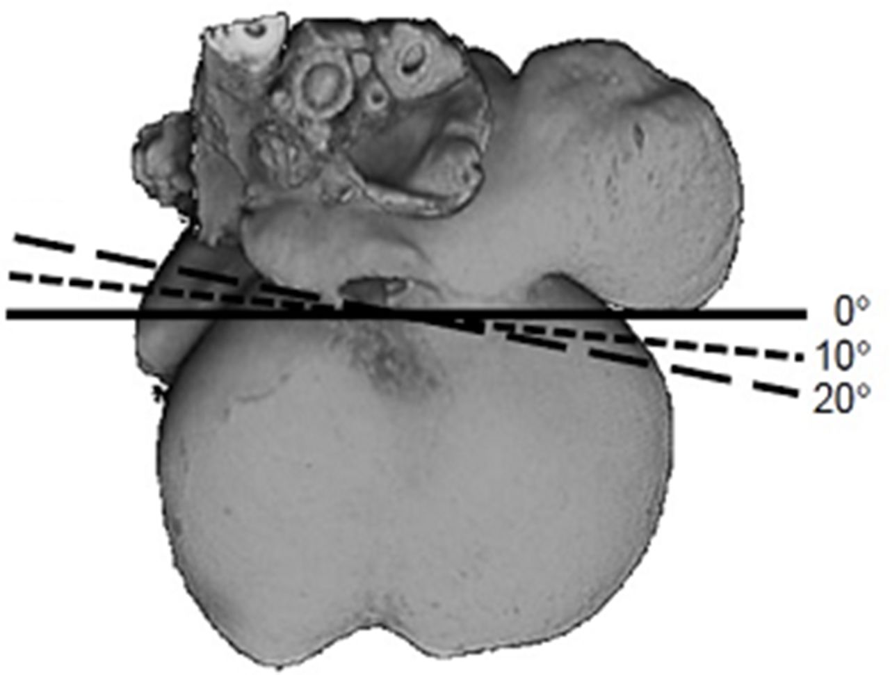


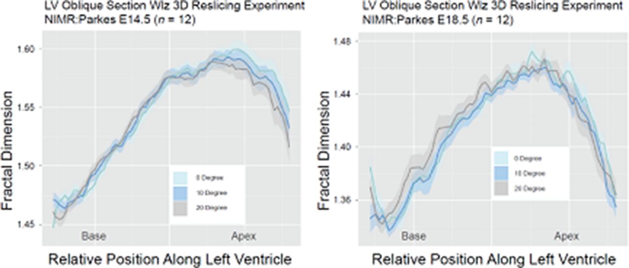
Reslicing Woolz experiments for the fractal method applied to HREM.

HREM reconstructions showing the morphological differences between WT and Mib1flox;ccTnT-cre littermates. The highly conserved NOTCH signalling pathway regulates cell-fate specification, differentiation and patterning. Altered signalling is known to cause human cardiovascular disease. Mib1 is a NOTCH pathway regulator. Myocardium-specific mouse Mib1 mutants develop LVNC resulting from a developmental arrest in trabecular maturation and myocardial compaction which are clearly shown in these HREM 3D reconstructions of the left and right ventricles. Stage E16.5 is represented here. Mib1 mutants (right) had a dilated heart with a thin compact myocardium and large, immature, noncompacted trabeculae (blue arrow heads) protruding toward the right and left ventricular lumen. The myocardium was well-compacted and less trabeculated in WT mice (left). Scale bars, 0.5 mm. Abbreviations as in Figure 1.
Results LV trabecular complexity showed a significant drop between E14.5 and E18.5 (Figure 2). Across all embryonic stages, the apical half of the LV retained the highest fractal dimensions (FD) when compared to the base. By E18.5 the myocardium was almost fully compacted registering the lowest FD. For the first time, we demonstrate that strain-specific differences in LV trabecular patterning exist in mouse because NIMR:Parkes compacts earlier than C57BL/6 (Figure 3). Reslicing experiments (Figure 4) and separate validation tests on Mib1 mutants and WT littermates (Fig.5) confirmed how the proposed methodology is a reliable and effective tool for the detection of mutagenesis-related differences in trabeculation.
Conclusion Reported here is a method in which sequential, 2D sections of mouse embryo hearts may be analysed using a fractal algorithm to calculate ventricular trabecular complexity – a technique so sensitive, that small inter-strain differences in somitogenesis are detectable in mouse pups.
Precise knowledge of the trabecular architecture as it presents itself in WT, is a prerequisite for the correct identification of pathological trabecular phenotypes in mouse models of cardiac disease, explaining the need for a quantitative fractal atlas of trabecular development.
Fractal mathematics in combination with HREM has the potential to answer to many developmental biology questions in the heart, with future applicability to other organ systems and to other species.
- Cardiac development
- Myocardial cardiomyopathy disease
- Structure