Article Text

Abstract
Background Pre-participation cardiovascular evaluation is being recommended by a growing number of sporting bodies and scientific organisations in an attempt to identify athletes harbouring quiescent cardiac conditions which put them at risk of exercise-related sudden cardiac death (SCD). However cardiac adaptation to exercise may itself result in ECG patterns that overlap with those observed in several cardiac conditions, including hypertrophic cardiomyopathy (HCM), the leading cause of SCD in athletes worldwide. This raises significant challenges for physician’s involved in ECG-based pre-participation cardiovascular evaluation of athletes with respect to distinguishing physiological from pathological ECG changes.
Data demonstrates that cardiac conditions such as hypertension may modulate the HCM phenotype. However the effect of exercise on the HCM phenotype remains unknown. We compared the ECGs and echocardiographic parameters of athletes with HCM to sedentary HCM patients and healthy athletes to establish whether physical activity modifies the phenotype in HCM.
Methods The presenting ECGs of 103 young (14–35) consecutive asymptomatic athletes with HCM identified through pre-participation or familial evaluation were compared to the presenting ECGs of 106 sedentary HCM patients (14–40) and 110 consecutive healthy young athletes. Echocardiographic characteristics indicating disease severity and patterns of left ventricular hypertrophy (LVH) were also compared.
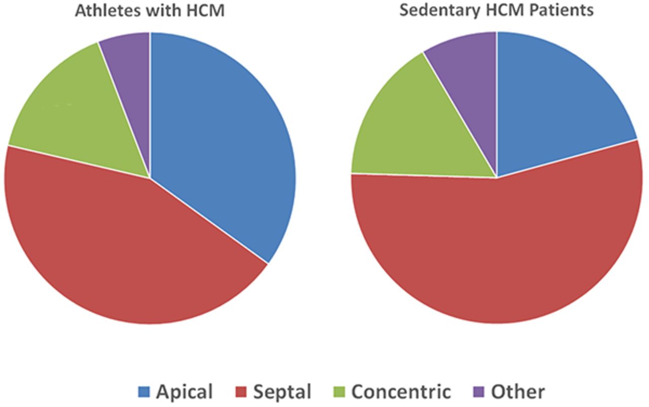
Results Sedentary HCM patients were older than the athlete groups (Table 1). All 3 groups contained a similar number Afro-Caribbean individuals (30.2% sedentary HCM vs. 33.0% athletes with HCM vs. 30.9% healthy athletes, p > 0.05), the remainder being Caucasian. Athletes with HCM exhibited significantly more repolarisation changes (specifically T-wave inversions [TWIs] and deep TWIs) in addition to voltage criteria for LVH compared to sedentary HCM patients and healthy athletes (p < 0.05 in each case). This was despite a milder phenotype on echocardiography (maximum left ventricular wall thickness 15.5 ± 2.9mm in athletes with HCM vs. 19.3 ± 5.3mm in sedentary HCM patients, p < 0.0001). No significant differences were found between athletes and sedentary patients with HCM with respect to other ECG changes including axis deviation, voltage criteria for atrial enlargement and pathological q-waves. Athletes with HCM exhibited a greater prevalence of the apical variant compared to sedentary HCM patients (35.0% vs. 20.8%, p < 0.05; Figure 1).
Conclusions Athletes with HCM exhibit more ECG repolarisation changes compared to sedentary HCM patients and healthy athletes, suggesting that exercise modifies the ECG phenotype in HCM. Virtually all athletes with HCM revealed TWIs, rendering softer ECG changes such as axis deviation and atrial enlargement less relevant to the detection of HCM in athletes. These results should be taken into consideration when devising criteria for interpretation of an athlete’s ECG.
- hypertrophic cardiomyopathy
- electrocardiography
- screening