Article Text

Abstract
Introduction Platydeoxia orthopnoea is a rare syndrome characterised by desaturation and dyspnoea on movement from standing to supine in the presence of an intra-cardiac shunt. The pathophysiology is poorly understood.
Methods We report a case with patent foramen ovale (PFO). Cardiac catheterisation and 4D-flow magnetic resonance (MR) were performed (Philips Achieva 3T, 6-channel array, retrospective ECG and respiratory gated TFE, spat res: 3 mm3, temp res: 50–55 ms, 20 phases). Pathline analysis was performed with GTflow (v2.0, Gyrotools).
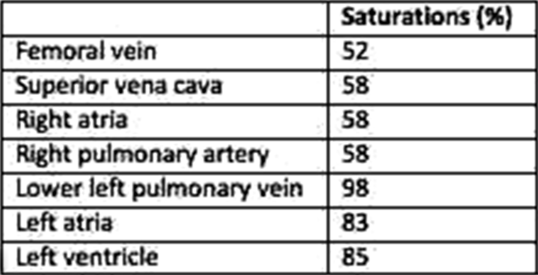
Results A 65 year-old man presented with desaturation during anaesthetic induction for anterior vitrectomy. He was asymptomatic. He had Type 2 diabetes mellitus, hypertension, hypercholesterolemia and centripetal obesity (BMI 38). Pulse oximetry showed saturations of 92% (standing) and 80% (supine). Supine pO2 was 5.9KPa. Spirometry was normal and CT excluded arterio-venous malformation. TOE demonstrated a tunnel-type PFO with spontaneous right to left shunt. At catheterisation (Table 1), right and left atrial pressures were normal (6 and 8 KPa). There was no pulmonary hypertension. On PFO balloon occlusion, pulse oximetry showed saturations of 97%. 4D-flow MR demonstrated reversal of the usual right atrial vortex. Inferior vena cava flow intersected with superior vena cava flow in a perpendicular fashion and was directed towards the PFO (Figure 1). The usual anticlockwise left atrial vortex was absent with a small clockwise vortex seen in the region of the right upper pulmonary vein, entraining the PFO flow. Amplatzer PFO device closure normalised saturations in both upright and supine positions.
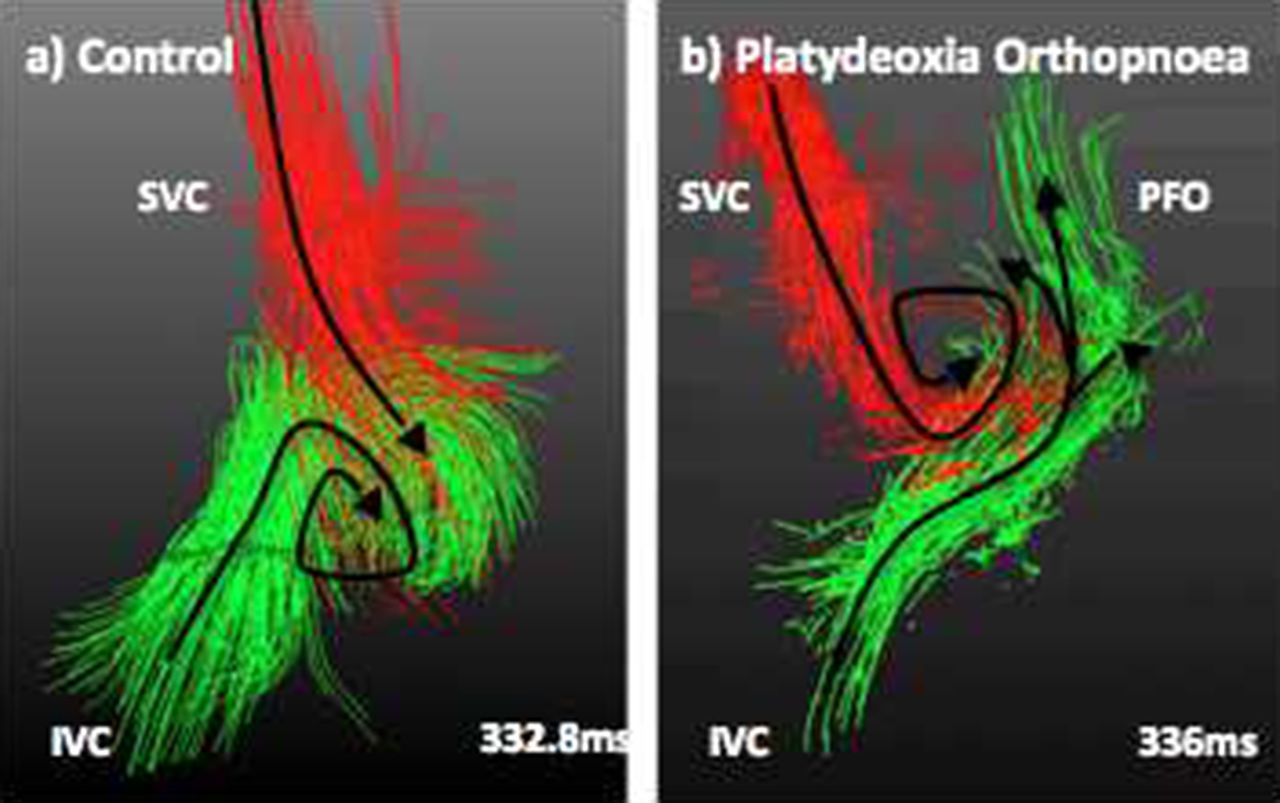
Right atrial flow (anterior aspect) in a) control showing usual forward turning right atrial vortex and in b) subject showing perpendicular arrival of IVC flow. Reversal of vortex and laree shunt across PFO
Saturations at cardiac
Discussion This case is one of a handful reported and the first to begin to define the pathophysiology of this condition. Despite suboptimal 4D-flow MR due to body habitus and breathing pattern, we demonstrate disturbance of the usual flow patterns that facilitate substantial shunting across the PFO. Whilst the positional component needs further investigation, we hypothesise that the relationship between the inferior vena cava and right atrium may contribute.
- Patent foramen ovale
- Magnetic Resonance Imaging
- Platydeoxia Orthopnoea