Article Text

Abstract
Background Large, population based studies have shown that diabetes mellitus (DM) of long durationis associated with increased incidence of heart failure, independent of underlying hypertension and coronary artery disease. Cardiovascular magnetic resonance imaging (CMR) and magnetic resonance spectroscopy (MRS) provide comprehensive, non-invasive, multiparametric measures of the functional, structural and metabolic status of the heart. We used magnetic resonance imaging and spectroscopy to determine whether subclinical functional, structural and metabolic alterations occur in a patient cohort of early-onset, type 2 DM.
Objective We aimed to assess the earliest features of diabetic cardiomyopathy in a cohort of uncomplicated, stable type 2 DM patients with short disease duration (<4 years) using multiparametric CMR and MRS.
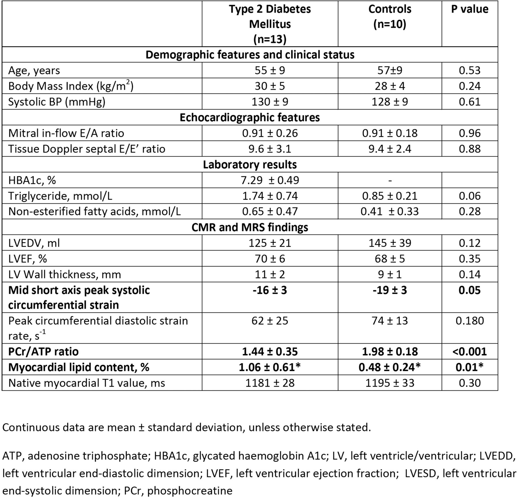
Methods 13 patients (7 male, 6 female, mean age 55 ± 9 years) with early-onset (median 3 [IQR: 1–4] years) type 2 diabetes and 10 healthy volunteers (5 male, 5 female, mean age 57 ± 9 years) were studied.Patients were either drug naive for diabetic therapy or on treatment with metformin monotherapy, HBA1c ≥6.7 and ≤8%, with no history of coronary artery disease or uncontrolled hypertension.Myocardial PCr/ATP ratios and lipid content were quantified using 31P and 1H-MRS, respectively.CMR included cine, tagging and native T1 mapping and was performed at 3.0 T. Left ventricular diastolic function was characterised using echocardiography.
Results Diabetic patients were well-matched with controls (Table 1).Myocardial energetics were decreased by 27% (PCr/ATP ratio: 1.44 ± 0.35 vs. 1.98 ± 0.18, p < 0.001) and myocardial triglyceride content increased 2.2-fold (1.06 ± 0.61 vs. 0.48 ± 0.24%, p = 0.01), despite relatively short disease duration.Subtle regional LV dysfunction was detected, indicated by reduced peak systolic circumferential strain (Table 1). However, left ventricular volumes, mass and ejection fraction, as well as echocardiographic indices of diastolic function were similar in both groups.Despite the myocardial metabolic abnormalities in patients with diabetes, there was no difference in T1 values, a measure of cardiac fibrosis, between diabetic patients and controls (1181 ± 28 ms vs. 1195 ± 33 ms, p = 0.30).
Conclusions Abnormal myocardial energy metabolism, steatosis and reduced LV strain are early manifestations of diabetic cardiomyopathy and precede the development of structural or other functional changes.CMR and MRS are sensitive, non-invasive tools for assessment of myocardial pathophysiology, and allow comprehensive phenotyping and staging of myocardial involvement in DM.
Funding The National Institute for Health Research Oxford Biomedical Research Council and Hoffmann-La Roche supported this work.
- Diabetic Cardiomyopathy
- Cardiac Magnetic Resonance Imaging
- Magnetic Resonance Spectroscopy