Article Text

Abstract
Background Giant cell arteritis (GCA) is the commonest primary systemic vasculitis affecting older people. Dilatation of the aorta may occur as a late complication and is believed to arise from damage to the aortic wall from inflammation.
Objectives To determine the prevalence of thoracic aortic dilatation and assess aortic stiffness by CMR in patients with GCA diagnosed at least 2 years previously.
Method Consecutive patients recruited to the UK GCA Consortium study were invited. 49 patients (median disease duration 4.5 years) underwent CMR at 3.0T (Philips Achieva TX). Cine images were acquired to measure the diameter of the ascending aorta (AsAo) and descending aorta (DsAo) at the level of the main pulmonary artery (MPA) and aortic arch from luminal edge-to-edge. Aortic stiffness was assessed by aortic distensibility (AD) and pulse-wave velocity (PWV). For AD, cine images (50 phases) were acquired in a plane transverse to AsAo at the level of MPA. Aortic contours were drawn manually at the times of minimal/maximal distension. For PWV, through-plane phase contrast velocity mapping was performed perpendicular to AsAo/DsAo at the level of MPA. Velocity-time curves were derived and the distance between the two locations measured to calculate PWV using the transit-time method.
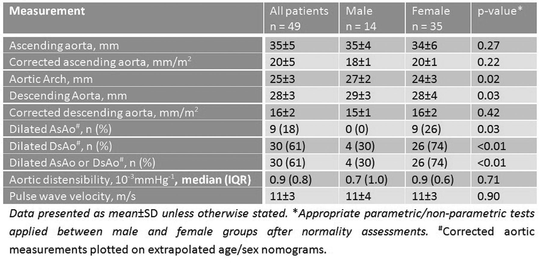
Results Patient characteristics: Mean age 73 ± 6 years, female gender 35 (71%), biopsy-positive 31 (63%), body surface area (BSA) 1.8 ± 0.2 m2, systolic blood pressure (BP) 148 ± 20 mmHg, pulse pressure 75 ± 21 mmHg. CMR measurements in Table 1.
CMR findings in GCA patients
30 (61%) patients had dilated thoracic aortas – corrected to BSA and applied to CMR nomograms [Davis et al . 2014. JCMR 16(1):9]. 5 (10%) patients had dilated AsAo at surgical intervention thresholds according to AHA/ACC guidelines. Aortic stiffness was increased with lower AD (median [IQR] 0.9 [0.8]10–3 mmHg-1) and higher PWV (11 ± 3 m/s) than normal ranges Aquaro et al.1Disease duration did not correlate with aortic measurements. 10/11 biopsy-negative and 17/31 biopsy-positive patients had dilated aortas. Age, body mass index (BMI) and BP were similar between dilated and non-dilated aorta groups. There was a female to male preponderance in the dilated group (26/35 vs. 4/30 respectively, P < 0.01). There was no gender difference with respect to patient characteristics.
Conclusion Dilatation of the thoracic aorta and arterial stiffness are common in patients with GCA. There is female preponderance in dilatation without differences in basic demographics. In biopsy-negative patients, under-treatment and/or variability in phenotype could explain increased aortic dilatation. Further investigation will be required to evaluate the effect of severity, treatment length/type, disease duration and cardiovascular risk factors on aortic morphology and function.
Reference
Aquaro, et al.Observational study of regional aortic size referenced to body size: productionof a cardiovascular magnetic resonance nomogram Davis et al. J Cardiovasc MagnReson 2014;16:9, doi:10.1186/1532-429X-16-9
- Giant Cell Arthritis
- Aortic Aneurysm
- Cardiac MRI