Article Text

Abstract
Cardiovascular and neuronal dysfunction have to a large extent been treated as separate disease categories from both research and clinical perspectives. However, there is now growing evidence that pathological changes in the shared circulatory system may be key drivers of both cardiovascular and neuronal dysfunction. It is speculated that compromised circulatory function, as can be seen in inflammatory vascular conditions such as atherosclerosis, may impact the regulation of cerebrovascular blood flow in response to dynamically changing neuronal metabolic demands also known as neurovascular coupling. With impaired neurovascular function being a pathogenic factor underlying cerebrovascular pathology, here we aim to establish if atherosclerosis elicits any alterations in the neurovascular function.
Paigen Diet fed ApoE -/- mice fitted with a stable cranial window over the right somatosensory cortex combined with state of the art multi-modal neurovascular imaging comprised of 2D-optical imaging spectroscopy (OIS) to measure evoked blood flow, volume, and oxygenation changes and electrophysiology to record neuronal activity. Any neurovascular breakdown will then be further investigated using high resolution multi-photon microscopy and immunohistochemistry to identify cellular deficits and potential molecular targets. Functional magnetic resonance imaging (fMRI) will also be used later to assess any sub-cortical effects that 2D-OIS and electrophysiology cannot detect, which will assist in combining highly detailed but invasive measurements using animal models and non-invasive MRI readouts with possibility of translation to humans. This research will thus bring together specific advantages of working in animal models using a complementary range of techniques in order to produce robust findings with maximal translational potential.
Results have currently demonstrated that Paigen diet fed ApoE-/- mice are stable under anaesthesia for long term studies using our multi-modal neurovascular imaging methods to produce haemodynamic and neuronal activity readouts. We now aim to continue to establish any alterations in neurovascular function using animal models of inflammatory cardiovascular disease with the hope of identifying potential biomarkers of cerebrovascular dysfunction. This may provide novel diagnostic and prognostic tools for detecting/predicting cardiovascular and cerebrovascular events such as stroke, vascular dementia, and manifestation of cognitive impairment. Future work will include investigating if targeting key inflammatory signalling can attenuate any atherosclerosis induced neurovascular breakdown, using drugs or genetic knockdowns. Work will also progress onto validating these potential findings in other models of atherosclerosis such as LDLR-/- and newly developed viral models.

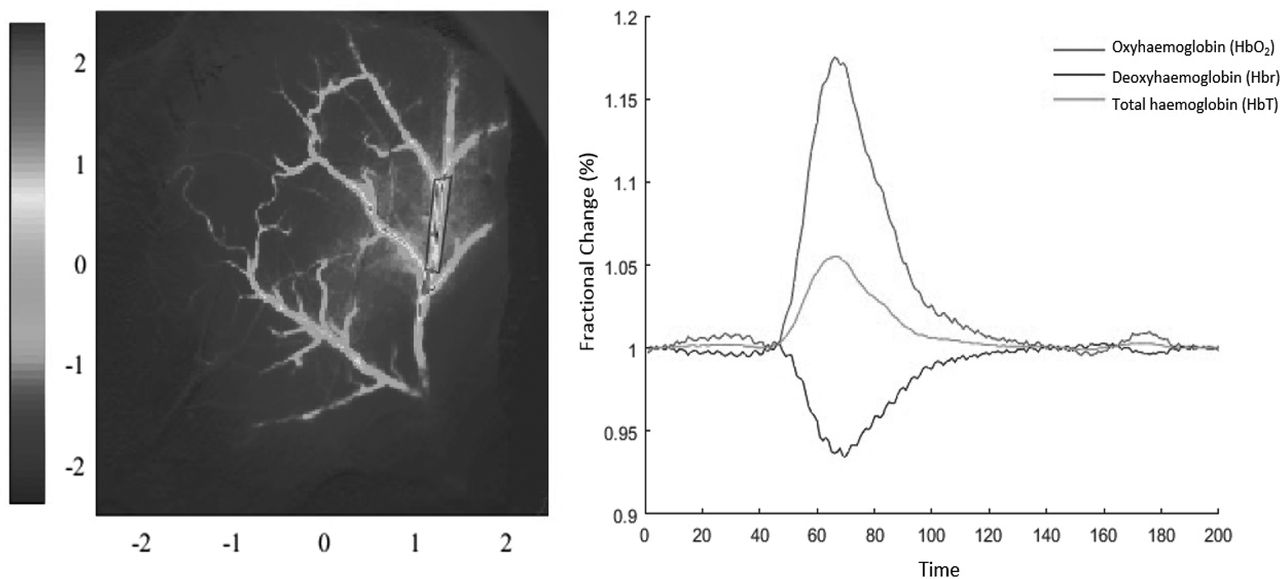
Spatial cortical haemodynamic responses to 16s stimulation in the somatosensory cortex of the ApoE-/- mouse following 8 week Paigen diet. Left panel showing the surface arterial vasculature overlying the somatosensory cortex show the location of cerebral arteries and averaged fractional changes in concentration of oxy- (HbO2), deoxy- (HbR) and total (Hbt) haemoglobin are shown in response to 16 seconds of stimulus evoked cerebral haemodynamics. Scale bar represents z-score of activated pixels. Right panel showing spatial haemodynamic response time series to whisker stimulation in regions of interested select as shown on the right of the arterial vascular compartment

Spatial cortical haemodynamic responses to 2s stimulation in the somatosensory cortex of the ApoE-/- mouse following 8 week Paigen diet. Left panel showing the surface arterial vasculature overlying the somatosensory cortex show the location of cerebral arteries and averaged fractional changes in concentration of oxy- (HbO2), deoxy- (HbR) and total (Hbt) haemoglobin are shown in response to 16 seconds of stimulus evoked cerebral haemodynamics. Scale bar represents z-score of activated pixels. Right panel showing spatial haemodynamic response time series to whisker stimulation in regions of interested select as shown on the right of the arterial vascular compartment
- Neurovascular
- Atherosclerosis
- Neuroimaging