Article Text
Image challenge
A 72-year-old male with recurrent syncope

Abstract
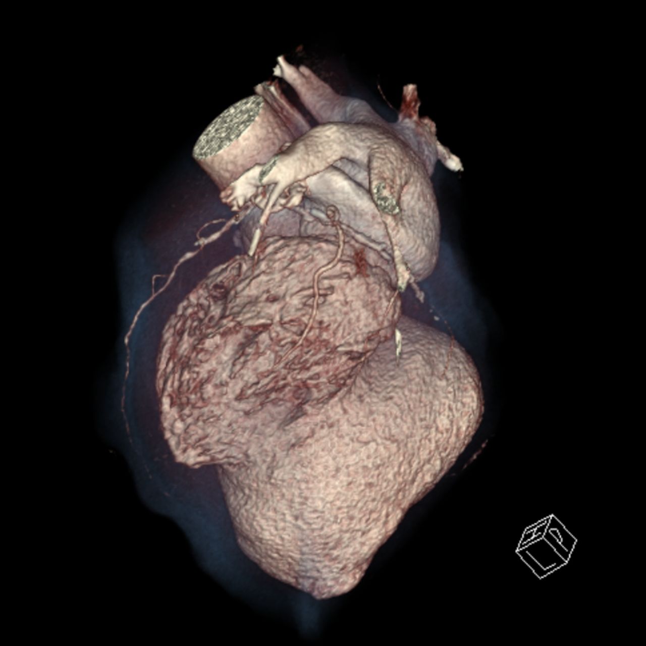
Clinical introduction A 72-year-old patient presented with recurrent syncope 1 year after a myocardial infarction. Two recent falls resulted in fractures to the femur. Serial troponins were negative and ECG demonstrated fixed inferior ST-segment elevation and pathological Q waves. A Holter monitor recorded non-sustained ventricular tachycardia. A subsequent echocardiogram was abnormal, and further investigation with a three-dimensional (3D) cardiac CT coronary angiogram was performed (figure 1).

Figure 1
Cardiac CT coronary angiogram—three-dimensional reconstruction.
Question What is the most likely diagnosis?
Cardiac tumour
Hypertrophic obstructive cardiomyopathy
Ventricular aneurysm
Ventricular diverticulum
Question