Article Text

Abstract
Clinical introduction A 51-year-old woman was referred to our hospital with a 4-month history of progressive dyspnoea on exertion (New York Heart Association Functional Classification III), chest heaviness, dry cough, weight loss and tiredness. She worked as cleaning woman and had no relevant medical history, apart from an Epstein-Barr Virus (EBV) infection 2 months before symptom onset. She did not smoke and family history was negative.
On examination, blood pressure was 104/80 mm Hg and heart rate was regular at 145 bpm. On auscultation, heart sounds were distant, muffled and there was no murmur. Minimal, bilateral pitting oedema was observed. Laboratory findings were unremarkable. During hospitalisation, cardiac monitoring revealed paroxysmal new-onset atrial fibrillation.
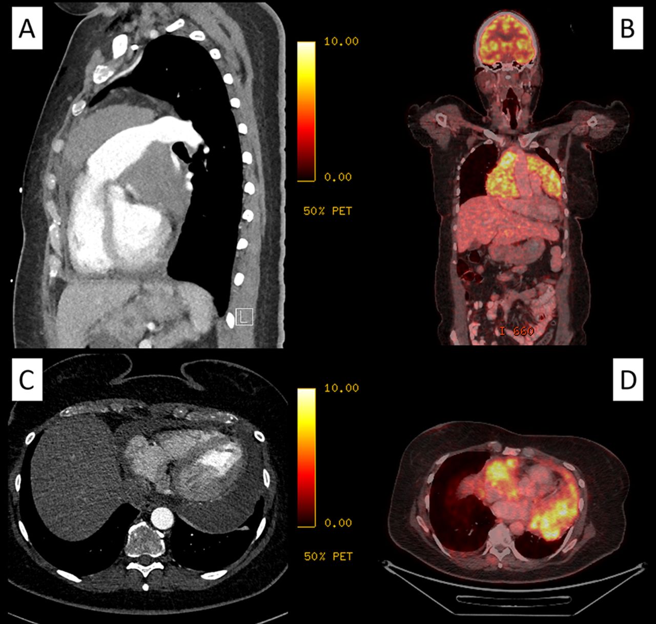
Chest radiography from a previous hospital had revealed cardiomegaly and subsequent echocardiography had shown pericardial effusion with diastolic dysfunction, for which she had received percutaneous pericardiocentesis. However, repeated echocardiography at our hospital showed recurrence of pericardial effusion with diastolic dysfunction and the presence of a pericardial mass. CT and Fluorine-18-fluorodeoxyglucose PET (18F-FDG PET) scanning were done (figure 1).
| Contrast-enhanced CT scanning and 18F-FDG PET scanning. (A) CT scan, sagittal view; (B) 18F-FDG PET scan, frontal view; (C) CT scan, axial view and (D) 18F-FDG PET scan, axial view.
Question Which of the following is the most likely diagnosis?
And based on patient history and imaging, are further diagnostics needed?
Benign pericardial lipoma
Fibrinofibrous pericarditis following EBV infection
Inflammatory pseudotumor
Primary cardiac lymphoma
Primary malignant pericardial mesothelioma
- Cardiac imaging and diagnostics
- Cardiac computer tomographic (CT) imaging
- Pericardial constriction
- Pericardial effusion
- Pericardial tamponade
Statistics from Altmetric.com
Footnotes
Contributors CVdB, LC and VS did clinical assessment and investigation. VS was responsible for care of the patient. CVdB wrote the draft of the report, and LC and VS revised the report. All authors approved the final version.
Competing interests None declared.
Provenance and peer review Not commissioned; externally peer reviewed.
Collaborators Marleen Praet; Eric Derom; Annelies De Paepe.