Article Text

Abstract
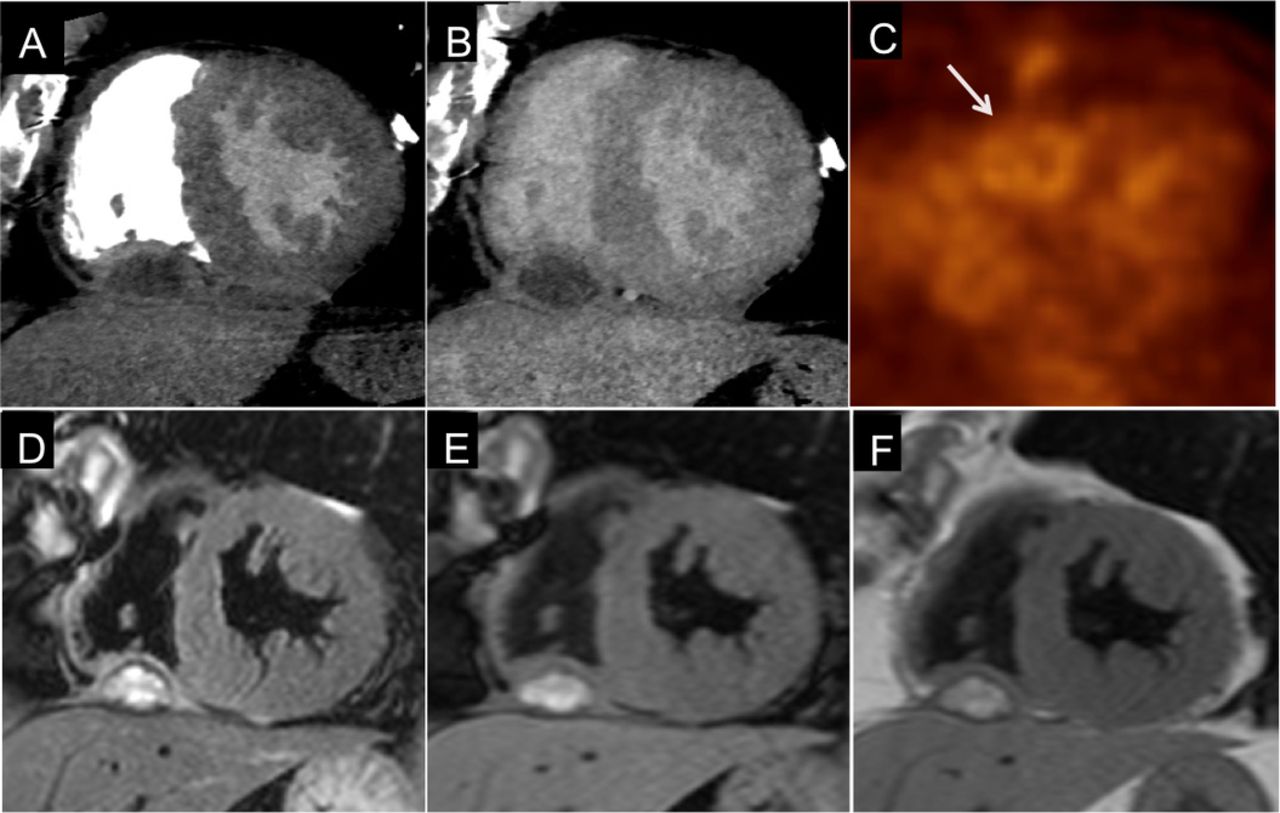
Clinical introduction A 71-year-old man, with a history of chronic aortic regurgitation and negative follow-up after bladder cancer resection 10 months before, had an aortic valve surgery. Two months after, a mass near the right side of the heart had been detected by transthoracic echocardiography performed for dyspnoea, without a cough or fever. The quality of ultrasound images did not allow for an appropriate evaluation due to the outcomes of the sternotomy and the presence of calcified pachypleurite. In order to evaluate this finding, coronary CT (CCT) (figure 1A,B) and positron-emission tomography with 2-[18F] fluoro-2-deoxy-D-glucose (FDG-PET) (figure 1C) were performed. Finally, a cardiac magnetic resonance (CMR) was requested (figure 1D–F, see online supplementary videos).
(A) Short axis image of early contrast enhancement phase coronary CT (CCT); (B) short axis of delayed phase of the same CCT; (C) lesion on positron-emission tomography with 2-[18F] fluoro-2-deoxy-D-glucose image (white arrow); CMR short axis (D) T2-weighted image with fat saturation; (E) T1-weighted image with fat-saturation; (F) T1-weighted image without fat-saturation.
Question Which of the following is the most likely diagnosis of the pericardial mass?
Primary pericardial tumour.
Pericardial metastasis.
Intrapericardial abscess.
Intrapericardial haematoma.
- cardiac imaging and diagnostics
- cardiac magnetic resonance (CMR) imaging
- positron emission tomographic (PET) imaging
- cardiac surgery
Statistics from Altmetric.com
Footnotes
Contributors GG: images and clinical date retrieval, manuscript organisation, manuscript preparation and envoy. LC: bibliography, manuscript and images revision. CN: bibliography, manuscript and images revision.
Competing interests None declared.
Patient consent Not required.
Provenance and peer review Not commissioned; externally peer reviewed.