Article Text

Statistics from Altmetric.com
- cardiac magnetic resonance (cmr) imaging
- valve disease surgery
- aortic stenosis
- transcatheter valve interventions
Learning objectives
To review the pathophysiology of fibrocalcific aortic stenosis, the myocardial response to pressure overload and current clinical guidelines concerning the timing of valve intervention.
To explore and to quantify the risks of earlier intervention in asymptomatic patients compared with the risks of a watchful waiting strategy.
To detail future potential strategies for deciding on timing of aortic valve intervention and current ongoing randomised controlled trials.
Introduction
Aortic stenosis (AS) is the most common valve disease requiring surgical intervention in high-income countries.1 It is characterised by progressive thickening, fibrosis and calcification of the leaflets leading to restriction and valve obstruction.2 The consequent increase in left ventricular afterload leads to a hypertrophic response of the left ventricle, normalising wall tension and maintaining cardiac output. However, with progressive valvular stenosis, this hypertrophic response eventually decompensates resulting in symptom development, heart failure and death.
With no medications proven to attenuate or reverse stenosis progression, the only available treatment is valve replacement. This should ideally be performed when the risks of the disease process (ie, sudden cardiac death, irreversible functional impairment and heart failure) outweigh those of intervention (ie, procedural risk, long-term complications and potential need for reoperation). However, we frequently lack robust evidence to make accurate assessments of such risk. Deciding on the timing of valvular intervention is therefore difficult in many patients, and contemporary clinical guidelines are often underpinned by historical observational data rather than high-quality randomised controlled trials. This article will review our current understanding of the pathophysiology of AS, describe and examine the evidence behind current guideline recommendations and explore potential future strategies to optimise the timing of valve intervention.
Pathophysiology of valvular stenosis and the hypertrophic response
Since the original description of AS by Mönckeberg in 1904, the decline in rheumatic fever and ageing population have led to a demographic transition towards fibrocalcific disease. For many years, fibrocalcific AS was viewed as a degenerative disease where progressive ‘wear and tear’ led to structural damage and passive valvular calcification. However, contemporary thinking is that fibrocalcific AS develops as part of a series of intricate and highly regulated inflammatory, fibrotic and osteogenic processes. The pathophysiological processes driving aortic valve stenosis can be divided into two phases.2 The initiation phase is characterised by endothelial injury accompanied by infiltration of lipids, lipid oxidation and proinflammatory response. Despite the clear similarities with atherosclerosis, three large randomised trials have failed to show any effect of statins on disease progression or clinical outcome. The propagation phase is characterised by the appearance of osteoblast-like cells that coordinate progressive valvular calcium and bone matrix deposition. This osteogenic phenotype involves many signalling molecules involved in bone formation and is both self-perpetuating and highly regulated.2 Advances in imaging now allow for non-invasive assessment of both the burden and activity of calcification in the valve3 4; however, the severity of aortic valve obstruction is still best assessed using echocardiography.5
Myocardial response
The traditional focus of AS assessments has been on the valve. However, the left ventricular myocardial response to pressure overload is equally important,6 particularly as the correlation between echocardiographic measures of AS severity and the degree of myocardial hypertrophy is moderate at best.7 While left ventricular hypertrophy maintains wall stress and cardiac output for many years, it eventually decompensates, with cell death and myocardial fibrosis identified as key processes.8 Many imaging and biomarker surrogates of these processes have been investigated providing significant prognostic information that will be discussed later in this article. Gender appears to have an important influence on both the LV remodelling response and patient outcomes,9 but detailed discussion is beyond the scope of this article.
Current guideline-recommended treatment strategies and their limitations
Broadly speaking, contemporary clinical guidelines recommend aortic valve intervention when stenosis severity is deemed severe and there is evidence of left ventricular decompensation, using either direct objective or surrogate symptomatic measures (figure 1 and table 1).10 11 Haemodynamic severity is best assessed using echocardiography but can be challenging when measures of severity are discordant or low-flow states exist. New recommendations for confirming AS severity are given in the 2017 ESC/EACTS guideline update.5 Detailed discussion of low-flow states is beyond the scope of this article but can be found elsewhere.12
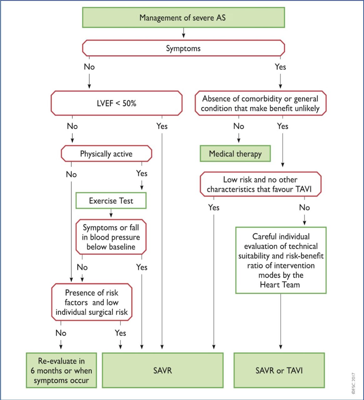
ESC/EACTS algorithm for management of severe AS (2017 guidelines). AS, aortic stenosis; LVEF, left ventricular ejection fraction; SAVR, surgical aortic valve replacement; TAVI, transcatheter aortic valve implantation.
Recommendations for Intervention in patients with severe AS (ESC/EACTS guidelines 2017)
Presence of AS-related symptoms
It is universally accepted that the development of patient symptoms (exertional dyspnoea, angina or syncope; table 2) serves as an indicator of left ventricular decompensation and a dismal prognosis without intervention. This was first described in the seminal paper by Braunwald and Ross in 196813 and forms the underlying framework of how we manage patients today. However, this finding was based on retrospective data from just 12 patients with a mixture of bicuspid and rheumatic valve disease and a mean age of death of 63 years. The changing demographics of AS make it difficult to interpret the current relevance of these historical data to the patients seen in current practice who are frequently in their eighth or ninth decades. Symptom assessment can be highly challenging in these patients due to the high prevalence of both comorbidity, which may cause symptoms to be falsely attributed to AS, and physical inactivity, which can conceal exertion-related problems.
Symptomatology of severe aortic stenosis
Exercise testing may help unmask symptoms in many patients and is safe when performed in stable patients.14 ESC guidelines recommend surgery in patients with severe ASand typical symptoms on exercise test (class I, level C) or a fall in systolic blood pressure at peak exercise (class IIa, level C). This recommendation is based largely on observational data demonstrating that a positive exercise test is a strong predictor of sudden death or symptom development.15 However, these data are limited, consisting of a series of relatively small observational studies with inherent risk of bias and heterogeneity as to what constituted an abnormal test. According to a recent meta-analysis, while the negative predictive value of stress testing for subsequent cardiac events is reasonable (79%), the positive predictive value is modest (66%).14 Exercise testing has other major limitations; up to 20% of patients will be unable to perform the test due to poor mobility, while pre-existing ECG abnormalities are present in up to 50% of patients confounding test interpretation.16 It is worth noting that exercise testing may also detect abnormalities caused by coexistent coronary disease, which is an important determinant of both management and prognosis.17
Impaired left ventricular ejection fraction
Development of left ventricular systolic impairment, as identified by a reduced left ventricular ejection fraction, is an inevitable consequence of progressive and untreated valvular stenosis assuming sudden death does not occur. Although the risk of perioperative mortality is elevated in the setting of reduced ejection fraction, these patients have a dismal prognosis without intervention and improved long-term outcomes with valve replacement earning a class I, level C recommendation in clinical guidelines.10 11
In clinical practice, patients with AS can develop a reduction in ejection fraction for a variety of reasons, and it remains important to consider the mechanism of this reduction and whether it is reversible. Reductions in ejection fraction occur as a direct response to increases in afterload and will reverse following valve replacement. By contrast, the ejection fraction does not improve in approximately 25% of patients10 11 18 19 who are more likely to remain symptomatic and who have adverse long-term outcomes (twice as likely to die over 5 years follow-up).20 In these patients, persistent systolic dysfunction appears related to the development of irreversible scar due to either myocardial infarction or decompensation of the hypertrophic response.21 In sick, frail patients, such information may govern whether valve intervention is likely to be of benefit.
Reductions in ejection fraction are therefore a late, non-specific and often irreversible feature in AS, leading to interest in alternative methods for detecting left ventricular decompensation7 22–24 as will be discussed.
Very severe AS
Patients with critical AS appear to have a particularly poor prognosis, similar to that of symptomatic severe AS.25 Indeed patients with peak aortic jet velocities of >5.0 and >5.5 m/s demonstrate a 2-year event-free survival of 43% and 25%, respectively, compared with 70% in those with Vmax 4.0–4.9 m/s.26 The ESC/EACTS guidelines therefore recommend consideration of aortic valve replacement in patients with Vmax >5.5 m/s if the estimated surgical risk is low (class IIa, level C). However, these observational studies mostly examined the composite endpoint of mortality and referral for aortic valve intervention with a strong risk of referral bias and event rates mainly driven by decisions to perform surgery.
Rapid haemodynamic progression
Although the average rate of progression (measured by peak aortic-jet velocity) is 0.24±0.30 m/s/year, this rate is highly variable.27 Moreover, it is subject to scan–rescan variation in peak velocity measurements, which can be high in clinical practice. Patients with rapid progression (>0.3 m/s/year) and significant valve calcification have a rate of symptom development or mortality of 79% at 2 years.28 As a result, referral for surgical intervention in these patients is given a class IIa, level C recommendation in the latest guidelines. However, again, this is based on limited observational data, and this strategy requires standardised high-quality echocardiography over several years to confidently determine rate of progression.
Elevation of B-type natriuretic peptide (BNP) levels
BNP is the first cardiac biomarker to be included in the decision-making algorithm for aortic valve replacement. Early studies investigating natriuretic peptides in AS showed promise but were criticised for their small size, observational nature and use of softer outcome endpoints.29 30 In addition, many patients were symptomatic, and the variation in normal BNP with age and sex were not accounted for. A more recent study of 565 patients with asymptomatic moderate-to-severe AS identified that a BNP ratio (measured BNP value divided by upper limit of normal for patient’s age and sex) of >1 was independently predictive of mortality and a ratio of >3 had an HR of 7.3 for survival in patients with asymptomatic severe AS.31 As such, the latest clinical guidelines reflect these data with a level IIa, class C recommendation for aortic valve replacement if the BNP ratio is persistently above 3 and overall surgical risk is low. However, BNP is a non-specific marker of cardiac dysfunction, and its utility, like each of the other parameters, has yet to be tested in a randomised controlled trial.
The recently published ESC clinical guidelines also removed two previous IIb indications for AVR in asymptomatic patients: an increase in mean aortic gradient of >20 mm Hg with exercise, or the finding of excessive LV hypertrophy in the absence of hypertension.
Balancing competing risks
There are clear limitations with many of our guideline-advocated strategies. Most are based on limited observational data and supported by level C recommendations. There is therefore a need for randomised controlled trials assessing the optimal timing of surgery and novel objective methods to guide this major clinical decision. Ideally, intervention would be performed in patients just as the left ventricle is starting to decompensate but before substantial irreversible damage has accrued and at a time when the short-term and long-term risks of the intervention are outweighed by the risks of not intervening (figure 2 and table 3). An understanding of these competing risks is therefore critical.

Optimising the timing of aortic valve intervention in progressive aortic stenosis. TAVI, transcatheter aortic valve insertion.
Estimates of clinical risks associated with watchful waiting or early intervention strategies
Risks of valve intervention
Surgical aortic valve replacement remains the standard of care for valvular intervention, with improvements in surgical and postoperative care driving perioperative mortality down to ~1%–3%. Other important perioperative complications include conduction disease requiring permanent pacemaker insertion (1.5%–8.6%32) and cerebrovascular accidents (2.4%–8.1%33–35). There is also the risk of cognitive decline (due to perioperative cerebral hypoperfusion microemboli or anaesthetic agent neurotoxicity35). An individual’s risk of these complications can be estimated using surgical risk calculators such as EUROSCORE II and the Society of Thoracic Surgeons score. An argument in favour of early surgery is that operative risk is lower in younger patients that are asymptomatic, have less comorbidity and have normal left ventricular function.
The emergence of minimally invasive transcatheter aortic valve insertion (TAVI) over the last 10 years has completely changed the landscape for decision making regarding valve intervention in symptomatic patients. Current trials show non-inferiority of this percutaneous technique compared with surgical intervention in both high-risk and intermediate-risk patients,34 36–39 and procedural risk may further reduce with increasing clinical experience and advances in prosthesis design and delivery. Indeed, major vascular complications have decreased substantially (from >10% to <5%40) as have stroke rates, which are between 2% and 3% in contemporary cohorts.40 However, the requirement for permanent cardiac pacing postprocedure remains consistently higher than surgical intervention at >10%40 and while TAVI allows for rapid patient recovery and mobilisation, the long-term durability of these bioprostheses has not been demonstrated.41 This will be key before their widespread use in younger or asymptomatic patient groups can be recommended.
Performing valve intervention introduces small but significant annual risks associated with the presence of a prosthetic valve. These risks are heavily influenced by valve type, with both anticoagulant related major bleeding (1.8%–2.6% per year) and thromboembolism (0.7%–1.0% per year) more frequent with mechanical valves.42 In addition, there is an increased risk of endocarditis (1%–3% during the first year then <0.5% per year43), which has a high associated morbidity and mortality. Whereas structural valve degeneration is exceedingly rare in mechanical valves, bioprosthetic valves have a limited lifespan which can be difficult to predict. In these patients, valve degeneration usually starts to occur 10 years following implantation44 and occurs more rapidly in younger patients.45 This is an extremely important issue if bioprosthetic valves are to be used in younger asymptomatic patients. Ongoing research into decellularisation techniques and tissue engineering may lead to improved bioprosthetic valve longevity, while advances in mechanical valve design might eventually eliminate the need for anticoagulation and associated bleeding risk. In addition, the use of a transcatheter valve inside a surgical bioprosthetic valve (so called valve-in-valve TAVI) may reduce the risk of future procedures should valve degeneration occur.
Risks of not intervening
The risk of sudden cardiac death in patients with asymptomatic severe AS managed conservatively is ~1% per year and occurs without preceding symptoms in 70% of cases.46–48 Once symptoms develop, further clinical deterioration can be rapid with a significant risk of sudden death while awaiting intervention (4% at 1 month, 12% at 6 months).49
Delaying aortic valve intervention until there is evidence of advanced left ventricular decompensation results in greater perioperative risks.48 Observational studies have quoted increased perioperative mortality (9%–19%10 20 50) in patients who have developed left ventricular systolic impairment and advanced myocardial fibrosis.51 Further risk stratification can be performed using stress echocardiography to assess myocardial contractile reserve, with lower perioperative risks if contractile reserve is present (5% vs 22%–32%11 52). However, given the dismal prognosis of untreated AS, even patients without contractile reserve have improved long-term survival if they survive the perioperative period.10 11
The highest burden in mortality and morbidity related to delaying valve intervention appears to occur in the months and years following AVR, particularly in those patients that have evidence of left ventricular decompensation. As discussed, patients with an impaired ejection fraction prior to AVR have a poor long-term prognosis,20 while in a recent study of AS patients with a high probability of LV decompensation, more than half were either dead or admitted to hospital with heart failure within 2 years.53 Both these observations may reflect the development of irreversible scarring in the myocardium while patients are waiting for surgery.
Possible future strategies
Several different strategies for optimising the timing of valve replacement in AS have been proposed, many of which are currently being evaluated within the context of randomised controlled trials (figure 3 and table 4). Many of these target asymptomatic patients, and it should be recognised that many patients that feel otherwise fit and healthy might not want to undergo major heart surgery.

Comparison of EARLY-TAVR and EVoLVeD randomised controlled trial designs. Currently, recruiting randomised controlled trials generally fall into two groups: those investigating valve intervention in all asymptomatic patients with severe AS (eg, EARLY-TAVR) and those looking to target intervention based on measures of left ventricular decompensation (eg, EVoLVeD). AS, aortic stenosis; CMR, cardiac magnetic resonance; EARLY-TAVR, Evaluation of Transcatheter Aortic Valve Replacement Compared to SurveilLance for Patients with AsYmptomatic Severe Aortic Stenosis; EVoLVeD, Early Valve Replacement Guided by Biomarkers of Left Ventricular Decompensation in Asymptomatic Patients with Severe AS; hs, high-sensitivity; LV, left ventricular; TAVI, transcatheter aortic valve insertion.
Current and planned randomised controlled trials investigating timing of aortic valve intervention
All-comers with severe AS
Historical teaching has been that ‘aortic valve replacement is the most common cause of death in patients with asymptomatic severe aortic stenosis’. However, improving outcomes following surgical and transcatheter valve replacement are challenging this doctrine. Performing valve intervention on all asymptomatic patients with severe AS is a simple and pragmatic solution that does not seek to identify the point at which left ventricular decompensation occurs. Although some patients will undergo intervention earlier than they may have required (and therefore be exposed unnecessarily to the problems associated with prosthetic valves), the risks associated with contemporary intervention techniques are low, and no patient should be left with irreversible left ventricular decompensation. This strategy is supported by evidence from the Japanese Contemporary outcomes after sURgery and medical tREatmeNT in patients with severe Aortic Stenosis (CURRENT AS) registry. Propensity-score matching was used to compare 291 asymptomatic patients who underwent early surgery with 291 patients who were managed conservatively. Those who received early AVR had a reduced all-cause mortality at 5 years (15%) compared with those who were initially managed conservatively (26%). Heart failure hospitalisation was also reduced in the early intervention group (4% vs 20%). However, propensity matching may not have accounted for all potential influences on outcomes and a significant proportion of the conservatively managed patients who developed symptoms were not referred for intervention, undoubtedly contributing to the worse observed survival in this group: confounding by indication. Three randomised controlled trials (AVATAR, ESTIMATE and EARLY-TAVR; table 4) are currently recruiting, which will examine whether valve intervention in unselected asymptomatic patients with severe AS can improve clinical outcomes.
Refined assessment of valve structure and function
An alternative strategy is to operate only in asymptomatic patients with very high peak aortic-jet velocities. Peak velocities >4.5 m/s are associated with increased referral for surgical intervention26 but also increased rates of perioperative death and cardiac death in a prospective cohort study with propensity matching.47 The RECOVERY randomised controlled trial will examine whether early aortic valve replacement in asymptomatic patients with velocities >4.5 m/s and a negative exercise test leads to improved patient outcomes compared with watchful waiting (table 4).
The total haemodynamic load seen by the left ventricle can also be quantified by calculating the valvuloarterial impedence (ZVa=(systolic blood pressure+mean AV gradient)/indexed LV stroke volume). This measure has consistently been shown to be an independent marker of adverse outcome in asymptomatic patients54 and warrants further study for its use in determining the timing of intervention.
Another approach is to quantify valvular calcium burden using CT calcium scoring. Validated, sex-specific thresholds for severe AS have been proposed (2000 Agatston units (AU) for men, 1200 AU for women),5 which provide powerful prediction of clinical events of incremental value to echocardiographic assessments.3 Performing valve intervention on the basis of severe valvular calcification on CT might therefore represent an attractive alternative strategy.
Imaging and biomarkers of left ventricular decompensation
Simple cardiac biomarkers beyond BNP are being investigated in AS as markers of LV decompensation. Cardiac troponin is a structural protein present in cardiomyocytes, which is released into the bloodstream during myocardial injury and can now be detected at very low plasma concentrations using high-sensitivity assays. In AS, troponin I concentrations are associated with a more advanced left ventricular hypertrophic response, replacement myocardial fibrosis and worse long-term patient outcomes in patients with AS.55 They are thought to reflect the cardiomyocyte death that drives progressive left ventricular decompensation alongside myocardial fibrosis.8 Elevation in cardiac troponin is not however specific to AS. By contrast, the presence of LVH and the strain pattern on the 12-lead ECG demonstrate high specificity (but low sensitivity) for left ventricular hypertrophy and myocardial fibrosis, respectively, and also provides prognostic information.56 As will be discussed, there is interest in using these simple and cheap biomarkers as screening tools to aid the detection of LV decompensation (figure 4).

Imaging and biomarker assessments of stage of valvular stenosis and myocardial response to increased afterload. Progressive haemodynamic obstruction as a result of aortic leaflet restriction is assessed using echocardiography. However, specific valvular pathologies such as fibrosis and calcification can be assessed using CT methods. Ejection fraction is a poorly sensitive marker of myocardial decompensation with abnormalities in Doppler measures, longitudinal strain and systolic function, which are all detectable prior to this. However, these measures, along with biomarkers such as troponin and B-type natriuretic peptide (BNP) are non-specific and may be abnormal as a result of coexistent myocardial pathology such as coronary heart disease. T1 mapping methods and late gadolinium enhancement are more specific for decompensation as a result of pressure overload.
What about imaging assessment to detect left ventricular decompensation? Despite its limitations, ejection fraction remains the current gold standard; however, several imaging techniques are under early investigation that can detect earlier abnormalities in left ventricular function. Echocardiography can detect alteration in various measures of diastolic and longitudinal systolic function in patients with AS, which appear related to the presence of myocardial fibrosis.7 Reduced left ventricular global longitudinal strain can be observed in asymptomatic patients with AS, acting as an independent predictor of mortality.23 However, several of these measures still require standardisation across vendor platforms and all suffer from significant overlap between results in healthy individuals and those with AS. Moreover, these imaging markers are not specific to valve heart disease and like symptoms might equally reflect comorbidity such as ischaemic heart disease.
Perhaps the most promising technique is cardiac MRI, which offers myocardial tissue characterisation and can detect the myocardial fibrosis that drives the development of left ventricular decompensation. Indeed, the late gadolinium enhancement technique allows direct visualisation of this fibrosis in a midwall pattern that can easily be differentiated from prior myocardial infarction. Midwall fibrosis is a direct and specific marker of left ventricular decompensation with close association with measures of left ventricular function, myocardial injury and functional capacity.7 Furthermore, multiple studies have confirmed that midwall fibrosis is a strong independent predictor of all-cause mortality and cardiovascular death.21 51 57 Increasing burden of midwall fibrosis correlates with a worse outcome,21 51 and this fibrosis appears irreversible following valve intervention.58 Midwall fibrosis therefore appears as a useful tool to identify the early stages of irreversible left ventricular decompensation. T1 mapping is an alternative technique that might allow detection of the preceding stage of reversible diffuse interstitial fibrosis.59 At present, there are issues with variations in T1 values at different magnetic field strengths and on different scanners and problems caused by the overlap of T1 values between different disease states. Further work is required, although recent studies have shown promising early results for T1 parameters that seek to measure the overall myocardial fibrosis volume.7
It is possible that using a multibiomarker strategy to identify LV decompensation may prove superior to any single biomarker in isolation. For example, in the EVoLVeD randomised controlled trial (table 4), patients are initially screened with high sensitivity troponin I and an ECG. Patients with a normal troponin (<6 ng/L) are deemed to have a normal heart with no further imaging required. Patients with an elevated troponin or the ECG-strain pattern proceed to cardiac magnetic resonance (CMR) and those found to have midwall fibrosis then randomised to either early valve intervention or routine clinical care. It is hoped that this strategy will target valve intervention to those patients who will derive greatest benefit.
Practical recommendations and processes of care
Care of the asymptomatic patient with AS can be complex and challenging. While additional evidence is awaited, we advise detailed clinical and echocardiographic assessment with exercise testing and advanced imaging (eg, stress echocardiography, TOE, CT and CMR) performed as indicated to clarify symptoms, severity of AS and myocardial health. This should be performed by heart valve specialists working as part of a heart team.5 Patient involvement in decision making is key where indications for intervention are borderline. Given delays between referral and procedure that are common in most healthcare systems, this could also be discussed with the patient and may inform decision making for earlier intervention. The recent ESC/EACTS guideline update also recommends the establishment of heart valve centres with access to advanced imaging modalities and contemporary interventional techniques supported by robust internal audit processes.5 Finally, patients managed conservatively should be educated as to the typical symptoms of AS (table 2) and the importance of prompt symptom reporting. Regular clinic surveillance is essential, and current guidelines recommend clinical assessment with echocardiography at least every 6 months.5
Conclusions
AS is a common condition; the only treatment for which is replacement of the aortic valve. The optimal timing of this valve intervention remains unclear with current guidelines based on observational data and expert opinion. However, multiple randomised controlled trials are currently underway investigating whether novel strategies might improve patient outcomes compared with current watchful waiting, heralding a new era of evidence-based medicine for patients with heart valve disease.
Key messages
Aortic stenosis is a disease of both the valve and the myocardium, characterised by fibrosis and calcification of valve leaflets, progressive left ventricular hypertrophy and myocardial fibrosis.
Although no randomised controlled trial data exist, current clinical guidelines recommend valve intervention when severe aortic stenosis is accompanied by evidence of left ventricular decompensation.
Timing of valve intervention is crucial. Too early and the patient will be unnecessarily exposed to risks of intervention and prosthetic valve complications; too late and irreversible myocardial damage can lead to persistent symptoms and risk of adverse events. Ideally valve replacement would be performed just as left ventricular decompensation is starting to develop.
Improved surgical methods and perioperative care, as well as transcatheter aortic valve implantation techniques have resulted in major reductions in procedural risk. As such, earlier valve intervention in asymptomatic patients could be contemplated, and randomised controlled trials are underway that will help inform our future management.
CME credits for Education in Heart
Education in Heart articles are accredited for CME by various providers. To answer the accompanying multiple choice questions (MCQs) and obtain your credits, click on the ‘Take the Test’ link on the online version of the article. The MCQs are hosted on BMJ Learning. All users must complete a one-time registration on BMJ Learning and subsequently log in on every visit using their username and password to access modules and their CME record. Accreditation is only valid for 2 years from the date of publication. Printable CME certificates are available to users that achieve the minimum pass mark.
References
Footnotes
Contributors RJE performed the literature search and drafted the article. Critical revisions were performed by M-AC, PP and MRD. All authors approved the final manuscript and MRD is the guarantor for the overall content.
Funding The work was supported by the British Heart Foundation (FS/14/78/31020 to MRD), the Sir Jules Thorn Charitable Trust (15/JTA to MRD), the Québec Heart and Lung Institute Foundation and Canadian Institutes of Health Research (FDN-143225 and MOP-114997 to PP and M-AC).
Competing interests None declared.
Patient consent Not required.
Provenance and peer review Commissioned; internally peer reviewed.
Data sharing statement Not applicable.
Author note References which include a * have been selected as key references for this paper.