Article Text

Abstract
Introduction Immune cells are known regulators of cardiac repair, modulating both beneficial and detrimental processes following myocardial infarction (MI). Harnessing the beneficial effects of macrophages remains an elusive and, as yet, untapped therapeutic option following MI. Zebrafish regenerate their hearts after injury in a macrophage dependent manner, offering a much-needed roadmap of optimal macrophage phenotype following MI.
Methods We used a model of heart laser-injury in the zebrafish embryo to characterise cardiomyocyte-macrophage interactions after cardiac injury. Real-time optically-gated light sheet microscopy permitted imaging of macrophages infiltrating the beating heart 0–48 hours after injury. Our in vivo imaging of cardiomyocyte-macrophage interactions has facilitated qualitative and quantitative assessment of inflammatory processes after cardiac injury.

Abstract 89 Figure 1
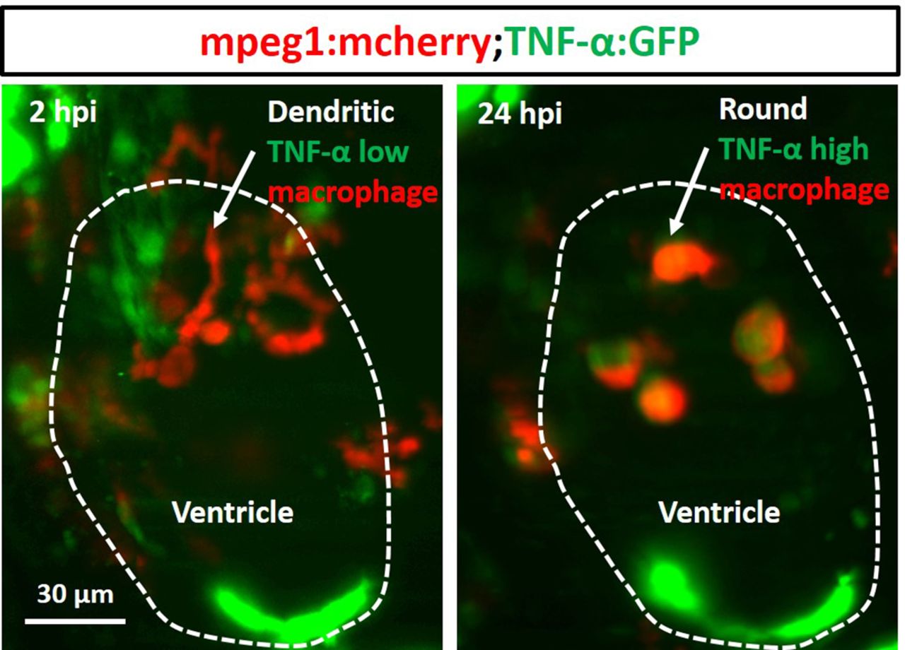
Results During the first four hours after injury, the injury-site expands by secondary cell death reaching 6.67% ±1.5 of total ventricular volume. The injury begins to regress from four hours post-injury (hpi), decreasing to 2.1% ± 0.69 of ventricular volume by 48 hpi. Wound-associated macrophages during the first 12 hours are motile, dendritic in morphology with TNF-α signal. As repair progresses these cells become less motile with rounded morphology and strong TNF-α signal. Cardiomyocyte proliferation occurs over approximately two days alongside budding and bridging of cardiomyocytes from the wound margins. Macrophages migrate to the necrotic wound border, peaking in numbers at 12 hours post-injury and remain at the site in elevated numbers for up 48 hours after injury.
Conclusions During heart regeneration individual macrophages undergo a phenotypic switch at the site of injury with upregulation of TNF-α, suggesting that wound-associated macrophages transition to an inflammatory activation state. This zebrafish injury model appears to replicate the secondary apoptosis of peri-infarct cardiomyocytes observed in human MI, offering a simple system in which to investigate the impact of interventions on macrophage function following cardiac injury.

Abstract 89 Figure 2