Article Text

Abstract
Background Pulmonary hypertension (PH) is debilitating disease characterized by a progressive increase in pulmonary arterial pressure (PAP) that leads to right ventricular (RV) failure and death. Mixed venous oxygen saturation (SVO2) represents the oxygen saturation of blood returning to the lungs before reaching the alveolo-capillary units. SVO2 is strongly associated with clinical outcomes in PH. The relationship of non-invasive CMR metrics to this prognostically relevant parameter in patients with PH are unknown.
Purpose This study sought to develop an early understanding of which CMR volumetric and flow parameters are most associated with SVO2.
Methods Eighteen (n=18) patients were prospectively recruited at a large tertiary PH unit. The SVO2 was measured during right heart catheterisation. All patients had CMR on the 1.5 T scanner (HDx scanner, GE Healthcare, Waukesha, Wisconsin, USA), using an 8-channel cardiac coil. Subjects were scanned in the supine position with electrocardiogram (ECG) gating. CMR protocol included long and short axis cines and through-plane pulmonary artery phase contrast acquisition. The velocity encoded images were analysed for the following: mean pulmonary artery (MPA) systolic velocity, MPA stroke volume, MPA wall shear stress (WSS) and wall shear rate (WSR). The 4 chamber cine was used to measure end-diastolic right atrial (RA) area. RV volumes were analysed using standard methods. Stepwise multiple regression model of significantly associated parameters (p<0.05) was developed.
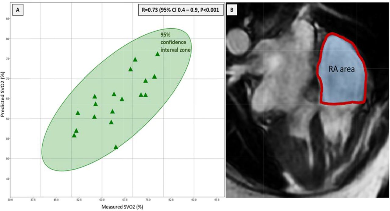
Results The left ventricular and RV volumetric functional parameters demonstrated no association to SVO2 (p>0.05) in any of the participants. However, a negative association was shown between RA area and SVO2 (R=−0.57, p=0.01). The only other parameter which correlated with SVO2 was MPA stroke volume (R=0.5, p=0.03). In stepwise multiple regression, both parameters demonstrated independent association to SVO2. The predictive values generated by a combined model demonstrated high correlation to measured SVO2 (R=0.73, p<0.001).
Summary of CMR parameters evaluated in the study

Panel A: scatter plot of measured and predicted SVO2. Panel B: Right atrial size was computed in 4-chamber view at end-systole (RV), just before the opening of the tricuspid valve
Conclusion(s) RA area and MPA stroke volume are independently associated with SVO2. A novel CMR prediction model comprised of these two metrics, demonstrates high association to the measured SVO2 by invasive haemodynamic study. Further work to determine reproducibility and reliability is needed before this method becomes an important prognostic tool for PH patients.