Article Text

Abstract
Introduction The apelin receptor is a class A GPCR that binds two endogenous peptide ligands, apelin and elabela/toddler, to regulate the cardiovascular system. Apelin peptide levels are downregulated in pulmonary arterial hypertension (PAH) but the receptor remains present and clinically targetable. The NIHR BioResource BRIDGE study, a prospective component of the Genomics England 100,000 Genomes Project, has performed case-control genomic analysis of ∼7423 patients with rare cardiovascular diseases including PAH, and has identified a number of mutations in the apelin receptor in this cohort. Of these, 11 were selected for further assessment using high content imaging, following previous saturation binding studies that showed mutational effects on receptor expression and ligand binding.
Methods The Opera PhenixTM High Content Screening System was used to generate high-throughput triple fluorescence confocal images in fixed CHO-K1 cells transiently transfected with eGFP-tagged wild-type or mutant apelin receptor. Cells were stained with wheat germ agglutinin-AlexaFluor-594 and Hoechst 33342 as membrane and nuclear markers respectively. Harmony High Content Imaging and Analysis Software was used to empirically quantify fluorescence intensities of the eGFP-tagged apelin receptor in the cytoplasm and membrane of transfected cells.
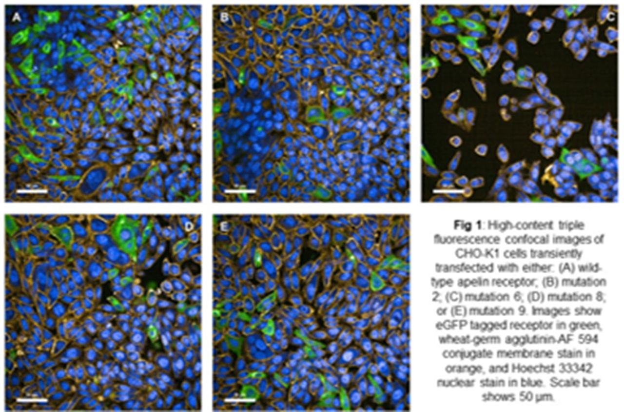
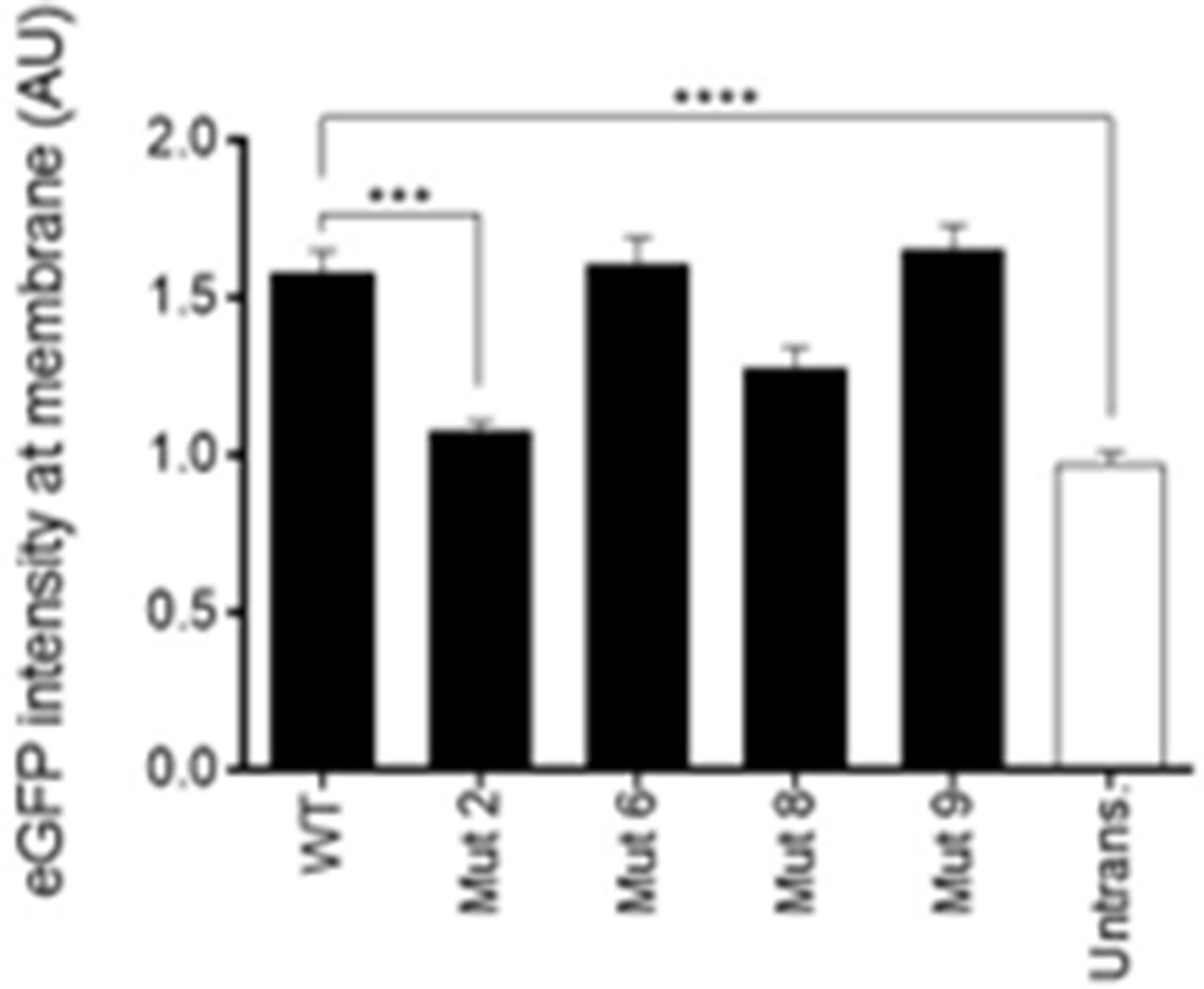
Results Images showed high expression of wild-type receptor with some degree of co-localisation with the membrane stain (fig 1A; fig 2). Mutation 2 showed visibly lower total protein and reduced membrane expression (fig 1B; fig 2). However, mutations 6 and 9 (Fig 1C and 1E respectively) showed similar expression and no significant difference in membrane co-localisation compared to wild-type (fig 2). Mutation 8 showed a trend for reduced membrane co-localisation (fig 1D) compared to wild-type. Note that membrane co-localisation differences closely matched differences in overall eGFP signal. Future work will aim to use machine learning to accurately isolate cell populations that show definite apelin receptor-eGFP signal at the membrane before re-running analysis.

Abstract BS46 Figure 1

Abstract BS46 Figure 2
Conclusion We report the successful use of high content imaging to quantify differences in expression and membrane co-localisation of the apelin receptor when naturally occurring mutations identified in the NIHR BioResource BRIDGE study are introduced into the protein. When considered with receptor affinities and densities in previous saturation binding data, this work suggests that certain mutations may exhibit an overall impact on receptor expression (i.e. mutation 2), whilst others may directly alter ligand binding without significantly influencing protein levels (i.e. mutant 8). This work may provide great insight on the effects of real human apelin receptor mutations that may be contributing to disease phenotypes.
Conflict of interest none