Article Text

Abstract
The mechanisms linking shear stress, endothelial physiology and plaque biology are currently poorly understood, but their elucidation could identify new strategies to reduce plaque growth and rupture. To address this, we use eNOS as high shear stress marker coupled to immunofluorescent staining, optical clearing and light-sheet microscopy, to develop a system for analysing the spatial distribution of proteins in murine plaques and correlate them with local WSS.
Results Confocal microscopy of concavity mounted slides revealed strong eNOS staining at the outer curvature of WT mice but significantly reduced staining at the inner curvature (N=5; P<0.001). This is consistent with high WSS induction of eNOS because the outer curvature corresponds to a HSS site (Suo et al) however precise correlations between eNOS and shear stress could not be made because the tissue geometry was lost during processing.
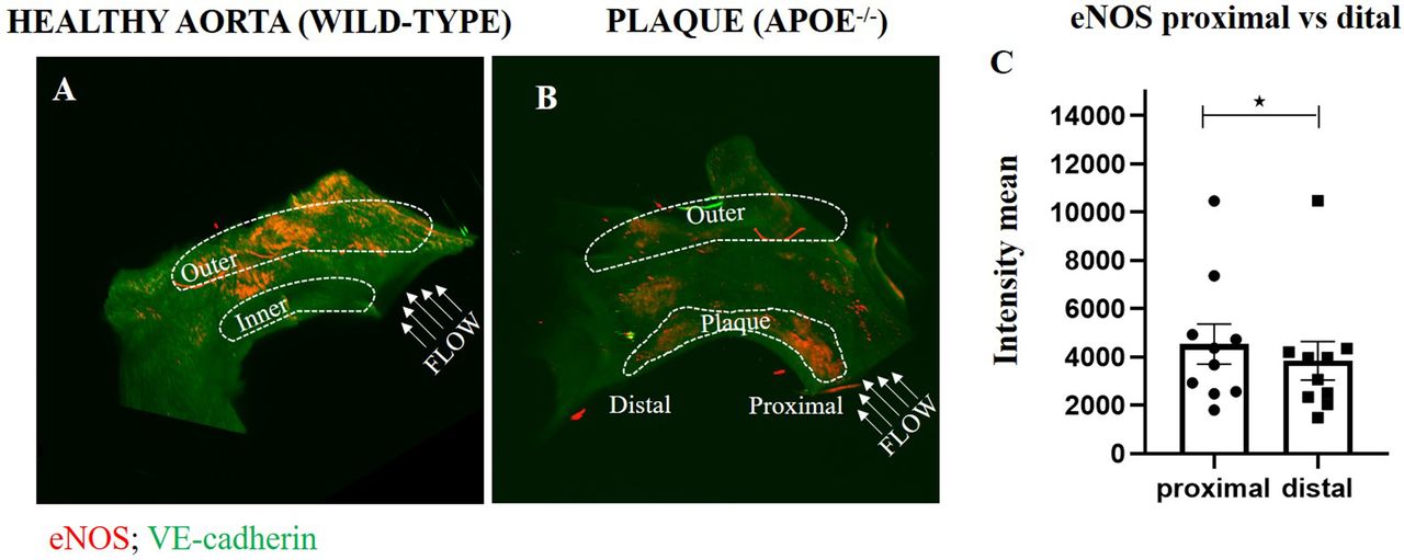
Light-sheet microscopy of cleared samples with preserved 3D structure confirmed elevated expression of eNOS at HSS regions of the outer curvature (figure 1A; N=5 WT; P<0.01). In aortic arches of ApoE-/- mice, eNOS was observed at the outer curvature but was also present at portions of atherosclerotic plaques (figure 1B). Further analysis revealed that eNOS expression was higher at the proximal (upstream) part of the plaque compared to distal (downstream) suggesting a potential correlation with WSS (figure 1C).
Conflict of Interest Atherosclerosis