Article Text

Abstract
Infective endocarditis (IE) is an evolving disease with a persistently high mortality and morbidity, even in the modern era of advanced diagnostic imaging, improved antimicrobial chemotherapy, and potentially curative surgery. Despite these improvements in health care, the incidence of the disease has remained unchanged over the past two decades and may even be increasing. Chronic rheumatic heart disease is now an uncommon antecedent, whereas degenerative valve disease of the elderly, mitral valve prolapse, intravenous drug misuse, preceding valve replacement, and vascular instrumentation have become increasingly common, coinciding with an increase in staphylococcal infections and those caused by fastidious organisms. The current understanding of this difficult condition is reviewed and recent developments in medical and surgical management are updated.
- ELISA, enzyme linked immunosorbent assay
- HACEK, Haemophilus species, Actinobacillus actinomycetemcomitans, Cardiobacterium hominis, Eikenella corrodens, and Kingella kingae
- ICE, International Collaboration on Endocarditis
- IE, infective endocarditis
- PCR, polymerase chain reaction
- infective endocarditis
- valve disease
Statistics from Altmetric.com
- ELISA, enzyme linked immunosorbent assay
- HACEK, Haemophilus species, Actinobacillus actinomycetemcomitans, Cardiobacterium hominis, Eikenella corrodens, and Kingella kingae
- ICE, International Collaboration on Endocarditis
- IE, infective endocarditis
- PCR, polymerase chain reaction
Come and look, Madame Mahler. Even I have not seen streptococci in such a marvellous state of development. Just like seaweed.—Gustav Mahler’s bacteriologist, Paris, 1911
Almost 100 years since the death of the great Bohemian symphonic composer from complications of the disease, infective endocarditis (IE) continues to surprise, frustrate, and perplex. Even in the modern era of advanced diagnostic imaging, improved antimicrobial chemotherapy, and potentially curative surgery, IE remains an evolving disease with a persistently high mortality and morbidity. Despite these improvements in health care, the incidence of the disease has remained unchanged over the past two decades at approximately 1.7–6.2 cases/100 000 patient years and may even be increasing.1 Almost all aspects of the disease, including its natural history, predisposing factors, sequelae, and causative organisms, are virtually unrecognisable compared with Osler’s original descriptions from the 19th century. In particular, chronic rheumatic heart disease is now an uncommon antecedent, whereas degenerative valve disease of the elderly, mitral valve prolapse, intravenous drug misuse, preceding valve replacement, and vascular instrumentation have become increasingly common, coinciding with an increase in staphylococcal infections and those caused by fastidious organisms. Furthermore, previously undetected pathogens are now being identified with the disease and multidrug resistant bacteria challenge conventional treatment regimens. This short article provides a concise review of current understanding of this difficult condition and an update of recent developments in medical and surgical management.
EPIDEMIOLOGY
A recent review of contemporary case series encompassing a total of 3784 episodes of IE between 1993 and 2003 found a median incidence of 3.6/100 000 population/year with a progressive increase in relation to age.2 The male to female ratio was 2:1 and median in-hospital mortality rate 16% (range 11–26%). Staphylococci and streptococci accounted for the majority of cases and notable trends included a rising prevalence of staphylococcal skin flora caused by iatrogenic nosocomial infection, Staphylococcus aureus affecting intravenous drug users, and Streptococcus bovis (mainly Streptococcus gallolyticus) in the elderly, often connected to underlying gastrointestinal neoplasia. These findings, particularly the increasing problem of IE affecting the elderly population, have been confirmed in other recent European series.3–5
Nosocomial infection
Nosocomial infection accounted for endocarditis in 22% of one recent series with a mortality greater than 50%.6 Predominant pathogens were staphylococci and enterococci, often related to intravenous catheters or surgical procedures, and fewer than 50% of patients had underlying structural heart disease. Particular risk groups in this category include the immunosuppressed with central venous catheters and those undergoing haemodialysis.
Intravenous drug users
Intravenous drug users predominate in series of young people and overall incidence of IE in this group is 1–5%/year.7 The tricuspid valve is infected in over 50% of patients and the majority have no known pre-existing cardiac disease. Repeated injections of impure material could, however, encourage cytokine production, valvar inflammation, and fibronectin deposition on previously healthy valve tissue, thereby predisposing to infection. S aureus species predominate, although unusual infections including Pseudomonas aeruginosa, fungi, bartonella, salmonella, and listeria may also be encountered, particularly in those who are HIV positive, where outcome is inversely related to CD4 count.8
Prosthetic valve endocarditis
Prosthetic valve endocarditis accounts for 10–15% of most series with an overall incidence of 0.1–2.3%/patient year.9 Cases may be classified as early or late depending on whether infection arises within one year of surgery or later, and both mechanical valves and bioprostheses appear equally susceptible. Early infection peaks two months after surgery and is often caused by Staphylococcus epidermidis or S aureus, whereas the spectrum of late infection mirrors that of native valve disease.
PATHOPHYSIOLOGY
A detailed discussion of the clinical features of IE is beyond the scope of this article and is covered elsewhere.1 Both acute and insidious presentations are common and classical clinical signs are often absent. Thus, a low index of clinical suspicion and early investigation of those at risk are decisive.
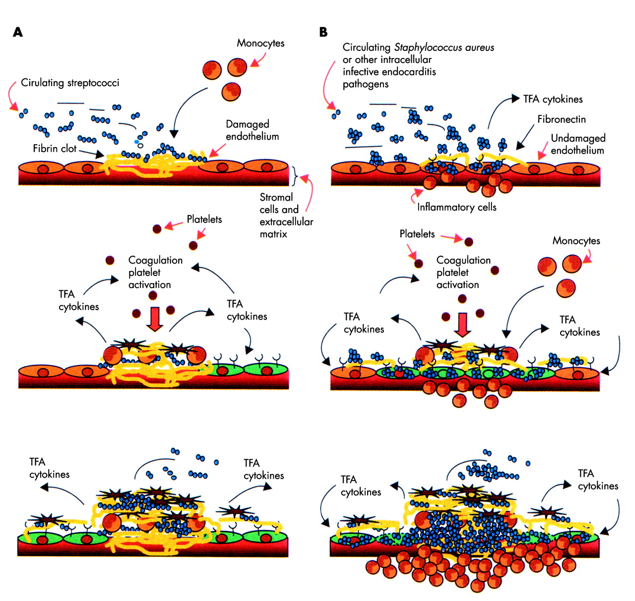
Recent advances in our understanding of the underlying pathophysiology, particularly in staphylococcal and streptococcal infection, provide insight into mechanisms of disease progression and offer the prospect of improved management and directed treatment. At the cellular level, mechanical and inflammatory lesions promote microbial adherence to injured endothelium during transient bacteraemia (fig 1). Parallel inflammation induced expression of β1 integrins by endothelial cells facilitates adhesion of pathogens that carry fibronectin binding proteins on their surface (for example, S aureus), thus providing a mechanism for the development of IE in patients without pre-existent valve disease. Endothelial disruption also permits contact of blood with subendothelial factors (extracellular matrix proteins, thromboplastin, and tissue factors) that promote coagulation. Pathogens associated with IE bind avidly to the resultant coagulum, initiating a cycle of monocyte activation and cytokine and tissue factor production, resulting in progressive enlargement of an infected vegetation. Subsequently, local extension and tissue damage may result in abscess formation and, ultimately, septic emboli may disseminate to remote organs, notably the brain, spleen, and kidney, with corresponding resultant clinical sequelae.
Early steps in bacterial valve colonisation. (A) Colonisation of damaged epithelium: exposed stromal cells and extracellular matrix proteins trigger deposition of fibrin-platelet clots to which streptococci bind (upper panel); fibrin adherent streptococci attract monocytes and induce them to produce tissue factor activity (TFA) and cytokines (middle panel); these mediators activate coagulation cascades, attract and activate blood platelets, and induce cytokine, integrin, and TFA production from neighbouring endothelial cells (lower panel), encouraging vegetation growth. (B) Colonisation of inflamed valve tissues: in response to local inflammation, endothelial cells express integrins that bind plasma fibronectin, to which microorganisms adhere by wall attached fibronectin binding proteins, resulting in endothelial internalisation of bacteria (upper panel); in response to invasion, endothelial cells produce TFA and cytokines, triggering blood clotting and extension of inflammation, and promoting formation of the vegetation (middle panel); internalised bacteria eventually lyse endothelial cells by secreting membrane active proteins such as haemolysins (lower panel). Reproduced from Moreillon and Que2 with permission.
DIAGNOSIS
Blood cultures
Positive blood cultures remain the cornerstone of diagnosis and provide live bacteria for susceptibility testing. The first two sets of cultures are positive in more than 90% of cases. The need for sampling before antibiotic administration is self evident, though surveys of contemporary practice show consistent failure in this respect.3,10 Although IE caused by anaerobes is uncommon, cultures should be incubated in both aerobic and anaerobic atmospheres to detect organisms such as Bacteroides or Clostridium species. If the patient has a history of antibiotic treatment, diagnostic yield is increased by use of sodium polyanetholsulphonate or a dedicated adsorbent resin, both of which inactivate antimicrobial effects. When cultures remain negative at five days, subculture on to chocolate agar plates may allow identification of an atypical organism. Prolonged culture is associated with rising likelihood of contamination, however, and alternative techniques (or an alternative diagnosis) should be considered at this stage.
Culture negative IE and atypical organisms
Blood cultures are negative in 2.5–31% of all cases of IE, often delaying diagnosis and the onset of treatment with a profound impact on clinical outcome. Negative cultures arise most commonly as a consequence of prior antibiotic administration, but an increasingly common scenario is infection by fastidious organisms with limited proliferation under conventional culture conditions or requiring specialised tools for identification.11 Such pathogens include Coxiella, Legionella, the HACEK group (Haemophilus species, Actinobacillus actinomycetemcomitans, Cardiobacterium hominis, Eikenella corrodens, and Kingella kingae), Chlamydia, Bartonella, Tropheryma whipplei, and fungi, including Candida, Histoplasma, and Aspergillus species, and Torulopsis glabrata. These organisms may be particularly common in IE affecting patients with prosthetic valves, indwelling venous lines, pacemakers, renal failure, and immunocompromised states. Table 1 summarises diagnostic techniques and treatment regimens in these difficult scenarios.
Investigation and management of rare causes of culture negative endocarditis
Echocardiography
Transthoracic and transoesophageal echocardiography are now ubiquitous and their utility in diagnosis and management of IE is clearly recognised.12 Transoesophageal imaging has superior sensitivity and specificity, is cost effective, and is recommended when clinical suspicion is high and a transthoracic study is negative, in all cases of prosthetic valve endocarditis, and when complications are suspected or likely, particularly before surgery. The utility of both modes of investigation is diminished when they are applied indiscriminately, however, and appropriate application in the context of simple clinical criteria improves diagnostic yield.13
Advances in imaging technology have had a minimal impact at the day to day clinical level. The use of harmonic imaging has improved study quality without altering sensitivity in the detection of vegetations, whereas the roles of three dimensional echocardiography and other alternative modes of imaging (computed tomography, magnetic resonance imaging, and technetium scintigraphy) have yet to be formally evaluated.
Diagnostic criteria and their limitations
The original von Reyn diagnostic criteria, based on clinical and microbiological features, have now been surpassed by the Duke criteria, which emphasise the role of echocardiography.14,15 Many studies have now shown the superiority of the Duke criteria and a scientific statement of the American Heart Association has concluded that these criteria should be adopted as the primary diagnostic schema when IE is suspected.16 Nevertheless, clear deficiencies remain and sensitivity is diminished in patients whose blood cultures are negative, those with infection affecting a prosthetic valve or pacemaker lead, and those with IE affecting the right heart (particularly drug misusing patients).17
Modified Duke criteria and new diagnostic techniques
In 1997, Lamas and Eykyn proposed a number of clinical amendments to the Duke criteria (“the St Thomas modifications”).18 Simultaneously, recognition of the role of Q fever—a worldwide zoonosis caused by Coxiella burnetti and a particularly frequent cause of IE in France—increasing prevalence of staphylococcal infection, and widespread use of transoesophageal echocardiography resulted in further modifications to the Duke criteria (table 2).19,20
Duke criteria for the diagnosis of infective endocarditis (IE) and proposed modifications
Histological/immunological techniques
Histological findings are included in the Duke diagnostic criteria and pathological examination of resected valve tissue or embolic fragments remains the reference standard for the diagnosis of IE. Pathological examination may also guide antimicrobial treatment if the causative agent can be identified by means of special stains or immunohistological techniques. Electron microscopy has high sensitivity and may help to characterise new microorganisms but is time consuming and expensive. C burnetti and Bartonella species may be easily detected by serological testing with indirect immunofluorescence or enzyme linked immunosorbent assay (ELISA).
Molecular techniques
The polymerase chain reaction (PCR), with nucleic acid target or signal amplification, alone or in combination with sequence analysis allows rapid and reliable detection of fastidious and non-culturable agents in blood and surgical material of patients with IE.21 It may also be of value when phenotypic characterisation is essential after isolation of two or more organisms in separate cultures (most commonly caused by contamination with skin commensals during sampling or polymicrobial infection in intravenous drug misusers). The utility of the technique has recently been validated in a series of patients undergoing valve surgery for IE.22 Its incorporation as a major Duke diagnostic criterion has been proposed with widespread support.23 Although the technique offers several advantages, including extreme sensitivity, there are inherent limitations including the risk of sample contamination, false negatives due to the presence of PCR inhibitors in clinical samples, and an inability to provide information concerning bacterial sensitivity to antimicrobial agents. Results therefore require careful interpretation and the technique seems unlikely to supersede blood cultures as a prime diagnostic tool. Future improvements include the possibility of quantification by real time PCR (eliminating the need for gel electrophoresis) with faster, more accurate results, and the investigation of common antimicrobial resistance genes enabling a targeted and cost effective approach to antibiotic treatment.
TREATMENT
Successful outcome depends on careful collaboration between the cardiologist, microbiologist, and cardiac surgeon. IE is an evolving clinical entity and careful scrutiny for progression of disease and development of complications is mandatory.24 Although randomised controlled trials providing an evidence base to guide treatment decisions are virtually non-existent, detailed international guidelines provide robust recommendations.16,25
Antimicrobial chemotherapy
Recommendations for the treatment of the most common causes of IE have been recently published and provide a detailed review of the multiple available antimicrobial regimens.2,25,26 Bactericidal antibiotics are essential and high serum concentrations are desirable to ensure diffusion into vegetations. Long term treatment for 4–6 weeks is usually necessary to kill dormant bacteria within infected foci, although shortened courses of combination treatment may be considered for those with sensitive organisms. Inpatient parenteral treatment is the traditional and preferred option, but outpatient treatment (ideally with once daily treatment regimens) may be appropriate for selected patients, particularly once the initial two week period (when risk of complications is highest) has elapsed. Adverse reactions to potent combinations of antibiotics are common during these prolonged courses of treatment and careful clinical and laboratory monitoring is required. There is no evidence to support the use of oral “follow on” treatment after completion of a course of intravenous treatment.
If IE is suspected after appropriate blood cultures have been performed, patients should start empirical broad spectrum treatment.25,26 Once the infecting organism is established, an optimal treatment regimen is determined based on antibiotic susceptibility testing and the minimum inhibitory concentration of principal drugs for the pathogen. Minimum bactericidal concentration is outmoded and no longer required. Newer antimicrobial agents for the treatment of Gram positive cocci (quinupristin/dalfopristin, linezolid, and daptomycin) show promise but require further study before their specific application in IE is clear.
Resistant pathogens
Bacterial resistance to conventional antibiotic regimens is increasingly recognised and presents a grave therapeutic challenge. Specialist advice is always necessary and early surgery may have a particular role.
Streptococci may resist penicillin and other β lactams due to decreased β lactam affinity of their membrane bound penicillin binding proteins. Intermediate resistance may be overcome by using a β lactam in synergy with an aminoglycoside, and highly resistant strains remain susceptible to vancomycin.
Methicillin resistant staphylococci remain widely prevalent in most hospital environments. Vancomycin resistance, mediated through chromosomal mutations affecting cell wall synthesis, is now an emerging problem. Innovative and often unlicensed combinations of old and new antibiotics may be required and outcome is invariably poor.
Similar problems arise in the treatment of multidrug resistant enterococci. Aminoglycosides have a potential role and streptomycin may be of particular value.
Special subsets
Intracardiac prosthetic material
IE may affect prosthetic valves, permanent pacemakers, or intracardiac defibrillators. Cases involving intracoronary stents or closure devices have been reported, though they remain extremely rare. A 4–6 week course of antibiotics is recommended and all infected material should be explanted when possible. Repeat surgery is recommended for all those with early prosthetic valve endocarditis and for the development of complications in patients with a late presentation.
Intravenous drug users
A methicillin sensitive S aureus is the causative organism in the majority of cases and antibiotic regimens should reflect this. Treatment will include either penicillinase resistant penicillins or vancomycin, depending on the likelihood of methicillin resistance. Polymicrobial infection is common, and P aeruginosa and Candida species should be considered for patients who do not respond to treatment. Short course combination treatment and oral regimens may be considered for those with IE localised to the right heart.
Antiplatelet and anticoagulant treatment
Despite experimental evidence to suggest a beneficial role of aspirin in reducing embolic complications and attenuating microbial virulence, a recent randomised trial in left sided IE found no significant benefits and increased risk of bleeding.27 Similarly, anticoagulant treatment carries significant hazard in IE and should be avoided unless essential.28
Surgery
Surgery for IE is potentially life saving and required in 25–30% of cases during acute infection and in 20–40% during convalescence.29,30 Assessment of the impact of surgery on outcome is difficult since patients referred for surgery are commonly those with severe complications related to virulent organisms. Conversely, the sickest patients (often the elderly with attendant co-morbidity) are often deemed unfit for surgery. Nevertheless, overall surgical mortality in active IE is 8–16%, with actuarial survival rates of 75% and 61% at five and 10 years, respectively.31
Clear indications for surgery include the following: (1) haemodynamic decompensation due to acute valvar regurgitation; (2) persistent fever and bacteraemia despite appropriate antibiotic treatment; (3) development of abscesses or fistulae caused by local spread of infection; and (4) involvement of microorganisms highly resistant to treatment (for example, fungi, Brucella, Coxiella species) or (5) with potential for rapid tissue destruction (for example, Staphylococcus lugdunensis).25 A low threshold for surgery is also recommended in early prosthetic valve endocarditis, particularly when associated with S aureus infection, and in those with complications arising from a late presentation.32 Surgery may be considered for patients with large vegetations of high embolic potential (notably those > 10 mm or on the mitral valve), those increasing in size despite antibiotic treatment, and those > 20 mm on the tricuspid valve after recurrent pulmonary emboli. In the difficult scenario where cerebral embolism causes neurological deficit, surgery should be considered early (within 72 hours) once cerebral haemorrhage has been excluded. If this is impractical, surgery should be deferred for 3–4 weeks in those with cerebral infarction and for longer in those with intracerebral haemorrhage.33
After complete excision of all infected tissue, valve replacement with a mechanical or biological prosthesis is required by the majority of patients. Use of a homograft has particular attractions in those with IE affecting the aortic valve, especially when complicated by abscess formation, though uptake in contemporary series was lower than anticipated, reflecting the need for particular surgical expertise and possible difficulties with valve procurement.3,34 Good results from conservative valve preservation techniques, particularly mitral valve repair and the Ross procedure, have also been reported in several series, though technical expertise is required and experience to date is limited.
Final outcome has little relation to the duration of previous antibiotic treatment and surgery should not be delayed when clearly indicated in the vain hope that a sterile operative field can be achieved.31 The duration of postoperative antibiotic treatment is determined by the results of valve culture. For patients with negative valve cultures, preoperative plus postoperative antimicrobial treatment should equal a full course of recommended treatment. Patients with positive valve cultures and most of those with prosthetic valve endocarditis should receive a full course of treatment after surgery. Survivors of surgery are a high risk group for recurrent IE and vigorous prophylaxis is essential in this group.
PROPHYLAXIS
The efficacy of antibiotic prophylaxis in the prevention of IE remains controversial. Case control studies indicate that prophylaxis prevents only a limited number of cases and randomised controlled trials have never been undertaken (nor are they likely), since the number of patients required would be excessive and ethical issues prevent use of a placebo group.35 Overall uptake of prophylaxis and levels of patient education are poor.3 Bacteraemia related to daily transfer of organisms from mouth to blood is more often implicated than dental or other surgical procedures.
Current recommendations therefore maintain the principle of antibiotic prophylaxis while limiting indications to cases with the highest ratio of individual benefit to individual and collective risk (table 3).25,36,37 General preventive measures (good dental care and skin hygiene, avoidance of unnecessary procedures and instrumentation) remain essential and recommended antibiotic regimens are widely available.
Summary of current recommendations for prophylaxis of IE
INTERNATIONAL COLLABORATION
To date, knowledge of the clinical features and natural history of IE has relied largely on small, uncontrolled, outdated studies; modern, well designed registries and trials reflecting current disease patterns are long overdue. The recently elaborated International Collaboration on Endocarditis (ICE) will contribute significantly to both our current and future knowledge of IE, allowing the development of new diagnostic and treatment strategies.38 Since the collaboration’s inception in 1999, 39 sites in 16 countries have become involved in this project headed by an international steering committee. The initial merger of existing databases has yielded a primary group of 2200 well characterised patients with definite IE by the Duke criteria, allowing the assessment of regional differences in presentation and outcome. Indeed, analysis of the dataset has already enabled valuable insight into emerging epidemiological patterns of the disease and its clinical presentation.39–42 Although databases from specialised units have the potential for referral bias and consequent overreporting of seriously ill patients and those with uncommon disease manifestations, the ICE infrastructure will allow prospective recording of all new cases of IE, including a minimum standardised clinical dataset, with reanalysis of microbiological samples and echocardiographic studies in core laboratory facilities. In future, this platform will provide the basis for sorely needed adequately sized randomised clinical trials in the management and treatment of IE.
NEW DEVELOPMENTS
Several exciting developments offer the prospect of improved prevention and treatment of IE. Vaccines targeted at specific bacterial adhesins may inhibit valve colonisation, and encouraging results have been obtained with antistreptococcal and antistaphylococcal vaccination in vitro and with haemodialysis patients in vivo.43,44 Newer antibacterial agents with novel effects may digest the essential Gram positive peptidoglycan by triggering of bacteriophage encoded bacteriolytic enzymes or they may attenuate the invasive properties of S aureus by reducing secretion of haemolysins and toxins.45,46 Lastly, modified biomaterials in development may reduce the risk of IE in patients with artificial heart valves or other intracardiac prosthetic material. Despite these advances, however, the changing face of IE seems set to challenge the endeavours of cardiologists, microbiologists, and cardiac surgeons for many decades yet.
REFERENCES
Footnotes
-
Published Online First 10 October 2005
Linked Articles
- Miscellanea
- Miscellanea
- Miscellanea
- Miscellanea
- Editorial