Article Text

Abstract
Objective: The purpose of the present study was to provide evidence regarding the safety of real-time flash-contrast echocardiography combined with dobutamine–atropine stress echo (DASE).
Background: The combination of perfusion assessment using myocardial contrast echocardiography (MCE) with DASE has shown very promising results for the diagnosis of coronary artery disease. Concerns have, however, been expressed regarding the safety of the use of echo-contrast agents in echocardiography.
Design: 5250 individuals (70.8% men, aged 64.6 years (SD 10.6)) were submitted to DASE, with concurrent MCE using a low mechanical index technique with the administration of high-energy impulses in order to assess replenishment time.
Results: No deaths or myocardial infarctions were observed. Sustained ventricular tachycardia (VT) or fibrillation requiring resuscitation occurred in two cases (0.04%). The incidence of other arrhythmic events was: sustained VT not requiring resuscitation, 10 (0.18%); non-sustained VT, 18 (0.34%); atrial tachycardia, 4 (0.08%); atrial fibrillation, 25 (0.48%). Other observed adverse events included: intense headache, 52 (1%); intense back pain, 26 (0.5%). Vagal reactions with marked systolic blood pressure falls were observed in 45 cases (0.9%). Hypersensitivity reactions were reported in 23 cases (0.44%), although no serious cases of hypersensitivity requiring hospitalisation were recorded. The sensitivity, specificity and overall accuracy of DASE/MCE were 92%, 61% and 85%, respectively.
Conclusions: This report of safety data regarding stress-contrast echocardiography in a large series of subjects suggests that this is an exceptionally safe technique, given that in 5250 studies no study-related deaths or myocardial infarctions were encountered, whereas serious adverse events requiring hospitalisation were extremely rare (one in 2625 studies).
Statistics from Altmetric.com
Dobutamine–atropine stress echocardiography (DASE) is an established modality for the diagnostic approach of coronary artery disease (CAD),1–3 with demonstrated safety in several settings.4–6 The advent of intravenous echo-contrast media signalled their use during stress echocardiography studies, initially for better endocardial border delineation7 8 and subsequently for myocardial perfusion studies.9–13 In particular, the concurrent use of real-time myocardial contrast echocardiography with stress (both exercise and pharmacological) echocardiography has shown high diagnostic accuracy in the detection of CAD.11–13 Despite reports concerning the potentially detrimental bio-effects of microbubble destruction in experimental settings,14 as well as in small series of patients,15 clinical studies have shown good safety and tolerance of contrast use during DASE, both for left ventricle opacification16 and myocardial perfusion imaging.17 There is, however, a paucity of data in large series of patients examined with DASE and myocardial flash-contrast echocardiography (MCE), which utilises a combination of low mechanical index imaging with high mechanical index impulses.
METHODS
Study population
A total of 5250 consecutive adult patients, referred for stress echocardiography by their attending doctors to the echo laboratory of the University of Athens 1st Department of Cardiology, were submitted to stress-contrast echocardiography, using the combination of DASE and MCE. All patients included (70.8% men, aged 64.5 years (SD 10.5)) were tested for the detection of ischaemia. Exclusion criteria were: unstable angina; an acute coronary syndrome in the previous 30 days; an episode of ventricular fibrillation or symptomatic ventricular tachycardia (VT) in the previous 3 months; a known history of hypersensitivity to any of the administered substances; haemodynamic instability and severe aortic stenosis. The use of echo-contrast agent (Sonovue) and the prospective collection of safety data were approved by the competent Institutional Review Boards in July and October 2002. The collection of diagnostic accuracy data in a subgroup of the total population was retrospective.
Stress-contrast protocol
We used an abbreviated dobutamine stress protocol in order to reduce patient exposure to the drug, based on previous reports regarding the feasibility and safety of accelerated dobutamine protocols.18 19 An intravenous line was placed and dobutamine was infused in four 2-minute (instead of the conventional 3-minute) stages: 10–20–30–40 μg/kg per minute. Atropine was administered at the end of the 8-minute period, as required (at a dose of 0.2–1 mg) to achieve the age-adjusted target heart rate (0.9 × (220 − age)), unless contraindicated (eg, due to severe prostate enlargement, the presence of glaucoma, etc). A short-acting beta-blocker (esmolol 20–40 mg) was administered upon test completion to all patients (except those with a known history of severe bronchospasm), in order to accelerate normalisation of the heart rate.
All studies were performed with a Sonos 5500 ultrasound machine (Philips Medical Systems, Andover, Massachusetts, USA). Echo-contrast agent (sulphur hexafluoride microbubbles; SonoVue, Bracco International BV, Manno, Switzerland ) was administered to all patients at baseline and at peak stress. The contrast agent was administered by means of an automated mechanical syringe pump over a period of 60 s (typical flow rate programming for the pump was 4 ml/min for the first 8 s and then 0.8 ml/min for the rest of the 60 s), during the baseline and peak phases of the protocol (if a study was terminated prematurely, contrast was administered at that time). Segmental wall motion was assessed using left ventricle opacification settings (mechanical index 0.5). Real-time MCE was performed, using a low mechanical index (0.1–0.2) technique (power modulation). Perfusion was assessed semi-quantitatively, by the rate of replenishment of each myocardial segment by the microbubbles, following their destruction by means of a high mechanical index impulse (flash, ie, a burst of ultrasound waves with a mechanical index of 1.7 lasting for four cardiac cycles; the number of cycles has been set in the software of the echocardiography instrument in order to achieve as complete a destruction of myocardial microbbubles as possible), as previously described.20 21 The 17-segment model of the left ventricle was used.22 Both wall motion and perfusion analyses were taken into account to characterise each segment as normal or ischaemic. Myocardial ischaemia was diagnosed if an ischaemic response was detected in two or more contiguous segments.
The stress protocol was terminated if: (1) the age-adjusted target heart rate was achieved; (2) four or more contiguous segments showed signs of ischaemia (either wall motion abnormality or perfusion defect); (3) the patient reported intense chest pain; (4) the electrocardiogram showed VT, ventricular bigeminy or trigeminy, or multiform premature ventricular complexes; (5) if any other grade >2 adverse event appeared.
Criteria of ischaemia
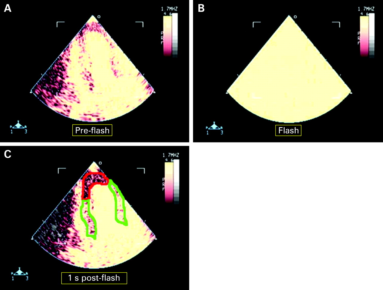
Myocardial segments were graded, according to their wall motion at baseline and at each stage of the protocol, as normokinetic (grade 0), hypokinetic (grade 1), akinetic (grade 2) or dyskinetic (grade 3). Any deterioration by one grade or more, from baseline to peak stress, was considered indicative of ischaemia. At baseline and at peak stress (during myocardial contrast imaging) segments were also graded according to perfusion, which was assessed by the microbubble replenishment time of the myocardium after destruction by the high mechanical index impulses. Failure of the contrast to replenish a given myocardial segment within 1 s after the flash at peak stress was considered a sign of ischaemia (fig 1). If a segment was found to be ischaemic by both wall motion and perfusion criteria or by perfusion criteria only (perfusion is affected earlier than wall motion, according to the ischaemic cascade), it was characterised as ischaemic.

Safety evaluation
Adverse events were recorded during the dobutamine stress echocardiography study and for the next 24 h, by contacting the patient or his/her attending doctor. In all patients positive for ischaemia and/or reporting chest pain during or following the DASE/MCE study, cardiac troponin I was measured using a bedside kit (Triage; Biosite, San Diego, California, USA), immediately and 6 h after the completion of the stress study. The final assessment of the recorded adverse events was based on the Cancer Therapy Evaluation Program Common Terminology Criteria for Adverse Events version 3.0 (CTCAE).23 24 Any unfavourable and unintended sign or symptom occurring during the echocardiographic stress study or in the next 24 h that might or might not be related to the procedure was considered an adverse event. Adverse event severity was graded using a five-grade scale: mild (grade 1); moderate (grade 2); severe (grade 3); life-threatening (grade 4); death caused by adverse event (grade 5) (see Appendix). Chest pain observed after a positive study was not considered an adverse event, because it is an expected consequence of a stress protocol causing ischaemia, as long as it was not associated with enzyme elevation or ECG alterations.
Evaluation of diagnostic parameters
The diagnostic accuracy of DASE/MCE was evaluated in 532 patients without a known history of CAD, who were submitted to coronary angiography in the catheterisation laboratory of our institution, within 4 weeks of the stress testing, without any intervening clinical event. The decision to submit a patient to coronary angiography was made by the attending doctor. These patients underwent coronary angiography and left ventriculography using the Judkins technique. A normal arterial segment was identified immediately proximally and distally to the lesion and was measured with an electronic calliper. The minimal stenosis diameter was also measured and severity was expressed as a percentage reduction of the normal diameter. A patient was considered as having clinically relevant CAD if a stenosis of at least 50% was found in one or more major epicardial arteries or major branches.
Statistical analysis
Parametric variables are expressed as mean ± standard deviation and were compared using the t-test. Non-parametric variables are expressed as percentages and were compared using the χ2 and the McNemar test as appropriate. The sensitivity, specificity, positive and negative predictive value and the overall accuracy of the combination DASE/MCE in detecting CAD (using angiography as the gold standard, with stenoses >50% considered clinically relevant) were calculated according to standard definitions. The analysis was made on a per-patient basis (true positive considered as a finding of at least one >50% stenosis in a major epicardial artery in a patient with a positive stress-contrast echo test), as well as on a per-coronary artery territory basis (true positive considered as a finding of at least one >50% stenosis in the corresponding epicardial artery in a patient with a positive stress-contrast echo test). All data analysis was performed using SPSS 12.0 for Windows. A p value of <0.05 was considered significant.
RESULTS
The basic characteristics of the 5250 patients submitted to DASE/MCE are shown in table 1.
Approximately half of the patients (48.2%) had a known history of CAD, 75% of whom had sustained a myocardial infarction in the past. These patients were referred for ischaemia testing with several indications: follow-up of revascularisation procedures (33% of examined patients with known CAD); finding of intermediate lesions on coronary angiography (19% of patients with known CAD); assessment of previously non-stenosed coronary arteries (28% of patients with known CAD); symptoms suggestive of cardiac ischaemia in patients with known CAD under medical treatment (20%). The average quantity of contrast agent used was 2.5 ml (SD 0.3) per study. Atropine was required to achieve the target heart rate in 72% of studies (mean quantity used was 0.6 mg (SD 0.2) per study). The studies were completed (age-adjusted target heart rate achieved or a termination criterion met) in 95% of cases.
Safety of DASE/MCE
No fatalities were recorded. There were no cases of myocardial infarction (either with or without ST-segment elevation) during the stress study or in the next 24 h. Elevation of cardiac troponin I was observed in three cases (0.02%), to a level lower than the cut-off considered consistent with myocardial infarction by the manufacturer of the assay.
Common side effects (observed at a frequency of >1%) were: dry mouth (19.8%) mainly observed in patients who received atropine; hypertension (2.1%); headache (6.3% of studies, one fifth of headache cases were severe in intensity); dizziness (7.4%); slight tremor (2.4%) and back pain (1.9%). All these adverse events were generally self-limiting, resolving a few minutes after the completion of the test, not requiring intervention.
Uncommon adverse events (observed at a frequency of 0.1–1%) were: hypersensitivity (0.44%); atrial fibrillation/flutter (0.48%); vasovagal episode (0.9%); ventricular bigeminy (0.2%); non-sustained VT (0.34%); sustained VT not requiring intervention (0.18%); mild-to-moderate confusion (0.2%); dyspnoea (0.9%) and urinary retention (0.8%). Hypersensitivity reactions consisted mainly of pruritic rash or urticaria and responded readily to antihistamines. All but three cases were observed immediately after completion of the study. The rest were observed several hours later and their causal link with the study procedure is uncertain; however, we had to include them due to the time association. There were no cases of life-threatening allergic reactions. Atrial fibrillation (new onset) occurred in approximately one in 200 studies and in almost all cases reverted spontaneously to sinus rhythm within 24 h. Vasovagal episodes, characterised by marked bradycardia and a fall in blood pressure, were mostly mild-to-moderate in severity, without loss of consciousness. In five out of 45 cases, the vasovagal reaction led to loss of consciousness during the recovery phase, within 30 minutes from the completion of the test. All these occurrences, however, responded well to atropine administration and placement in the recumbent position. Urinary retention was an uncommon adverse event, exclusively observed in patients who required the administration of atropine. In all cases, it resolved without the need for intervention.
Life-threatening situations were extremely rare, occurring in just two instances in 5250 studies. They consisted of the appearance of sustained VT/ventricular fibrillation, with acute haemodynamic failure. In both cases, standard resuscitation procedures, including electrical shock, were followed. Both patients had severe three-vessel CAD, with impaired left ventricular function (their left ventricular ejection fraction was lower than 0.25) and had to be hospitalised in order to be stabilised and revascularised. Table 2 lists the observed adverse events, along with their grade of severity and frequency.
Patients with significantly impaired (lower than 0.40) left ventricular function (N = 1153, 22.0%) showed a higher frequency of cardiac arrhythmias of any kind (9.5%) compared with patients with an ejection fraction higher than 0.40 (5.4%, p<0.001). This difference was significant after adjustment for gender and age. In fact, no episodes of sustained ventricular arrhythmia or ventricular fibrillation were observed in patients with normal (⩾0.55) ejection fraction.
Diagnostic accuracy of DASE/MCE
Among the 532 patients for whom angiographic data were available, 413 (78%) had significant CAD on angiography. DASE/MCE had identified as positive for ischaemia 378 among them (sensitivity 92%). The positive predictive value of DASE/MCE was 89% (378 of the 425 patients with positive DASE/MCE were indeed diagnosed with significant CAD on coronary angiography). On the other hand, the specificity and negative predictive value were 61% and 67%, respectively. The overall accuracy was 85% (on a per-patient basis). Table 3 summarises the diagnostic parameters of DASE/MCE, with coronary angiography as reference, per patient and per coronary artery territory. It appears that, although the overall accuracy was similar in all coronary artery regions, sensitivity was higher in the segments supplied by the left anterior descending artery, compared with the right coronary artery and the left circumflex territories.
DISCUSSION
Although myocardial perfusion imaging during DASE with the use of echo-contrast agents has been shown to be safe and effective for the diagnosis of CAD,12 17 the safety of flash MCE has not been demonstrated in a large number of patients. To our knowledge, this is the first study to assess the safety and efficacy of flash-contrast echocardiography (combined with DASE) in a large series of subjects; its principal finding is that the combination of an abbreviated protocol of DASE with MCE, for CAD diagnosis in 5250 real-life studies, caused no fatalities or myocardial infarctions and only rare serious complications. All mild or moderate adverse effects were generally self-limiting and transient in nature.
Ultrasound contrast agents have long been used to enhance ultrasonographic imaging of various organs and in several settings. In echocardiography, after their first use for Doppler signal enhancement,25 their applications have expanded to include left-ventricle opacification and the study of myocardial perfusion.26 Pharmacological echo-stress testing, on the other hand, is an established method for CAD diagnosis1 2 as well as for viability assessment,27 whose safety has been demonstrated in several settings.4–6 The use of increasingly aggressive dobutamine–atropine protocols allowed an impressive rise in the proportion of diagnostic tests, without a marked increase in complications.28 The combination of DASE with the study of myocardial perfusion using echo-contrast agents (MCE) offers considerable theoretical advantages, because it should allow the earlier detection of ischaemia, targeting an earlier step of the ischaemic cascade. This theoretical advantage has been translated into clinical efficacy, as shown in several studies combining a range of stress modalities with MCE, both in the detection of CAD11–13 29,30 and the assessment of myocardial viability.31–33
Concern has, however, been expressed over the safety of echo-contrast agents used in echocardiographic applications. These gas-filled microbubbles, with diameter and rheological properties similar to erythrocytes, resonate when exposed to ultrasound beams. When the energy conveyed by the beam, and thus the acoustic pressure, increases, the microbubbles expand and contract excessively, which leads to their destruction, a process called cavitation.34 35 In experimental isolated heart animal models, the interaction of ultrasound with echo-contrast microbubbles was shown to have significant biological effects when the acoustic energy of the beam exceeded a threshold, including microvascular damage, a transient decrease of contractile performance and increased lactate production.14 As these observations were made on isolated animal hearts, however, without the protection of the thoracic wall, with mechanical indexes and delivered acoustic energies well above the levels used for low mechanical index MCE, their clinical implications in humans are difficult to define.
Although the use of echo-contrast agents has been associated in experimental and small clinical studies with the appearance of premature ventricular complexes,15 35 Tsutsui et al17 showed in 1486 patients that myocardial perfusion imaging with low mechanical index real-time contrast echocardiography combined with DASE caused no deaths or myocardial infarctions and was generally well tolerated. We sought to elaborate these observations, using a combination of DASE with MCE, but with two important points of difference: first, instead of a totally low mechanical index technique, such as the one used by Tsutsui et al,17 we used a low mechanical index technique, with high mechanical index impulses, administered for four cardiac cycles, in order to destroy microbubbles and assess the myocardial replenishment velocity (flash-contrast echocardiography). In theory, this technique combines the advantages of low mechanical index imaging (real-time imaging, less mechanical energy delivered to tissues, less destruction of microbubbles and, therefore, less cavitation and less contrast used) with the ability of high mechanical index modalities to offer the opportunity to assess myocardial perfusion dynamically, by the replenishment of the myocardial microvascular bed with microbubbles after destroying them. The use of high mechanical index impulses, even for as brief a period of time as a few seconds (four cardiac cycles correspond to ∼1.5–2 s at peak stress), might result in an increased incidence of adverse events compared with totally low mechanical index techniques. In a recent publication, Vancraeynest et al36 reported an increase in coronary sinus troponin I after high mechanical index insonication of the heart (contrary to low mechanical index imaging), using perfluorocarbon-enhanced sonicated dextrose albumin as the echo-contrast medium. In that study, however, as well as in that of Borges et al,37 no increase in peripheral blood biomarkers was observed, which is in agreement with our findings (we found no significant elevations in troponin I). It should also be noted that in their study Vancraeynest et al36 used high mechanical index imaging for substantially longer intervals than those commonly applied in a low mechanical index/high mechanical index technique (such as flash-contrast echocardiography), which we employed.
Our results regarding adverse events are generally in agreement with previous reports and, in particular, the largest series of stress-contrast echocardiography studies published by Tsutsui et al.17 Although the technique of MCE that we applied involved the use of high mechanical index ultrasound impulses, as opposed to the exclusively low mechanical index mode used by Tsutsui et al,17 which in theory could lead to increased cavitation phenomena, we did not encounter an increased frequency of adverse events. This could be due to: (1) stringent patient selection (eg, we excluded patients with documented VT in the previous 3 months, patients with evidence of an acute coronary syndrome in the previous 30 days, etc); (2) less exposure to dobutamine (based on previous reports on accelerated stress protocols, we applied a protocol largely similar to the conventional one consisting of four 3-minute stages, abbreviating each stage to 2 minutes, thus reducing the total dobutamine dose by 33%); (3) the minimal use of echo-contrast agent by using perfusion imaging for short periods of time, only at baseline and at peak stress.
Regarding the relative contribution of each of the two combined techniques (DASE and MCE) to the appearance of adverse effects, although no immediate conclusions can be drawn from the present study, it becomes evident by comparison of the present data with previous studies of DASE safety, that the use of MCE does not add significantly to the serious adverse event profile of stress echocardiography. For example, in the study by Tsutsui et al,17 the 1012 patients submitted only to DASE (no MCE) showed an incidence of VT of 1% and sustained ventricular tachyarrhythmia of 0.20%. The rates in our study (DASE plus MCE) were 0.56% and 0.22%, respectively. Similarly, in the study of Timperley et al,16 the rate of VT was 0.60% in the 332 patients submitted to only DASE; again very close to our findings, despite the additional use of contrast.
Finally, a known problem of safety studies has been the use of a pleiad of different terms to identify the wide range of potential adverse events and, maybe even more importantly, a non-uniform way of describing the severity of these events. Although initially developed in the context of the Cancer Therapy Evaluation Program,23 CTCAE have also been used for non-cancer treatment-related studies.38 39 Using CTCAE, we were able to take advantage of the specific definitions of the severity of each type of adverse event provided in this document, thus avoiding presumptuous assessments of the degree of severity of the adverse events encountered. We believe that the use of a common terminology basis can contribute substantially to a more uniform approach to safety assessment of cardiovascular diagnosis and management procedures.
Study limitations
The principal aim of the present study was to assess the safety of the DASE/MCE combination, as a whole, in real-life conditions and thus it did not include a group of patients submitted to DASE without the use of contrast agents. Therefore, we cannot determine the specific contribution of dobutamine stress or echo contrasts to the occurrence of adverse events. Regarding the evaluation of the diagnostic parameters of DASE/MCE, it should be noted that in all such studies pre and post-test reference bias exists, because the result of the test is taken into account in deciding whether to catheterise a patient. In addition, the grounds on which each patient’s individual doctor decided to proceed with arteriography could not be uniform in these 5250 patients. These two factors may lead to under or overestimation of both the sensitivity and specificity of the method and may explain the unusually low specificity reported. Moreover, these were real-life studies, ie, they were not performed under ideal experimental conditions (eg, operators and image reviewers were not blinded as to the clinical status of the patients). This should also be taken into account when comparing the observed diagnostic accuracy with past reports.
CONCLUSIONS
The combination of DASE with myocardial perfusion study using flash-contrast echocardiography is an efficient and accurate modality for the evaluation of patients with known or suspected CAD. Moreover, our experience in 5250 studies indicates that, when careful patient selection is applied and dobutamine and contrast agent are used with moderation, it is a well-tolerated and exceptionally safe technique, with generally self-limiting adverse effects.
APPENDIX
REFERENCES
Footnotes
Competing interests: None.
Ethics approval: Ethics approval was obtained.
Patient consent: Patient consent was obtained for publication of fig 1.