Article Text

Abstract
Background Glycogen synthase kinase-3 (GSK-3) is a highly conserved serine/threonine kinase implicated in developmental myocardial growth and in the adult heart response to hypertrophic stress and adverse ventricular remodelling. Two isoforms exist and are expressed in equal measure within the heart, GSK-3α and GSK-3β. Phosphorylation and inactivation of GSK-3β occurs in both pharmacological and afterload modulation (aortic banding) models of cardiac hypertrophy. Strategies that maintain GSK-3β active appear protective. Conversely, GSK-3α activation is associated with a pro-fibrotic response to stress and inactivation in this setting may therefore be protective.
Objective We set out to characterise the chronic myocardial remodelling response to LAD ligation in mice expressing inactivation-resistant GSK-3α/β isoforms.
Methods Experiments were performed in knockin mice (KI), with targeted ser-21/9-ala mutations encoding ubiquitous expression of inactivation-resistant GSK-3α/β kinases, and their wild type (WT) counterparts. Age and weight-matched male adult mice were subjected to permanent LAD ligation or sham procedure. At 4 weeks echocardiography and invasive pressure-volume measurements were performed for a comprehensive interrogation of cardiac structure and function. Hearts were then excised, fixed in formaldehyde at set end-diastolic pressure and sectioned (700 μm) for morphometry.
(1) Morphometry Infarction resulted in significant increases in HW/BW ratio and left ventricular volume in both genotypes at 4–weeks compared to sham animals. This was associated with a significant reduction in mean LV thickness measured at the mid-papillary level, driven entirely by scar formation, and an increase in LV cavity dimensions (Abstract 017 Table 1) n=6/group, *p<0.05.
Morphology and P-V loop data
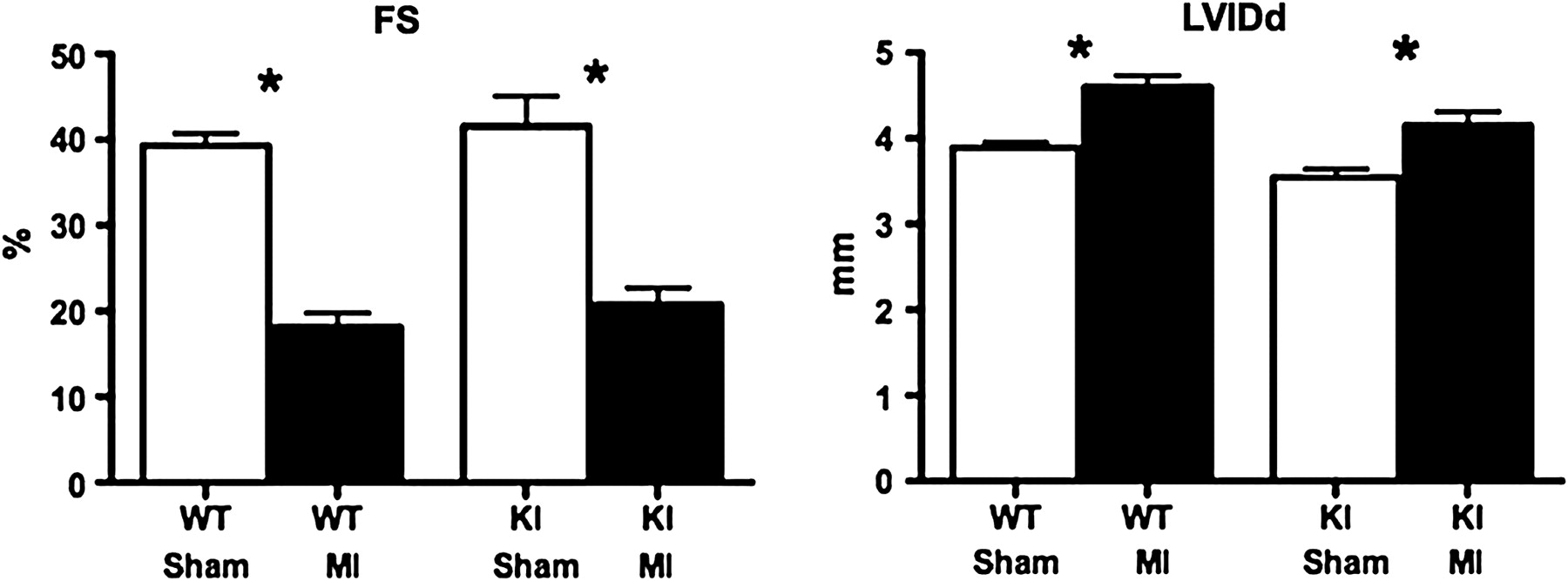
(2) Echocardiography Remodelling was evident in hearts of both genotypes at 4-weeks after infarction. There were significant reductions in fractional shortening, associated with increases in internal diastolic and systolic LV dimensions (Abstract 017 Figure 1) n=7/group, *p<0.05.
Echocardiography—Fractional shortening (FS) and LV internal diastolic dimensions (LVlDd) in WT and KI hearts at 4-weeks after LAD ligation (MI) or sham. (n=7, *p<0.05 vs within genotype sham).
(3) P-V loop assessment Corresponding reductions in ejection fraction were evident in infarcted hearts from invasive interrogation. End-systolic and -diastolic volumes were increased together with a significant increase in end-diastolic pressure compared to sham animals (Abstract 017 Table 1) n=10/group, *p<0.05.
Conclusion Inactivation-resistant GSK-3α/β expression in hearts of KI mice does not prevent chronic adverse ventricular remodelling in response to LAD ligation. At 4 weeks there were comparable increases in ventricular dilatation, volume loading and contractile dysfunction as that demonstrated in infarcted WT hearts. Accordingly, GSK-3 inactivation is not a necessary signalling mediator in post-infarct ventricular remodelling.
- infarction
- remodelling
- GSK-3