Article Text

Abstract
Background Myocardial haemorrhage after myocardial infarction (MI) frequently goes undetected. Since the paramagnetic effects of oxidised iron may result in signal loss on T2* and T2-weighted MRI, we investigated the diagnostic accuracy of T2-weighted MRI in experimental MI.
Methods Acute MI was created in swine (43±9.5 kg) by occluding the left anterior descending coronary artery (n=10) or circumflex (n=5) for 90 min followed by reperfusion for ≤3 days (n=2), 10 days (n=7) or 60 days (n=6). Cardiac MRI was performed at 1.5 T using T2-prepared steady-state free-precession (T2P-SSFP) and gadolinium enhanced (CE) MRI. Left ventricular (LV) sections were visually inspected, photographed and stained for histology. Gross images and histology were scored for myocardial haemorrhage by an experienced cardiac pathologist blinded to all other data. Regions of low signal intensity on T2-weighted and CE-MRI were independently determined by three cardiologists blinded to the pathology results.
Results Eighty ventricular slices of pathology were matched with MRI (n=68 for first pass CE-CMR). All sections exhibited hyperintense zones consistent with oedema on T2-weighted, scar on CE-MRI and pathologic evidence of MI. Myocardial haemorrhage occurred in 49 LV sections (61%) and corresponded with signal voids on 48 T2-weighted (98%) and 26 CE-MRI (53%). Alternatively, signal voids occurred in the absence of haemorrhage in three T2-weighted (90% specificity) and five CE-MRI (84% specificity). On first pass CE-MRI, 27/43 perfusion defects corresponded with haemorrhage (63% sensitivity) while 5/25 defects occurred in the absence of haemorrhage (80% specificity). The positive and negative predictive values for pathological evidence of haemorrhage were 94% & 96% for T2-weighted, 84% & 53% for CE-MRI, and 84% & 56% for first pass perfusion.
Conclusions T2-weighted MRI has high diagnostic accuracy for myocardial haemorrhage. Heterogeneity of signal intensity associated with acute MI on T2-weighted MRI is partially due to intramyocardial haemorrhage.

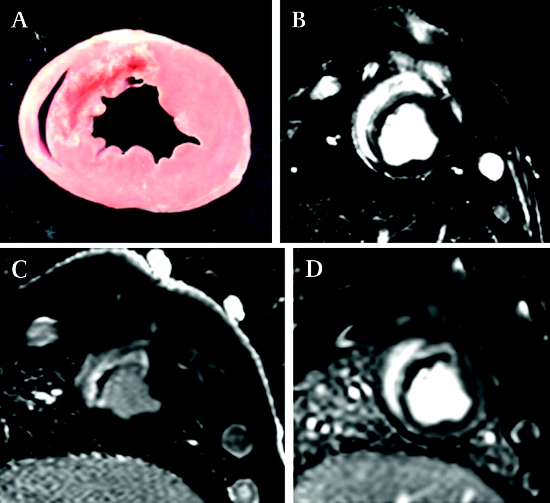
Left ventricular short axis section showing haemorrhagic anterior myocardial infarction (A). Corresponding cardiac MR images (B) T2-weighted SSFP (C) PSIR delayed gadolinium enhancement (D) first pass perfusion.
- haemorrhage
- T2
- MRI