Article Text

Abstract
Introduction Chronic total coronary artery occlusion (CTO) remains a significant challenge for percutaneous coronary intervention (PCI), a frequent cause of angina pectoris and a common reason for referral for coronary bypass surgery. The purpose of this study was to investigate a potential alternative treatment strategy for promotion of angiogenesis and collateral blood flow. To this end we investigated the effect of the prolyl-4-hydroxylase inhibitor di-methyl oxalyl glycine (DMOG) on collateral vessel formation in an entirely endovascular porcine model of coronary occlusion.
Method DMOG was loaded onto a polymer-coated coronary stent. A percutaneous model employing copper-coated stents was used to produce CTO in 20 Yorkshire white pigs. Coronary occlusions were present in all animals at 28 days. DMOG stents were implanted at day 28 and angiographic and physiological data collected on distal coronary and collateral flow. At day 56 the animals were sacrificed and histological analysis performed.
Results A complete total coronary occlusion was present in all animals at day 28 following implantation of a copper stent. (Abstract 104 Figure 1) At 56 days there was a trend to a greater increase in angiographic collateral volume in the DMOG group (10±4.1 mm2 increase vs 3.6±1.5 mm2; 84.5% increase vs 16.5%, p=0.057). There was no difference in collateral flow index between the groups at day 56. Histology revealed an increase in total number of collateral vessels seen around the site of occlusion in the DMOG group (29.9±2.6 vs 18.4±3.1, p=0.01) with similar distal collateral vessel density. (Abstract 104 Figure 2)

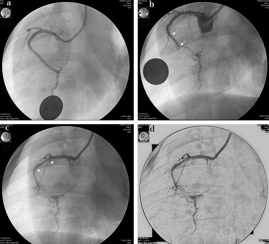
Angiographic images. (A) Initial RAO 30° view of RCA at day 0. (B) Day 28 showing CTO distal to copper stent (solid arrows). (C) Day 56 following implantation of DMOG stent (dashed arrows) with increased antegrade collaterals. (D) Digital subtraction image for image analysis.

A&B. Histology of the occluded pig coronary artery (H&E, H&E) 28 days after implantation of copper stent. M1 and M2=microvessels within neointima (NI) with larger collateral vessels (C). I (E) EL=internal (external) elastic lamina. Panels C&D show histology following DMOG stent. Endothelial cells are seen lining adventitial microvessels (H&E and factor VIII-related antigen stains).
Conclusions Implantation of a copper stent provides a reliable endovascular method of producing a CTO. DMOG increases the number of collateral vessels seen at the site of vessel occlusion in this endovascular model of CTO but did not increase neovascularisation in distal tissue. At baseline distal myocardium was subtended by extensive antegrade collaterals. The effect of DMOG in increasing neovascularisation appeared to be restricted to ischaemic tissue.
- Angiogenesis
- chronic total occlusion
- coronary stents