Article Text

Abstract
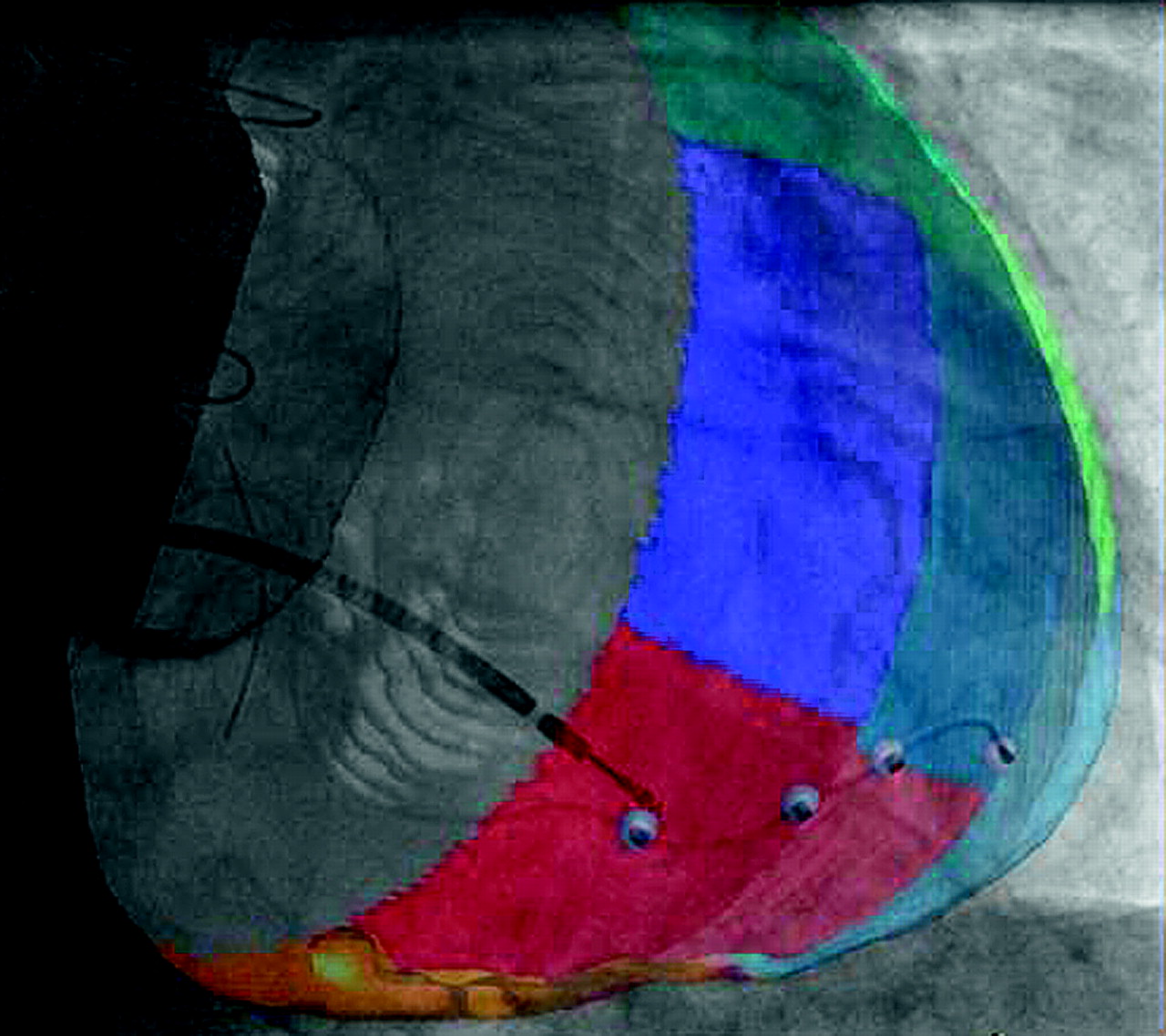
Introduction Optimal left ventricular (LV) lead placement via the coronary sinus (CS) is a critical factor in defining response to cardiac resynchronisation therapy (CRT). Using novel semi-automated image acquisition, segmentation, overlay and registration software we set out to guide lead placement by avoiding scar and targeting the region of the LV with the latest mechanical activation.
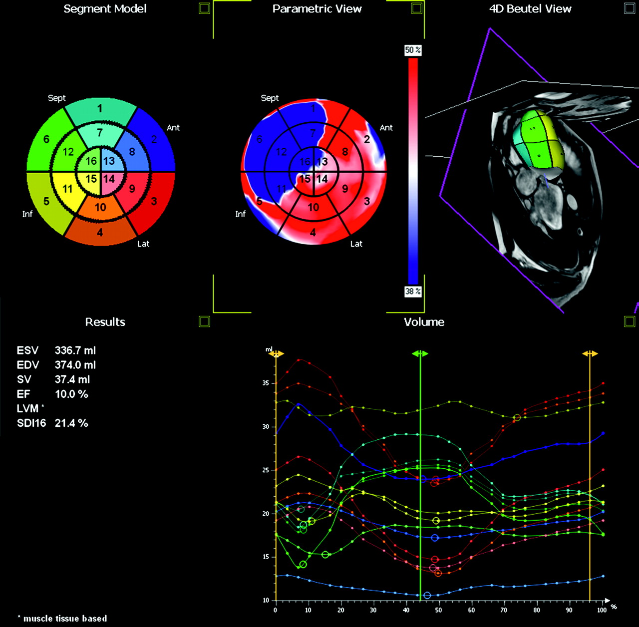
Methods 17 patients underwent cardiac magnetic resonance (CMR) scans. 3D whole heart images were segmented to produce high fidelity anatomical models of the cardiac chambers and coronary veins. 2, 3, 4 chamber and short axis cine images were processed using Tomtec software to give a 16 segment time volume-dyssynchrony map. In patients with myocardial scar the late gadolinium enhancement images were manually segmented and registered to the anatomical model along with the dyssynchrony map. The 3 latest mechanically activated segments with <50% scar were identified and this information was overlaid at CRT implant on to live fluoroscopic images using a prototype version of the Philips EP Navigator software. Subsequently, the x-ray C-arm and table could be moved freely while automatically maintaining a registered roadmap. We used a high fidelity pressure wire to assess the acute haemodynamic response to pacing in different regions of the overlaid 16 segment model. All dP/dt measurements were compared to baseline AAI or VVI (for those patients in AF) pacing at 5–10 beats/min above intrinsic rate.
Results 15 of the 17 patients underwent successful placement of a LV pacing lead via the CS with satisfactory pacing parameters and no phrenic nerve stimulation at implant. The mean time from insertion of the CS guide catheter into the venous sheath to successful cannulation of the CS was 1.3±1.0 min. In 2 patients we were unable to place a LV lead successfully in any branch of the CS. We paced in at least one of our 3 target segments in 11 patients. 67% of patients were responders as defined by a 10% increase in +dP/dt over baseline. The mean change in +dP/dt for the best lead position vs baseline+dP/dt was 15.9±11.3% for DDDLV pacing. This compares to a mean change in+dP/dt of 14.9±12.3% when the CMR dyssynchrony-map defined target region was paced DDDLV. The region of best+dP/dt response was postero-lateral, lateral or posterior in all cases.
Conclusion We have shown it is feasible to acquire, overlay and accurately register cardiac MR data on to fluoroscopic images at the time of CRT implant. Our data suggest that it is also possible to identify and place the LV lead in at least one target region in most patients. This appears to give close to the best acute haemodynamic response that can be achieved in any branch of the CS. The initial results of this pilot study suggest that a MR dyssynchrony guided approach to LV lead placement may allow ideal LV lead positioning (Abstract 152 figures 1 and 2).

- CRT
- MR
- dyssynchrony