Article Text

Abstract
Heart and circulatory diseases affect more than seven million people in the UK. Non-invasive cardiac imaging is a critical element of contemporary cardiology practice. Progressive improvements in technology over the last 20 years have increased diagnostic accuracy in all modalities and led to the incorporation of non-invasive imaging into many standard cardiac clinical care pathways. Cardiac imaging tests are requested by a variety of healthcare practitioners and performed in a range of settings from the most advanced hospitals to local health centres. Imaging is used to detect the presence and consequences of cardiovascular disease, as well as to monitor the response to therapies. The previous UK national imaging strategy statement which brought together all of the non-invasive imaging modalities was published in 2010. The purpose of this document is to collate contemporary standards developed by the modality-specific professional organisations which make up the British Cardiovascular Society Imaging Council, bringing together common and essential recommendations. The development process has been inclusive and iterative. Imaging societies (representing both cardiology and radiology) reviewed and agreed on the initial structure. The final document therefore represents a position, which has been generated inclusively, presents rigorous standards, is applicable to clinical practice and deliverable. This document will be of value to a variety of healthcare professionals including imaging departments, the National Health Service or other organisations, regulatory bodies, commissioners and other purchasers of services, and service users, i.e., patients, and their relatives.
Statistics from Altmetric.com
Introduction
Heart and circulatory diseases affect >7 million people in the UK. Furthermore, they cause 170 000 deaths each year, which equates to more than a quarter of all deaths. The resulting annual healthcare costs amount to £9 billion. The ageing and increasing population together with improved survival rates from heart and circulatory diseases mean that the importance and impact of these conditions will only rise in the future.1
Non-invasive cardiac imaging is a critical element of contemporary cardiology practice. Progressive improvements in technology over the last 20 years have increased diagnostic accuracy in all modalities and led to the incorporation of non-invasive imaging into many standard cardiac clinical care pathways. Cardiac imaging tests are requested by a variety of healthcare practitioners and performed in a range of settings from the most advanced hospitals to local health centres. Imaging is used to detect the presence and consequences of cardiovascular disease, as well as to monitor the response to therapies. For example, non-invasive imaging through CT coronary angiography (CTCA) has largely replaced invasive coronary angiography as first-line testing for stable chest pain of possible cardiac origin. The field is fast-moving: increasing microprocessor speed and the use of artificial intelligence is likely to see further rapid evolution in the next decade.
The previous UK national imaging strategy statement which brought together all of the non-invasive imaging modalities was published in 2010. At that time, CTCA was not a fully mature technology nor was cardiovascular magnetic resonance (CMR) widely used in mainstream clinical practice.
The changes in practice and resulting large increases in demand for advanced cardiac imaging since the document’s publication have highlighted a number of issues and challenges relating to cardiac imaging in the UK which include:
Wide geographic inequalities in provision.
Disparate clinical delivery settings.
National variability in infrastructure.
Multiple overlapping indications.
Multiple approaches to training and competence assessment.
For the purposes of this document, non-invasive imaging modalities are divided into four—echocardiography (echo), cardiac CT (CCT), nuclear imaging and CMR—on the basis of their underlying technology (figure 1).

The rich diversity of cardiac imaging—overview of cardiac imaging. CMR, cardiovascular magnetic resonance.
The purpose of this document is to collate contemporary standards developed by the modality-specific professional organisations which make up the British Cardiovascular Society (BCS) Imaging Council, bringing together common and essential recommendations. The development process has been inclusive and iterative. Imaging societies (representing both cardiology and radiology) reviewed and agreed on the initial structure. A writing group representing the four specialist societies (British Society of Echocardiography (BSEcho), British Society of Cardiovascular Magnetic Resonance (BSCMR), British Society of Cardiac Computed Tomography (BSCCT), British Nuclear Cardiology Society) generated an initial set of standard headings. Modality-specific content was generated by each society. The writing group then divided this content into common themes and modality-specific guidance. Finally, a panel of senior clinical, National Health Service (NHS) and patient representatives reviewed and critiqued the document for applicability. The final document therefore represents a position, which has been generated inclusively, presents rigorous standards, is applicable to clinical practice and deliverable.
This document will be of value to a variety of healthcare professionals including:
Imaging departments, who wish to ensure that they are meeting contemporary best practice standards.
NHS or other healthcare providers that wish to evaluate the quality of the services they provide.
Regulatory bodies, which in the UK include the Care Quality Commission (CQC),2 and initiatives like Getting It Right First Time,3 which evaluate a service against an agreed set of standards.
Commissioners and other purchasers of services, who wish to ensure that purchased services are properly configured and meet agreed professional standards.
Service users, that is patients and their relatives.
These standards are applicable directly to departments within the UK, and although there are many differences between UK practice and practice elsewhere in the world (outlined in table 1), we believe many of the standards applicable to healthcare systems in general are not limited by national boundaries.
How imaging in the UK differs from other countries
Cardiovascular imaging modalities
Echocardiography
Echo uses ultrasound to create two-dimensional (2D) and three-dimensional (3D) images of the heart (and aorta), which can be used to assess anatomy, function and blood flow. Images are most commonly non-invasively—transthoracic echo (TTE). Higher definition images can be acquired by the insertion of an echo probe into the oesophagus, transoesophageal echo (TOE). Echo machines range from high-end equipment, which has all current echo capabilities through to simple hand-held devices, which are limited to the acquisition of moving images and assessment of blood flow. This diversity makes clinical standards critically important to ensure consistent quality services.
Echo has the advantage over other cardiac imaging techniques of high spatial and temporal resolution, which means that it can visualise small and fast-moving structures. It is, therefore, widely used to assess ventricular and heart valve function, with other tests of cardiac function, notably CMR, reserved to answer questions concerning cardiac structure or function, which are unresolved by echo. Machines are highly portable, so imaging can be delivered in virtually any healthcare setting, whether hospital-based or in the community. Hand-held echo devices facilitate emergency bedside scanning for rapid diagnosis or exclusion of conditions such as cardiac tamponade and severe left ventricular systolic dysfunction. Echo does not involve the use of ionising radiation.
Echo is limited by low-quality images in a minority of scans due to poor acoustic windows (related either to the chest wall, soft tissue or lung tissue interfering with the ultrasound beam). This means that there is a higher degree of variability in image quality between studies than with other imaging modalities in some cases.
Cardiac CT
Technological developments in detector design and microprocessing have transformed the clinical applications of CCT. The technique uses contrast-enhanced ECG-gated CT scanning to depict the heart and its related structures (including the aorta) motion-free with a high degree of geometric accuracy. CTCA cannot only visualise severe coronary artery obstruction, it is also capable of detecting early stage, non-obstructive atheromatous disease. CCT image quality is susceptible to artefacts related to irregular heart rhythms and respiration, and it is less accurate in patients with heavily calcified coronary arteries.
CCT has been adopted by the National Institute for Health and Care Excellence (NICE)4 as the entry investigation for the detection of coronary artery disease in stable patients. This has resulted in challenges for national implementation, with a shortage in both appropriate CT scanners and the personnel to operate them.
Nuclear cardiology
Nuclear cardiology is a group of investigations, which uses radio-isotopes to elucidate different aspects of heart function. The method of imaging can be divided into ‘conventional’ single photon emission CT (SPECT) and positron emission tomography (PET). SPECT has a long history and strong evidence base for the investigation of coronary artery disease, while PET is becoming an important part of the management of a range of cardiac diseases. Specific clinical applications of these investigations include the detection of myocardial infarction and/or inducible myocardial hypoperfusion/ischaemia, assessment of left ventricular function (less relevant these days following the widespread availability of echo and increasing availability of CMR) and the detection of myocardial inflammation. All nuclear cardiology techniques expose patients to ionising radiation and they lack spatial resolution when compared with other imaging techniques.
Cardiovascular magnetic resonance
CMR is an advanced form of MRI, which uses ECG-gated image acquisition to minimise cardiac motion blurring. It allows the assessment of cardiac anatomy, function and viability of the myocardium. It is unique among imaging techniques in its ability to provide detailed information about tissue composition. CMR can be used to detect inflammation, fibrosis, infarction, inducible hypoperfusion/ischaemia and to assess congenital heart disease, heart valve dysfunction and the presence of inherited myocardial diseases. Access to CMR is increasing, but it remains a relatively specialist technique so the funding streams (in England) are through specialised commissioning. While there is currently a limited number of centres which have the infrastructure and caseload to deliver CMR to an acceptable quality, improvements in technology should help to enable its wider dissemination.
Cardiac imaging—integration into the patient journey
There is extensive overlap between modalities in their ability to answer the same clinical questions, for example, whether or not there is inducible myocardial ischaemia. The selection of the most appropriate imaging technique is a balance between the specific functionality of the modality, the aptitude of the local service, availability, waiting times, radiation dose and patient preference. It is important that the needs of the patient remain at the centre of the diagnostic journey and guide service development.
Assuring quality across cardiac imaging modalities
Each imaging department and modality will adopt processes individualised to its local circumstances, there are areas of commonality, which must form part of any quality framework. There is universal agreement that quality assurance must be a central part of the operation of an imaging department and that this should be reflected in the job planning of those concerned.
The essential components which all departments should be able to demonstrate are:
the attainment and demonstration of appropriate skills;
continuing professional development (CPD) in order to maintain and enhance skills;
audit and quality improvement;
learning from discrepancy incidents;
peer-to-peer evaluation (eg, systematic second over-read audit by second reporter).
This document supports departments adopting an Imaging Quality Framework along the lines of the originally developed process for echo by the BSE and described as the Echo Quality Framework (EQF, figure 2 ).5 This brings together many of these themes, and links them in a continuous cycle aimed at improving all aspects of the service including echocardiographic imaging quality, reproducibility, education and training and customer (patient or referrer) satisfaction. The EQF process is applicable to many imaging disciplines.

Imaging Quality Framework (adapted from the British Society of Echocardiography Echo Quality Framework (EQF).13
The Royal College of Radiologists (RCR) provides comprehensive standards6 including the 2019 document on Quality Standards in Imaging (QSI),7 as well as guidelines for learning from events and discrepancy. QSI is recognised by NHS England and the CQC. The 2020 Radiological Events and Learning Meetings8 guideline provides a roadmap for how departments should learn from events, errors and discrepancies, including outlining the frequency of meetings, and the expected rate of attendance for individual radiologists. It mandates an anonymous discrepancy review process, as well as the evaluation of the output of peer-to-peer evaluation processes (systematic over-reads). The role of the chair of the meetings is clearly defined, and guidance is provided for the conduct of both the chair and meetings in order to promote a ‘blame-free culture’. The process outlines the relationship between peer review9 and duty of candour,10 the latter being a General Medical Council (GMC) imperative, which may be triggered as a result of peer-to-peer or discrepancy processes.
Credentialing of practitioners
In the UK, all medical practitioners must comply with the overarching framework of the GMC for specialty training (through the Joint Royal Colleges of Physicians Training Board11 or the RCR,12 according to specialty) and with maintenance of professional standards through revalidation.
Many bodies including the British Society of Echocardiography (BSE), the European Association of Cardiovascular Imaging (EACVI), the British Society of Cardiovascular Imaging (BSCI)/BSCCT and the Society for Cardiovascular Magnetic Resonance (SCMR) offer accreditation in individual imaging modalities. Such accreditation is a convenient way for an individual to demonstrate the achievement and maintenance of agreed standards, but this should not be regarded as the only way to demonstrate competence or regarded as mandatory for clinical practice in the field. The GMC is currently exploring the use of a ‘credentialing’ process in areas where there is (a) a need to ensure patient protection which cannot be met through other means; (b) a demonstrable service need; (c) feasibility of developing and maintaining the credential and (d) support for the credential from a recognised authoritative body in the field.13
General service requirements
This section describes requirements that are applicable to all four imaging modalities. Each imaging modality has its own intrinsic characteristics (table 2). More detailed and modality-specific recommendations are described in subsequent sections.
Advantages of different imaging modalities
Indications
A locally agreed list of common indications should be available with recommended diagnostic pathways, which include imaging. A local system to ensure appropriate triage should be available.
Access
Timely access to appropriate diagnostic modalities should be available within an organisation or else provided by a planned regional network solution. Where an investigation forms part of an urgent or emergency care pathway, it should be available either on a 7-day per week or 24-hour a day basis as required.
Equipment
All equipment used for cardiac imaging purposes should be set-up appropriately for cardiac work including availability of necessary hardware and software modifications.
Imaging equipment typically has a 7–10 years lifespan. There should be a defined replacement cycle, ideally with a planned 7-year replacement schedule, and no device should be in use for >10 years.
There should be appropriate arrangements for the timely servicing and care of all equipment. For equipment essential to out-of-hours diagnosis, service contracts should include the potential for weekend repair.
Software for reviewing and reporting cardiac imaging studies such as Radiology Information Systems (RIS), Picture Archive and Communication Systems (PACS),14 etc, should be integrated so that imaging and associated reports for other modalities can be referenced (eg, echo images and reports should be available when reporting CMR scans).
The organisational imaging solution should permit the viewing of primary images in any area where clinical decisions incorporating the imaging are being made (eg, multidisciplinary team meetings (MDMs)).15
Reporting solutions, including tools for quantification of cardiac imaging studies, should be integrated into a seamless reporting workflow (ie, they should not require separate workstations and image transfers), and the results of functional evaluations, etc, should be transferable into RIS/PACS and the electronic patient record.
Workforce descriptors
Healthcare professionals undertaking and/or reporting cardiac imaging should be able to demonstrate appropriate training and maintenance of knowledge and skills through regular practice and validated CPD.
There should be an agreed expectation regarding the number of cases performed during an imaging session. This should be individualised according to the complexity of cases encountered within the relevant unit.
There are no statutory board examinations in imaging in the UK. For some practitioners, especially cardiologists, accreditation and revalidation is the basis for demonstrating competence. Radiologists may cover many organ systems and continuing competence in the cardiac imaging modality should be part of their CPD, and their annual appraisal and 5 yearly revalidation processes.
There should be a designated clinical lead for the service who ensures a system is in place for reviewing requests and reports, audit, quality control, protocols for imaging and urgent clinical review in response to abnormal findings.
Departmental clinical standards
There should be a standard operating procedure (SOP) against which all aspects of service delivery can be compared and audited.
The management of the department should be governed by written documentation, which, at a minimum, should include staff training, emergency procedures, radiation protection and escalation (according to local rules).
There should be an agreed and verifiable audit programme. Suggested audit topics include the eight ‘best practices’ relating to radiation exposure identified by the International Atomic Energy Agency and their radiation-related quality index indicating the number of best practices used by a laboratory,16 satisfactory stressing, downstream testing and correlative imaging.
A clear system for ensuring the appropriateness of imaging referrals should be in place.
A clear system for the escalation of important clinical findings should be in place.
MDMs form an essential part of the delivery of patient care and of the quality assurance process within an imaging service. MDMs are a forum for the communication of imaging findings, clinical interaction, innovation, good practice and CPD. MDM preparation and participation requires appropriate job planning. Documents from the BCS and the RCR describe the requirements for consultant imaging specialists and imaging departments to maximise the benefit to patients of imaging discussed at MDMs.15
There should be a ‘Learning from Discrepancies Meeting’ (LDM). LDMs play a crucial role in clinical governance. Alongside other related processes, and as part of a quality assurance programme, the LDM facilitates improvement in the quality of service delivery, is an important source of shared learning and contributes to improved patient safety.
Imaging department management meetings
These operational meetings should review:
capacity, activity and demand statistics;
workforce and staffing;
clinical governance;
patient and user feedback;
strategy and issues to escalate;
radiation protection.
Radiation protection
It is important that the administered radiation activity for each individual exposure is optimised to obtain optimum diagnostic information with the lowest dose. The Ionising Radiation (Medical Exposures) Regulations 2017 (IR(ME)R 2017) requires that employers regularly review, and make available to operators, diagnostic reference levels (DRLs).
All procedures should be undertaken in accordance with departmental written protocols. Local DRLs should be specified in the written protocols.
National DRLs for exposures used during SPECT and PET/CT imaging have been published and are available online.17
All staff involved in the organisation and administration of investigations, which use ionising radiation should be familiar with Departmental Local Rules and SOPs.
Infection control
Services in conjunction with local infection control departments should be able to demonstrate appropriate measures are in place to prevent person-to-person transmission of disease or other healthcare-related infection.
Reporting (including teleradiology)
All images should be reported in accordance with agreed local protocols and standards by competent staff, who are working within their defined scope of practice.
There should be an agreed approach in collaboration with radiology to the review and reporting of extracardiac findings where a cardiologist is the primary reporter.
Where imaging is delivered by an extra-organisational providers, clinically agreed protocols should be in place for the conduct and reporting (including any teleradiology arrangements).
The NHS Constitution pledges that patients (in England) should wait no longer than 6 weeks for a diagnostic test. Pathways should be developed which meet this standard.
Imaging for more urgent clinical indications (for instance, diagnostics for patients seen in a rapid access chest pain clinic) should be delivered by pathways, which are individualised to the relevant (2 weeks, in this case) time scale.
Departments should have key performance indicators (KPIs) for the turn-around of reports, which are structured around the urgency, the referral source and/or the type of investigation.
Teleradiology (remote review and reporting) is an increasing part of normal processes of all cardiac imaging departments. It provides a high degree of flexibility and can improve departmental robustness and clinician well-being. There should be an agreed schedule including appropriate equipment, monitor resolution (which will vary between modalities), processor and internet speeds. Reporting remotely should be performed to the same standards as departmental reporting.
Data management
All departments must have an electronic report database with facilities for storing and retrieving images and reports. Standards regarding imaging databases can be found at in the RCR standards for confidentiality and RIS and PACS18 and the RCR standards for the communication of radiological reports. 19
Compliance with guidance and legislation on patient confidentiality must be fully observed.20–23
All departments should ensure that their data/image management and storage is compliant with General Data Protection Regulations (GDPR).
There should be GDPR-compliant methods for the secure transfer of primary clinical imaging data (usually DICOM files) between institutions.
Strategic considerations for delivery and service development
When one of a number of different imaging modalities could reasonably be used in a clinical pathway in accordance with best practice, the imaging modality used should be determined by local availability and expertise. With increasing cost pressures across the NHS, the economic and resource impact of local practices should be considered when setting up or reviewing local services and/or patient pathways. Periodically national and best practice guidance will change with major implications for service configuration. A recent example of how such considerations impact national and local services can be seen in the implementation of the NICE guidelines for ‘recent-onset chest pain of suspected cardiac origin: assessment and diagnosis’24 and the associated resource impact analysis.25 Where such challenges exist or develop, these should be assessed and resolved at a cardiac network level taking into account existing skills and services and allowing strategic, integrated service development.
Providing a comprehensive patient-centred service
Many units or hospitals will not provide all cardiac imaging modalities. When the required imaging modality is not provided by the local hospital, it should be available to patients as part of a network-wide imaging strategy. Robust referral pathways should form part of the network arrangements. Patient involvement in the design of investigative pathways should be actively encouraged. There should be robust methods to receive and respond to patient comments, complaints and suggestions. Departments should be compliant with the NHS national complaints policy.26
Echocardiography
Principal indications
Due to its availability, portability and widespread availability, the indications for TTE are broad and the threshold for requesting the investigation is low. For principal indications, see table 3 and figure 3.
Principal indications for echocardiography
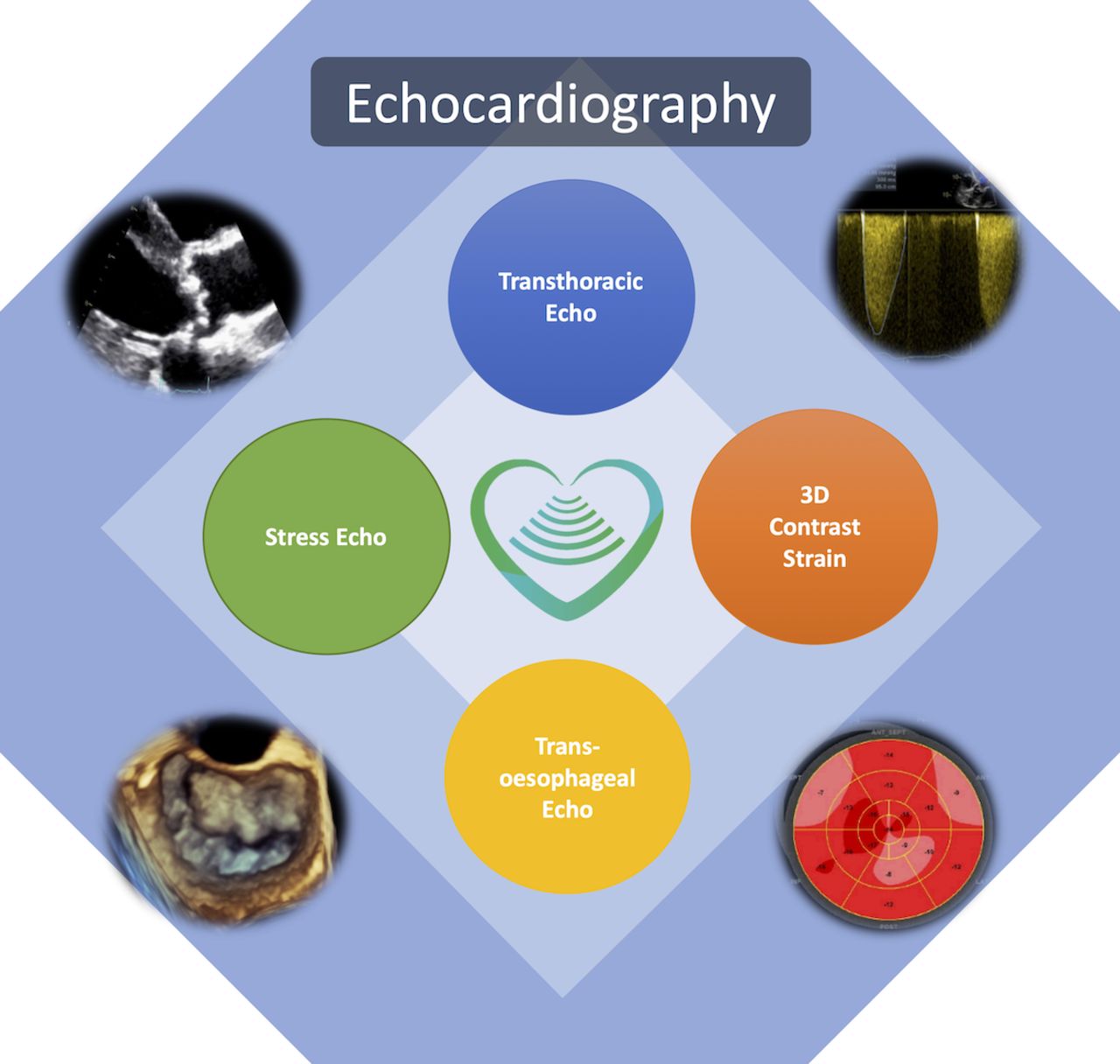
Overview of echocardiography. 3D, three-dimensional.
Appropriateness and recognition of risk
Echo does not involve the use of ionising radiation so it is frequently the first choice investigation for the assessment of cardiac structure and function.
Sixteen per cent of all echocardiograms performed are inappropriate.27 A triage system should therefore be in place in order to ensure adherence to appropriateness criteria.28
Due to the national shortage of sonographers, the scans can be performed by relatively junior cardiology trainees or other professionals which constitutes a risk from errors in image interpretation and reporting.29
These risks should be mitigated through the provision of appropriate clinical supervision, teaching and training for all staff groups acquiring and reporting echo images.
TTE is non-invasive and the procedure itself is not associated. Stress echo is also non-invasive. It is associated with a very low risk of ischaemia-related complications. By contrast, TOE is invasive and carries a 1 in 1000 risk of serious oesophageal traumatic injury.30
Access to investigation and timeliness
Where a hospital has an acute medical take, echo should be available 24 hours a day.3 31
This poses a significant challenge and organisations should mitigate this risk by investment in training, diversification of roles and by working in collaboration to provide network solutions.32
The challenges posed by remote communities may also require cooperation across healthcare organisations at a regional level, particularly when advanced imaging (TOE and stress) forms a core element of patient pathways which are recommended in guidelines from national bodies such as NICE or the Scottish Intercollegiate Guidelines Network.33
Equipment specification and replacement cycles
Echo machines must be equipped with the appropriate phased-array transducers and cardiac-specific imaging software. Stand-alone continuous wave Doppler and tissue Doppler capability are also recommended for all Doppler capable devices.
3D capability is recommended on at least one machine in the department.
An echo machine can normally be used for a maximum of 2500 studies per year, but this figure may be lower if it is used for a significant number of ward-based or complex studies.
Elective replacement planning should be at 5 years with no machine in regular clinical use being over 10 years old.
Appropriate couches are required to facilitate patient positioning for the acquisition of high-quality images.
Full recommendations for equipping an echo department can be found in the BSE’s description of its process for BSE departmental accreditation in echocardiography.
Workforce descriptors, accreditation, job planning and education and training
All echo departments should have a designated Head of Department (HoD). The HoD may be either a specialist registered medical practitioner or a specialist healthcare scientist. A cardiologist lead should be on the GMC’s Specialist Register for cardiology and should preferably hold individual TTE BSE/EACVI accreditation (or equivalent).
A healthcare scientist lead must hold similar accreditation and be graded band 7 or above. They should spend at least five sessions per week performing activities directly related to echo.
Any sonographer or cardiac scientist who performs and reports studies unsupervised should have individual TTE BSE/EACVI accreditation (or equivalent) and will usually be graded band 6 or above.
Sonographers or cardiac scientists should have access to study leave and related funding to undertake CPD activities in order to maintain and enhance their echo skills.
Departmental quality approval
All echo departments should have a verifiable process for ensuring ongoing quality assurance. Furthermore, processes should be in place that encourage staff engagement with quality assurance and which improve patient care through feedback mechanisms.
Demonstrating adherence to appropriate quality standards is best achieved by a department completing the process for BSE departmental accreditation in echo.34
Echocardiography reporting recommendations
All studies should be performed using an agreed minimum dataset (usually the BSE minimum standards dataset).
The average time required to undertake and report a comprehensive echo, however, is about 45 min. Sufficient time should be allocated in elective echo lists to enable the recording and reporting of an echo study in accordance with BSE standards.
Focused studies may be appropriate in some circumstances (eg, a level 1 emergency echocardiogram study, interval follow-up studies which should still be performed in line with the BSE minimum standards dataset), but the goals and scopes of such studies should be agreed in advance and be auditable.
Reports for routine studies should be issued within 24 hours of the examination.
A full or preliminary report should be issued within 1 hour for urgent or inpatient studies.
There should be agreed permissions stating which staff members can report echo studies and who can sign off reports.
A standard echo-specific electronic format must be established for reports. Furthermore, reports should be readily retrievable from the electronic archive and integrated into the electronic patient record.
Integrating organisational echo services and interdependencies
Improvements in echocardiogram technology have been accompanied by expanded indications for echo and by its increased use as a point-of-care test in the assessment of acutely unwell patients. In addition, TOE is performed during cardiac surgery, during percutaneous cardiac interventions and in guiding treatment on the intensive care unit. This has led to the dissemination of echo skills from cardiac scientists and cardiologists to cardiac surgeons, anaesthetists, intensive care specialists and emergency care physicians.
The relationships between cardiology imaging and intervention, intensive care, emergency medicine, pharmacy, audit departments, research and manufacturers should be clearly mapped and available for review.
The same standards of equipment and training are applied in these different environments and staff groups, respectively. Some of these issues have been addressed in detail through the BSE Level-1 Individual Accreditation process and the joint BSE/Intensive Care Society Emergency echo departmental accreditation standards.34
Echo should be delivered collaboratively, supported by the principal echo department within the organisation.
Accredited echocardiographers are core attendees at valve and structural heart disease MDMs. Their attendance at these meetings should be recorded.
Super-specialist echocardiography services
There is a variety of super-specialist echo services, including specialist TOE, stress echo and echo for congenital heart disease in children and adults. These fall outside the scope of this document, but the applicable standards have been published by the BSE. These services are often provided for a wide clinical network, and engagement with the network around appropriate configuration is essential.
Echocardiography-specific risks
Echo should be performed in an environment, which is suitable for maintaining patient dignity and for the acquisition of high-quality images. This should include appropriate privacy, space and lighting.
Additional space is required to perform TOE and stress echo compared with TTE.
A standard TTE is not formally considered to be an intimate examination, nevertheless, its performance requires patient sensitivity and all departments should have an approved chaperone policy.
There should be awareness of health and safety issues, especially relating to back and eye problems among staff, and adequate liaison with occupational health and risk management departments.35
Detailed safety standards for TOE and stress procedures fall outside the scope of this document. However, national safety standards for invasive procedures should be followed. Furthermore, local policies should comply not just with the requirements for imaging, but incorporate appropriate SOPs for the following areas:
Pharmacological stress;
Ultrasound contrast administration;
Probe decontamination (TTE and TOE);
Safe sedation;
Advanced life support;
Management of complications arising from TOE.
Echocardiography-specific audit and governance
Regular, ideally weekly, echo meetings, must be held to review unusual, challenging or otherwise difficult cases.
The BSEcho Quality Framework5 provides a template for the delivery of such meetings. Attendance should include cardiac scientists and consultant echo specialists as a minimum. There must be an established process for issuing revised reports, when relevant, following echo meeting discussions.
Additionally, departments are encouraged to participate in local and national audit processes.
An escalation policy for highlighting serious and unexpected findings should be in place.
Challenges to the delivery of echocardiography
Demand for echo has increased as the indications for its use have expanded and become better recognised. In combination with well-documented shortages in cardiac scientists and an ageing population among whom conditions, which require echo, such as valve disease and heart failure, are more prevalent, this has led to problems in the delivery of echo in a timely manner for some patients.
This problem may be compounded in some regions by long travel times, which have been linked to low uptake of diagnostic testing in cancer screening programmes. These challenges are associated with inequalities in access to healthcare in rural communities.
Where gaps in provision occur, a network approach to service delivery is recommended, with institutions working across organisational boundaries to provide the required services.
Remote review and telemedicine solutions may help to bridge some of these gaps, but the governance around such solutions must be clearly described.
Cardiac CT
Principal indications
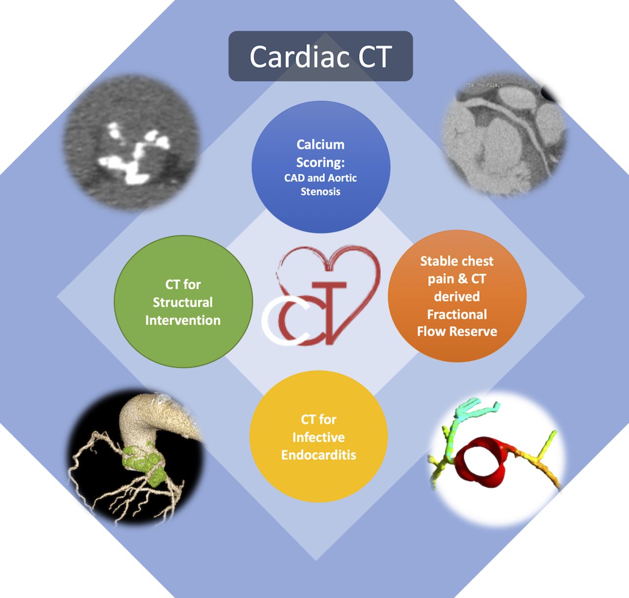
For principal indications see table 4 and figure 4.
Principal indications for cardiac CT

Overview of cardiac CT imaging.
Appropriateness and recognition of risk
Cardiac CT is non-invasive (but for the requirement for a venous cannula for the administration of X-ray contrast) and very safe.
Cardiac CT involves a relatively low dose of ionising radiation (which is similar to the dose from invasive coronary angiography36). Cardiac CT may be considered appropriate:
if it is likely to provide an accurate diagnosis;
if it is likely to identify patients who may benefit from specific interventions (such as medical therapy or myocardial revascularisation) or guide delivery of therapy (such as transcatheter aortic valve implantation (TAVI));
if it is likely to provide information relating to prognosis.
All requests should be triaged for appropriateness and priority according to an locally agreed set of indications.
Access and timeliness
There should be 24-hour, 7 days a week access to emergency motion-free CT imaging of the thoracic aorta for patients with suspected aortic trauma or acute aortic syndrome.
Provision of cardiac CT for structural heart disease and coronary artery evaluation is required within working hours.
Provision of CTCA services as per NICE CG95 (2016)37 represents a significant challenge both in workforce and equipment terms. However, services at a network level need to be configured to deliver investigation and report within 2 weeks.
Equipment specification and replacement cycles
The minimum technical requirements for cardiac CT imaging can be provided by a general purpose CT scanner which has a detector array of at least 64 rows and which has been ‘cardiac enabled’ with ECG-gating (safe practice).38 However, more advanced CT technology should be available, or at least accessible through local arrangements on a regional basis, when imaging patients who are difficult to scan (eg, those with arrhythmias) and when planning provision of large volume CTCA services (NICE DG3).39
The life of a cardiac CT scanner is around 7 years, according to manufacturers’ maintenance recommendations. Life expectancy is, however, influenced by scan volumes. A programme which prospectively plans scanner replacement around this expected lifespan should be in place.40
Specialist analysis software
Minimum requirements
Coronary artery calcium score calculation.
Coronary artery visualisation and stenosis quantification including automated centerline extraction for the major epicardial coronary arteries.
Additional software may include
Cardiac chamber volumetric and functional quantification.
Thoracic aorta assessment tools (for intervention planning, eg, TAVI).
Myocardial perfusion assessment including quantitative perfusion maps.
Estimation of fractional flow reserve by CT for non-invasive assessment of the functional significance of coronary artery stenoses (NICE MTG32).41
Other equipment
Resuscitation facilities (including defibrillation/oxygen/suction).
An emergency trolley which includes the storage of drugs to treat reactions to contrast agents and beta-blockers.
Monitoring equipment such as a non-invasive blood pressure machine and a pulse oximeter.
Phantoms for CT calibration and quality control.
Workforce descriptors, accreditation, job planning and education and training
Clinicians who supervise and report cardiac CT scans (usually radiologists or cardiologists) should be trained in cardiovascular CT to the equivalent of BSCI/BSCCT level 2 standard or higher.42 Radiographers who perform cardiac CT should be competent to do so through subspecialty training, which can be delivered in-house, by manufacturers and supplemented by online learning and by attendance at dedicated study days (Society of Radiographers).43
Guidance regarding the CPD needed for maintenance of skills and re-accreditation in cardiac CT is available through the BSCI/BSCCT.42
For job planning purposes, a programmed activity of direct clinical care delivering cardiac CT might include the supervision and reporting of between 4 and 8 scans depending on case-mix.
Departmental quality approval
There is currently no minimum requirement for departmental accreditation for cardiac CT, which will normally be provided by general CT departments.
It is recommended that departments which undertake cardiac CT dedicate a minimum of one session per week to cardiac scanning in order to justify the service and maintain competencies.
Regular (weekly) departmental cardiac imaging MDMs which include cardiac CT are recommended.
Cross-modality review meetings and regional network meetings are recommended.
Quality assurance activities should include contributions to case review, participation in local and national audit of radiation dose and downstream testing, discrepancy reporting and maintenance of CPD to enable re-accreditation and revalidation.
Cardiac CT reporting recommendations
No specific UK reporting guidelines exist. It is recommended, however, that reporting is undertaken according to standardised local policies.
The SCCT provides a reporting system in the form of ‘CAD-RADS’,44 which can be particularly useful for audit and follow-up purposes.
Approved reports should be available within 24 hours for outpatients and on the same day as the study for urgent inpatient scans.
There should be an established discrepancy reporting process.
The dose length product (DLP), which allows an estimate of the radiation dose, should be included in the report.
Interdependencies with other services
The relationships between radiology, cardiology imaging and intervention, medical physics, pharmacy, audit departments, research and manufacturers should be clearly mapped and available for review.
In particular, there should be a well-described process for the review and reporting of non-cardiac findings.
Accredited cardiac CT experts are core attendees at structural heart disease MDMs. Their attendance at these meetings should be recorded.
Super-specialist cardiac CT services
Super-specialised services, such as adult congenital heart disease services which require CT should be provided as part of an agreed network process as all units will not have the required skill mix.
Cardiac CT-specific risks
IR(ME)R 2017 governs the delivery of cardiac CT radiation doses according to the ‘as low as reasonably practicable’ (ALARP) principle (IRMER).45 Specific training of staff as to modes of ECG-gating and the relevance in ensuring that scans comply with the ALARP principle is recommended.
All staff involved in the delivery of cardiac CT should be trained in basic life support, with immediate access to staff who are trained in advanced life support. Appropriate equipment to facilitate advanced life support should be readily available. Cardiac CT staff should be familiar with the treatment of anaphylaxis and have immediate access to the necessary drugs.
The potential risks of intravascular administration of contrast agents must be weighed against the potential benefits. Withholding agents may deprive patients of the benefits of valuable diagnostic information or necessary therapy. Further guidance can be found on dedicated guidelines on safe use of contrast agents.46
An agreed protocol to manage the risks of contrast induced nephrotoxicity should be a place.
Cardiac CT-specific audit and governance
For the safe practice of cardiac CT in adult patients in UK imaging departments:
There should be a contrast extravasation policy in place, which is supported by appropriate patient literature.
Cardiac CT radiation doses should be audited regularly by means of the recorded DLP. Centres are encouraged to submit these data to BSCI/BSCCT for their national dose audits.
Cardiac CT centres are encouraged to participate in local and national audit processes of downstream testing.
Challenges to delivery of cardiac CT
It is estimated that 350 000 CTCAs are required per annum in the UK to deliver the NICE diagnostic pathway for recent-onset, stable chest pain of possible cardiac origin, NICE CG95.37 Provision for the assessment of structural heart disease, aortic disease, etc, requires more capacity still.
The specific challenges include delivering cardiac CT services (CTCA services, specifically) in the wake of NICE CG9537 are outlined in ‘challenges in delivering CTCA as the first-line test for stable chest pain’.47
Nuclear cardiology
Principal indications
For principal indications see table 5 and figure 5.
Principal indications for nuclear cardiology

Overview of nuclear cardiology. PET, positron emission tomography; SPECT, single photon emission CT.
Appropriateness and recognition of risk
Nuclear cardiology investigations are non-invasive, but for the requirement for a venous cannula for the administration of the appropriate radio-pharmaceutical and stressor agent. It is therefore very safe.
Radiation doses differ between different nuclear cardiology and choices (where available) should be made to keep the radiation dose as low as possible.
All requests should be triaged for appropriateness and priority according to an locally agreed set of indications. Choice of investigation and technique should be guided by patient characteristics.
If it is likely to provide an accurate diagnosis.
If it is likely to identify patients who may benefit from specific interventions (such as the needs for ischaemic revascularistion) or guide delivery of therapy (such as the removal of infected prosthetic material).
If it is likely to provide information relating to prognosis.
Access to investigation and timeliness
Access to nuclear cardiology services should be available at a local and networked level.
Where nuclear cardiology forms part of a diagnostic pathway (for instance, for the evaluation of chest pain, diagnostic workup for infective endocarditis, diagnosis of cardiac amyloidosis), it should be reliably delivered within the appropriate 2-week or 6-week time frame, respectively.
Where this is not possible for an individual organisation, a networked solution should be in place.
Where nuclear cardiology forms part of an acute care pathway, it should be available on a 7/7 basis.
Equipment specification and replacement cycles
Radiological equipment has a finite lifespan, beyond which there is reduced image quality and increasingly frequent malfunction.48
Equipment replacement cycle should be no more than 10 years. (Operating costs for older equipment are high when compared with newer equipment. Maintenance may not be possible or provided for very old equipment.)
Every healthcare institution or authority should have a plan for rolling upgrade or replacement of medical imaging equipment.
This plan should look forward a minimum of 5 years, and be updated on an annual basis.
Workforce descriptors, accreditation, job planning and education and training
Administration of Radioactive Substances Advisory Committee (ARSAC)45 requires employers and practitioners to hold a licence for the administration of radioactive substances for a specified purpose at any given medical radiological installation.
Each employer is required to hold a licence for each administration at each medical radiological installation for the purpose of the administration of radioactive substances to humans.
Every practitioner is required to hold a licence in order to justify the administration of radioactive substances to humans.
The majority of licences are issued for 5 years.
At a medical radiological installation where administrations of radioactive substances are undertaken, it is expected that there will be a multidisciplinary team involved with service provision. This will include practitioners, medical physics experts, radiopharmacists (if appropriate) and other healthcare professionals (eg, radiographers, technologists and nursing staff) with appropriate training and experience.
Practitioners are clinicians (nuclear medicine physicians, dual-accredited radiologists/nuclear medicine physicians, radionuclide radiologists and/or cardiologists).
Applicants for licences who have not undertaken any of these structured training programmes are required to demonstrate equivalent training, experience and competence pertaining to the procedures they wish to undertake (eg, EACVI/European Association of Nuclear Medicine 49 or American Certification Board for Nuclear Cardiology50).
Practitioners who wish to justify exposures as part of a PET/CT service will require training and experience additional to that required for conventional nuclear medicine procedures.
Practitioners are expected to undertake appropriate continuing medical education (CME) associated with the procedures for which they are licensed as part of the appraisal and revalidation processes, and to confirm this at the time of revalidation.
For job planning purposes, a programmed activity of direct clinical care delivering nuclear cardiology might include the supervision of eight stress tests and the reporting of between 8 and 10 scans.51
Departmental quality approval
ARSAC requires employers to hold a licence for each administration at each medical radiological installation for the purpose of the administration of radioactive substances to humans.
There is no specific departmental accreditation available for nuclear cardiology, but departments should be able to provide evidence of engagement with broader accreditation, for example,
the British Nuclear Medicine Society (BNMS) organisational audit tool.52 It is suggested that nuclear medicine departments aim to complete the use of the audit tool twice in a 5-year cycle.
The United Kingdom Accreditation Service is piloting assessments under its Medical Physics and Clinical Engineering scheme in the area of nuclear medicine.53
Nuclear cardiology-specific risks
All forms of nuclear cardiology result in ionising radiation exposure to patients, and each medical radiation exposure must therefore be justified and the exposure should be as low as possible while maintaining diagnostic quality. Staff are at risk of exposure to ionising radiation in certain circumstances, exposure to ionising radiation therefore requires monitoring with staff protection governed by occupational health standards. All procedures should be undertaken in accordance with departmental written protocols. Local DRLs should be specified in the written protocols (ARSAC Notes for Guidance).54 National DRL for CT exposures used as part of SPECT/CT and PET/CT procedures have been published and are available online.55
Nuclear cardiology reporting recommendations
The American Society of Nuclear Cardiology has published guidelines for performing nuclear cardiology procedures, which includes recommendations regarding standardised reporting.56
Interdependencies with other services
Nuclear medicine, and thereby nuclear cardiology, is a multidisciplinary specialty which involves nuclear medicine physicians, radiologists, cardiologists, medical physicists, radiopharmacists, nuclear medicine technologists and radiographers and nurses. Availability of radiopharmaceuticals often is subject to local, national and international relationships.
It is recommended that practitioners should participate in coronary, endocarditis and heart muscle disease MDMs.
Super-specialist services
Provision of certain aspects of nuclear cardiology is limited by resource constraints and minimum volume requirements, for example, myocardial perfusion PET, fluorodeoxyglucose PET/CT for infective endocarditis and deoxypyridinoline scintigraphy. Where these cannot be provided within a single organisation, a network solution or formal referral process to a suitable unit should be in place.
Nuclear cardiology-specific audit and governance
Nuclear cardiology departments should participate in regular external peer review, such as the BNMS Organisational Audit Programme.52
They should participate in local audit programmes. This should include both effectiveness audits around clinical/prognostic accuracy and environmental audits.
It is recommended that departments should hold regular LDMs.
Challenges to delivery of nuclear cardiology imaging
Nuclear medicine training is now part of the clinical radiology curriculum. Access to training centres and recruitment remains a challenge. This, along with other issues, including reduced numbers of trained staff coming to the UK from the European Union (EU) and retirements has led to a workforce shortfall.
The future of radiopharmaceutical supply remains uncertain following the UK’s exit from the EU. Problems in the supply chain could significantly affect the provision of nuclear cardiology, however this issue is not specific to nuclear cardiology.
Other challenges include: the advanced age of equipment throughout the UK and the ability to replace and upgrade due to heterogeneity of replacement policies; continuing concerns about radiation exposure and delivering ALARP; developing skill-sets to include independent nurse or physiologist stressing through a Patient Group Directive57 and non-medical reporting and ensuring that appropriate governance arrangements are in place to facilitate these developments.
Cardiovascular MRI
This is a concise summary of the full British Society of CMR Clinical Standards Document 2022.
Principal indications
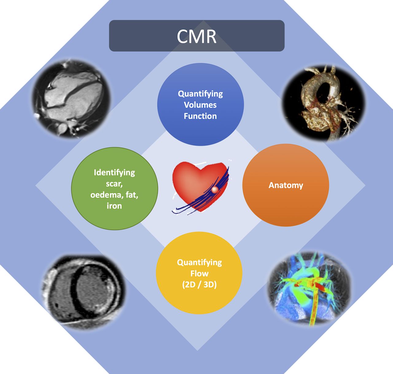
For principal indications see table 6 and figure 6.
Principal indications for cardiac magnetic resonance
Overview of cardiac magnetic resonance (CMR). 2D, two-dimensional; 3D, three-dimensional.
A concise guide to the categories of indication for CMR is included here and are available as a set of comprehensive indications online58 and to the expert consensus panel document.59
Appropriateness and recognition of risk
In general, CMR is considered appropriate when it can provide additional information which has the potential to alter the clinical diagnosis (including disease severity) and/or management of the patient. CMR is often used after previous testing, such as echo and/or coronary artery imaging, whether by CTCA or invasive coronary angiography, when important clinical questions remain unanswered or new clinical questions have arisen as a result of the initial test. It is not appropriate to use CMR purely to confirm the findings from previously used imaging modalities.
CMR is very safe. There are no known long-term health risks from the high magnetic fields or radiofrequency (RF) pulses used during testing. Precautions are, however, required regarding ferromagnetic objects, which may cause injury to the patient, staff and/or equipment if subject to the high magnetic field in the MR scanner room. Some patients with certain types of implants or embedded metal (eg, shrapnel) may, therefore, need to be excluded from undergoing CMR scanning.
Patients with cardiac pacemakers/defibrillators may be scanned under the following circumstances60:
The device and leads system are MR conditional (ie, safe to scan under certain conditions), and appropriate programming of the device has occurred prior to the scan. Processes should be in place to enable patients with MR conditional devices to be scanned in all MRI centres, particularly if MR conditional devices are implanted in that centre. These patients do not require referral to specialist centres.
The device and leads system is not MR conditional (‘ legacy’ devices),61 but the information from the scan is crucial to the patient’s management and outweighs the risks of scanning. This is a specialist area of practice, and in general is carried out in specific centres with the relevant experience and expertise (MRImyPacemaker.com).62
Access and timeliness—expanding to 7/7 and 24/7 working
CMR is not available in every hospital, as it requires specialist staff, equipment, expertise and regular practice to maintain skills. It is generally performed in larger hospitals with an established cardiac centre (and should be accessible by every cardiothoracic surgical centre in the UK). Where CMR is not available or its availability is limited, there should be a clear network pathway in place to deliver the service.
CMR is most commonly performed as an outpatient service (~95% of scans). There are no clear indications for emergency CMR as other cardiac imaging modalities, in particular echo, CT and invasive coronary angiography, are more appropriate in this setting. Twenty-four-hour provision is therefore not required. Urgent inpatient scans should ideally be performed within 48 hours. Elective outpatient scans should ideally be performed within 6 weeks but limited capacity in many areas means that waiting times are often longer than this.
Equipment specification and replacement cycles
Equipment specification
MRI scanner
A fully maintained, shared or dedicated MRI scanner (minimum 1.5 T) with cardiac capability.
Sufficient magnet access to achieve minimum annual unit (see NHS England National Service specifications).63 ECG gating, patient monitoring (including blood pressure, pulse oximetry), contrast pump.
For a new CMR installation, the BSCMR recommends the following specifications:
RF receiver: 16 or more RF channels (torso/body/cardiac receiver array with multiple elements).
Minimum gradient specifications: 30 mT/m, 150 mT/m/ms.
Artefact-resistant ECG hardware/software (eg, vectorcardiogram).
Further details on cardiac-specific sequences, specialist software for analysis and other essential MR compatible equipment can be found in BSCMR Standards Paper.64
Replacement cycles
BSCMR recommends replacement of MR scanners which are >10 years old, in line with the NHS England and NHS Improvement report from November 2019.65 Current guidance from the Biomedical Engineering Advisory Group recommends such equipment should be replaced after 7 years.66
Workforce descriptors, accreditation, job planning and education and training
A unit performing CMR should have:
A named director/clinical lead who is on the GMC’s Specialist Register for cardiology, radiology, nuclear medicine, accredited in CMR, and able to demonstrate ongoing appropriate CME/CPD in CMR.
A named lead CMR radiographer. Ideally, this would be their main or only role. The position should provide sufficient time to ensure that high-quality clinical care is delivered to patients in a safe environment which is supported by appropriate clinical governance processes. The postholder will also be responsible for equipment management and maintenance.
Appropriately trained medical and technical staff to deliver the service. They will be familiar with the local rules and procedures for safe working practice in the MR environment.
Arrangements for scientific and technical input from a medical physics expert who is trained in CMR methods.
Arrangements for appropriate staff development, including education, training, accreditation, CPD and revalidation. Further details can be found at BSCMR.org.
Current or planned (3 years) activity sufficient to maintain individual accreditation.63
Further recommendations for departmental quality control and minimum numbers of scans for a CMR centre can be found in BSCMR Standards Paper.64
Departmental quality assurance
All CMR departments should have a verifiable process for ensuring ongoing quality assurance. Furthermore, processes should be in place that encourage staff engagement with quality assurance and which improve patient care through feedback mechanisms.
Cardiovascular MRI reporting recommendations
All clinical CMR sessions should be supervised by a named consultant who is responsible for generating a formal scan report.
CMR reporting should be integrated with the clinical scenario and should conform to appropriate international standards.
CMR practitioners should participate in clinical MDMs in which CMR imaging plays an important part in informing patient management, in order to provide appropriate expertise in the interpretation of the CMR scans. Such MDMs include heart muscle disease, MDMs, structural heart disease MDMs and valve MDMs.
A unit performing CMR should have a clinical review meeting at least fortnightly, at which CMR scans can be reviewed and/or colleague advice sought.
CMR practitioners reporting in isolation is discouraged. Reporting physicians should have ready access to appropriately skilled imaging peers for advice regarding complex or ambiguous scans.
The BSCMR recommends that inpatient CMR scans should be reported by the next working day, and outpatient scans should ideally be reported within two working days.
Interdependencies with other services
Lower volume centres should establish links with high volume CMR reference centres for reporting of more complex and/or borderline scans (eg, some cardiomyopathy or myocarditis cases), with arrangements for image transfer and joint CMR review sessions. This could be established as a ‘hub-and-spoke’ model to reduce patient journeys to reference centres.
Super-specialist services
Adult congenital heart disease
Patients with simple congenital heart disease (eg, bicuspid aortic valve, straightforward coarctation) can be scanned locally if the relevant expertise is available. All other patients with ACHD should generally be imaged in a CMR centre which has specific experience in ACHD imaging. This is often in a designated ACHD centre. If a patient is found to have previously unrecognised complex congenital lesion(s) during a scan at a non-ACHD centre, the scan should be completed as far as possible, and further imaging performed if necessary in the future at an ACHD centre.
Non-MRI-conditional cardiac devices
CMR in patients with older ‘legacy’ non-MRI-conditional cardiac devices should be performed in centres experienced in scanning such patients. This service is generally provided on a regional basis.
CMR-specific risks
The main element of staff and patient safety specific to CMR is the area of magnet safety, referred to above and covered in detail elsewhere.67
A set of standard operation procedures should be in place to ensure a safe environment and quality.
Robust training and safety protocols around the magnet are mandatory. Detailed guidance on patient safety can be found in BSCMR Standards Paper.64
Emergency procedures should be reviewed and audited at regular (at least annual) intervals, and should cover cardiac arrest, fire and flooding, magnet quench, decreased oxygen level and loss of power and lighting.
Gadolinium-based contrast agents are remarkably safe, with a lower adverse event rate for both allergic type reactions and nephrotoxicity than iodine-based contrast media.68 The RCR recommends the following in patients with chronic renal impairment (in alignment with the European Medicines Agency)69:
The European Medicines Agency (EMEA) suspended the linear agents thought to be highest risk from intravascular use.
It previously concluded that cyclical products (those with lowest risk, called group II agents) can be used for patients with severely reduced renal function when a contrast-enhanced MRI scan is clearly the best method of examination.
A local protocol should be in place to manage care of patients with impaired renal function balancing the risks of gadolinium-induced complications and the clinical requirement for the investigation reflecting these guidelines.
MRI-specific audit and governance
CMR departments should participate regularly in local audit programmes, which should include both effectiveness audits around clinical and prognostic accuracy audits.
Challenges to delivery of cardiovascular MRI
The BSCMR recommends that every cardiology department has access to a CMR service, and that the NHS England diagnostic target of 6 weeks from referral to availability of scan report should be used as the benchmark KPI across the UK.
The BSCI/BSCMR made a detailed projection of the likely need for CMR in the UK which was followed by NHS England 2013/14.63
Access to CMR imaging is variable across the UK (BSCMR 2019 survey).70 The BSCMR is keen to promote greater availability of CMR in every region of the UK, sufficient to meet the NHS England 6-week target for outpatient diagnostics.
Conclusion
The major characteristics of high-quality, contemporary cardiac imaging services are described in this document. Such departments should be able to demonstrate these qualities through their audit and governance processes.
Further detailed recommendations regarding current national standards and position statements are contained in the appendices. It is inevitable, however, that advances in technology such as storage and processing power, together with new applications of precision medicine, artificial intelligence and virtual reality will fundamentally change the practice of non-invasive cardiac imaging over the next decade. In light of this, we recommend that this document is reviewed in 5 years.
Ethics statements
Patient consent for publication
Ethics approval
Not applicable.
Footnotes
Twitter @#ThomasTreibel, @NWAFT_Cardiac
Contributors We confirm that: (1) this paper is not under consideration by another journal, or elsewhere; (2) all authors have read and approved the manuscript and (3) full disclosure of any potential conflict/s of interest has been made.
Funding The authors have not declared a specific grant for this research from any funding agency in the public, commercial or not-for-profit sectors.
Competing interests None declared.
Provenance and peer review Commissioned; externally peer reviewed.