Article Text

Statistics from Altmetric.com
Clinical introduction
Early recurrence of focal ST-segment elevation after acute ST-elevation myocardial infarction (STEMI) usually indicates myocardial reinfarction. However, it can be seen in threatened myocardial rupture, a situation in which administration of potent antithrombotic and thrombolytic therapy may be catastrophic. Herein, we present the case of an 84-year-old woman with STEMI undergoing urgent stenting who had a recurrence of chest pain with cardiogenic shock and new ST elevation in the infarct-related leads mimicking myocardial ischaemia. A patent stent showed by repeated angiography, presence of pericardial effusion and absence of recurrent rise in cardiac marker suggest the diagnosis of a contained left ventricular rupture. The experience of any clinician in dealing with this complicated situation is likely to be limited.
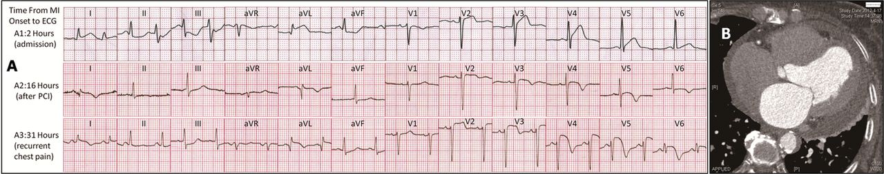
An 84-year-old woman with a history of diabetes mellitus and hypertension was admitted to our institute after experiencing the sudden onset of chest pain. ECG at admission showed ST segment elevation in leads I, aVL and V2–5 (Figure 1A1). Urgent coronary angiography revealed acute thrombotic occlusion of the proximal left anterior descending (LAD) artery. Balloon angioplasty and subsequent stenting of the LAD lesion was performed with good angiographic results. Her chest pain and ST-segment changes resolved (Figure 1A2), and she received aspirin, clopidogrel and low-molecular-weight heparin. One day later, she had a recurrence of severe chest pain with cardiogenic shock and new ST elevation in the infarct-related leads (Figure 1A3). Repeated emergency coronary angiography revealed the stent was patent. Furthermore, persistent elevated ST segments were observed. However, recurrent rise and fall of cardiac injury markers was absent. Eleven months later, a CT scan was performed (Figure 1B).

Images of serial electrocardiogram (A) and cardiac CT scan (B).
Question
What is the most likely cause of recurrent ST-segment elevation?
-
Coronary spasm
-
Transient embolic coronary occlusion
-
Pericarditis
-
Left ventricular rupture
-
Left ventricular apical aneurysm
For the answer see page 39
For the answer see page 29
Answer: D
After the second angiography, a bedside transthoracic echocardiogram was performed, revealing mild pericardial effusion. The findings suggest the diagnosis of a contained left ventricular rupture.1 – 3 Contained myocardial rupture may probably be roofed by visceral pericardium that could offer some protection against free bleeding into the pericardium and the immediate occurrence of cardiac tamponade.3 Answers A and B could be excluded due to persistent elevated ST segments even with patent LAD coronary artery. Answer C is also incorrect as no characteristic ECG findings and no fever occurred. As for answer E, ventricular aneurysm was not documented at the time of echocardiogram examination. Moreover, it usually grows at a relatively slow pace and could not cause pericardial effusion.
Risk factors associated with left ventricular free wall rupture (LVFWR) are female gender, old age, hypertension, small transmural infarction and first time infarction with no collaterals.4 Most of these risk characteristics were observed in our case. It has been demonstrated that new ST-elevation in infarct-associated leads may suggest the ‘stuttering’ type of LVFWR.2 It is likely to be erroneously attributed to ischaemia. In this situation, administration of thrombolytic therapy may be catastrophic. Unlike patients with classic LVFWR, patients with an initially contained rupture may survive.1 – 4 The key to diagnosis is the finding of a pericardial effusion on transthoracic echocardiography or with other imaging modalities.2 ,3 ,5
Footnotes
-
Competing interests None.
-
Patient consent Obtained.
-
Provenance and peer review Not commissioned; externally peer reviewed.