Article Text

Statistics from Altmetric.com
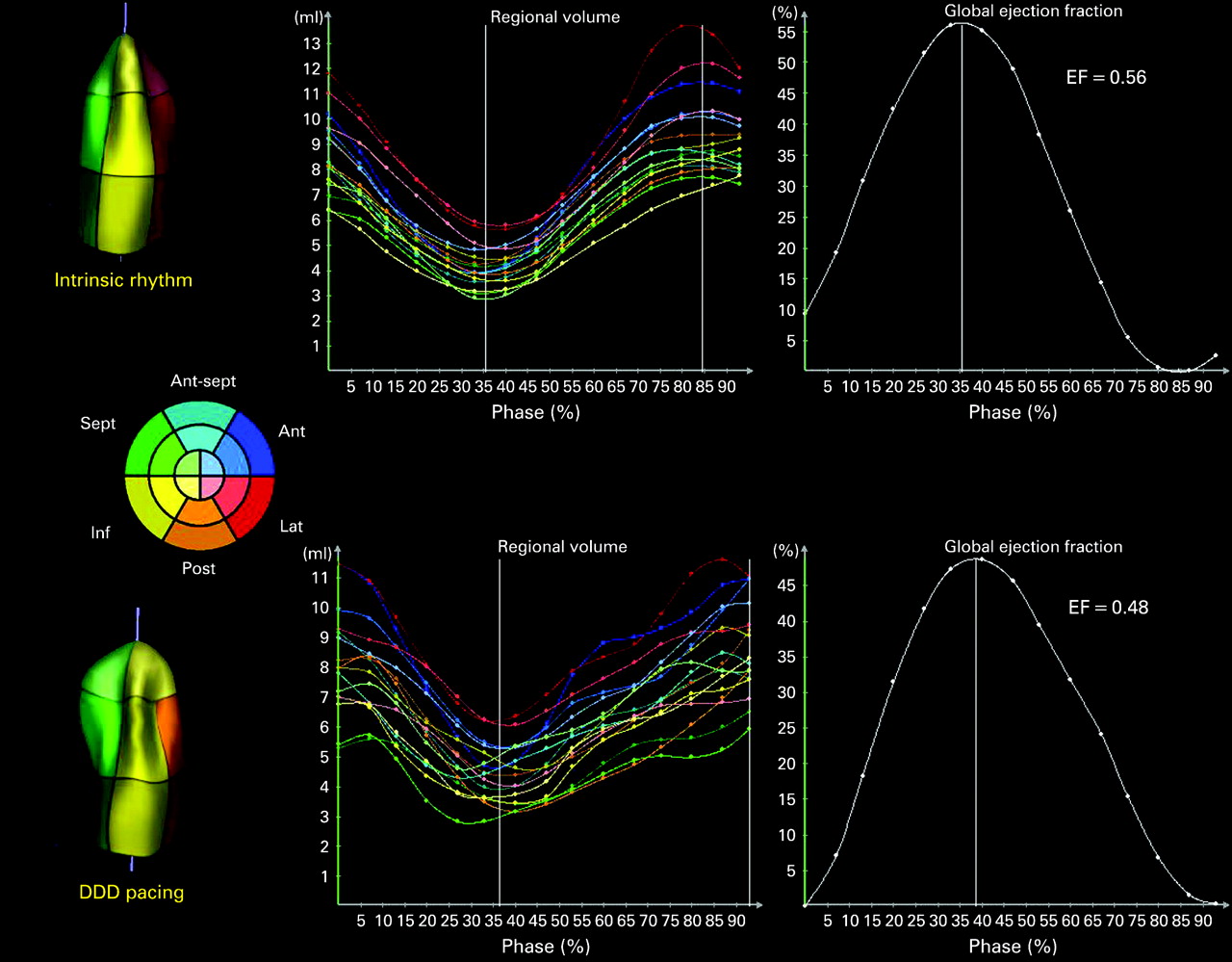
A 78-year-old man with sick sinus syndrome received a dual-chamber pacemaker, with the right ventricular lead implanted at the apex. A real-time, transthoracic, three-dimensional echocardiogram was performed using a Sonos 7500 Live 3D echocardiograph. Full volume acquisitions of the left ventricle were obtained and processed offline using the TomTec 4D LV analysis v1.1 software (TomTec, Unterschleissheim, Germany). The panel depicts regional volume curves and global left ventricular systolic function, during intrinsic ventricular rhythm (top) and immediately afterwards during DDD pacing (bottom). Note the dyssynchrony resulting from right ventricular apical pacing shown by the regional volume curves, with a reduction in global left ventricular ejection fraction. Animated sequences of the reconstructed left ventricular casts are available on the journal website, and illustrate dyskinesia of the interventricular septum during pacing (shown in the bottom left of the panel, with bulging of the septal segments in the end-systolic frame). Dedicated device algorithms may be programmed to favour intrinsic atrioventricular conduction, thereby avoiding adverse effects of ventricular pacing and improving clinical outcome.
Supplementary materials
web only media 94/12/1533
Files in this Data Supplement: