Article Text

Abstract
Objective Calcium metabolism has long been implicated in aortic stenosis (AS). Studies assessing the long-term safety of oral calcium and/or vitamin D in AS are scarce yet imperative given the rising use among an elderly population prone to deficiency. We sought to identify the associations between supplemental calcium and vitamin D with mortality and progression of AS.
Methods In this retrospective longitudinal study, patients aged ≥60 years with mild-moderate native AS were selected from the Cleveland Clinic Echocardiography Database from 2008 to 2016 and followed until 2018. Groups were stratified into no supplementation, supplementation with vitamin D alone and supplementation with calcium±vitamin D. The primary outcomes were mortality (all-cause, cardiovascular (CV) and non-CV) and aortic valve replacement (AVR), and the secondary outcome was AS progression by aortic valve area and peak/mean gradients.
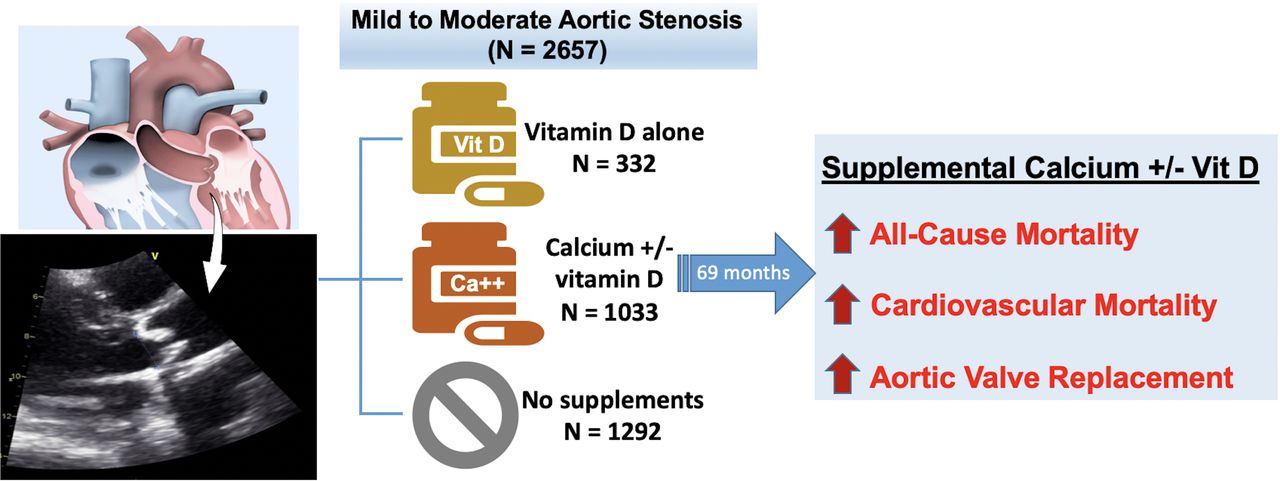
Results Of 2657 patients (mean age 74 years, 42% women) followed over a median duration of 69 months, 1292 (49%) did not supplement, 332 (12%) took vitamin D alone and 1033 (39%) supplemented with calcium±vitamin D. Calcium±vitamin D supplementation was associated with a significantly higher risk of all-cause mortality (absolute rate (AR)=43.0/1000 person-years; HR=1.31, 95% CI (1.07 to 1.62); p=0.009), CV mortality (AR=13.7/1000 person-years; HR=2.0, 95% CI (1.31 to 3.07); p=0.001) and AVR (AR=88.2/1000 person-years; HR=1.48, 95% CI (1.24 to 1.78); p<0.001). Any supplementation was not associated with longitudinal change in AS parameters in a linear mixed-effects model.
Conclusions Supplemental calcium with or without vitamin D is associated with lower survival and greater AVR in elderly patients with mild-moderate AS.
- aortic valve stenosis
- echocardiography
Data availability statement
Data are available upon reasonable request.
Statistics from Altmetric.com
Introduction
Aortic stenosis (AS) is the most common adult valvular disease in the developed world with a prevalence commensurate with age in 2% among those over 65 years and 4% over 85 years.1 Previously described as a progressive extension of aortic sclerosis, the condition is marked by severe calcification causing leaflet immobility and outflow obstruction, a process that occurs over a mean duration of 8 years.2 AS commonly manifests a prolonged asymptomatic period but portends dramatically increased mortality if left untreated once symptoms develop.3 With no proven pharmacological therapy, aortic valve replacement (AVR) is the sole treatment modality. Notwithstanding, prior efforts have targeted markers of AS progression to delay or reverse the process,4 with particular focus on the therapeutic effects of statins which have been inconsistent and especially disappointing in prospective trials.5 6 These studies highlight an active, inflammatory process similar to vascular atherosclerosis, beyond simple ‘passive’ age-related degeneration.2 7 Although a disease of the elderly, multiple modifiable risk factors have also been identified, including hypercholesterolaemia, tobacco use, and importantly, increased serum ionised calcium, parathyroid hormone and vitamin D3.1 7–9
The relationship of dietary and supplemental calcium or vitamin D with cardiovascular (CV) disease risk and mortality is controversial.10 Yet, in light of published guidelines, the supplemental use of each has notably risen in recent years, particularly among post-menopausal women susceptible to osteoporosis and bone fractures.11 12 Although dysregulated calcium metabolism has long been implicated in aortic valvular calcification, and AS progression is akin to pathways of bone remodelling and formation, data on the valvular effects of supplemental calcium or vitamin D are restricted to animal models or limited subjects.13–16 Studies defining the long-term safety and haemodynamic impact of supplementation are scarce but imperative with an increasing use among an elderly population prone to deficiency. In this retrospective longitudinal analysis of patients with mild to moderate AS followed over a median duration of 69 months, we aimed to describe the associations between oral supplementation and all-cause, CV, and non-CV mortality, need for AVR, as well as AS progression.
Methods
Study population
The Cleveland Clinic Echocardiography Database was queried from January 2008 to June 2016 for patients with AS. Patients aged 60 years or greater with mild to moderate AS, defined as aortic valve area (AVA) ≥1.0 cm2 and <2.0 cm2, on index echocardiogram, were selected and followed until December 2018. Those without follow-up echocardiogram at minimum 2 years apart were excluded. For patients with more than two studies, all available echocardiograms were included in the analysis to increase the duration of follow-up. Patients with normal aortic valves or severe AS on index echocardiogram or with prior valve replacement were excluded. Moreover, we excluded patients who underwent aortic valvuloplasty (n=53) but included those who underwent AVR during the follow-up period.
Study variables
Baseline characteristics including demographics, comorbidities, medication use and laboratory data were obtained by thorough review of the electronic medical records by two study investigators based on data at or nearest to the index echocardiogram date. Medication start and end dates were confirmed by manual prescription review, including the use of supplementation. Start dates were adjudicated to assure that subjects supplemented for at least 1 year, with confirmed active use either at or after the index echocardiogram and with initiation at least 1 year prior to the last echocardiogram. Among all subjects, the dosage of calcium supplementation varied from 500 to 2000 mg/day and vitamin D3 supplementation was at any available over-the-counter dose in market.
From our echocardiographic registry, we evaluated left ventricular ejection fraction (LVEF), AVA, aortic peak gradient (PG) and mean gradient (MG), and left ventricular end diastolic and systolic diameters. AVA was interpreted on two-dimensional echocardiogram using the continuity equation. PG and MG were calculated using pulse and continuous wave Doppler assessment of the aortic valve. Echocardiographic and Doppler data were obtained by an experienced sonographer and adjudicated by an expert echocardiography reader and board-certified cardiologist.
Outcomes
Comparative groups were stratified into: (1) no calcium or vitamin D supplementation, (2) supplementation with vitamin D alone and (3) supplementation with calcium±vitamin D.
Outcomes analyses were further performed in those who supplemented with calcium alone, however, this cohort was not chosen as a study group owing to the small sample size. The primary outcomes were all-cause, CV and non-CV mortality, as well as AVR. In order to ascertain cause of death and AVR status, a comprehensive review of institutional electronic medical records was performed by two independent study investigators. The secondary outcome was AS progression as quantified by progression into severe AS and by temporal change in AVA, MG and PG from index to last follow-up echocardiogram. The last follow-up echocardiogram prior to AVR was used for those who underwent AVR.
Statistical analysis
Continuous variables are expressed as mean±SD or median (IQR), and categorical variables as numbers (percentages). Baseline characteristics were assessed using analysis of variance or Kruskal-Wallis tests, as appropriate, for continuous variables and by the Χ2 statistic or Fisher’s exact test, as appropriate, for categorical variables. Statistical significance was set at p<0.05.
The primary outcomes were assessed through survival analyses with the Kaplan-Meier non-parametric method. In order to adjust for differences in baseline characteristics and calculate survival estimates, a multivariable Cox proportional-hazard model was employed with the proportional hazards assumption evaluated, and HRs with CIs were computed after 100-fold bootstrapping for internal validation. This model was preferred over propensity matching analysis owing to the extent of baseline clinical differences between groups, by which adequate covariate balance, as measured by standardised mean difference, was unable to be achieved between the calcium±vitamin D supplementation and no supplementation groups. Similar analyses were performed in patients who supplemented with calcium alone, as well as by subgroups including between men and women, those with and without osteoporosis, and those who did and did not undergo AVR. For CV and non-CV mortality, we performed a competing-risk survival regression analysis, using either outcome as the competing event.
The secondary outcome was evaluated through longitudinal data analyses of echocardiographic data at 3, 5 and 7 years after the index echocardiogram, as most patients lacked follow-up imaging beyond 7 years. Linear mixed-effects models with random intercept and interaction term with time were used to estimate differences in the temporal changes in individual echocardiographic parameters, including AVA, MG and PG, across the follow-up period. Beta coefficients were calculated to estimate the between-subjects effect of supplementation on echocardiographic parameters in the linear mixed-effects model. All longitudinal analyses were stratified by AVR status and performed after adjusting for potential confounders including demographics, comorbidities, medications, and laboratory and echocardiographic data. Regression diagnostics were performed to assess the assumptions of the linear mixed-effects model. All analyses were conducted with STATA V.13.0 (StataCorp, College Station, Texas, USA) and R studio (V.1.3.1073).
Patient and public involvement
No participants were involved in the design, conduct, reporting, or dissemination plans of the research question or outcome measures.
Results
Baseline characteristics, laboratory data and index echocardiographic features are shown in table 1. Among the final cohort of 2657 patients (mean age=74.3±7.5 years; 42.4% female), 1292 (49%) did not supplement, 332 (12%) took vitamin D alone and 1033 (39%) supplemented with calcium±vitamin D, 115 of whom took calcium alone. The median duration of supplementation was 67 (45–94) months and 70 (49–101) months for the calcium and vitamin D groups, respectively, with a median follow-up of 69 (48–100) months for the entire cohort. Overall, the initial mean AVA was 1.3 cm2, MG was 16.2 mm Hg and PG was 30.2 mm Hg. Among those who underwent AVR, the median time from initial echocardiogram to AVR was 50 (35–73) months overall, 65 (43–90) months for calcium±vitamin D vs 70 (49–102) months for vitamin D alone vs 70 (51–103) months for no supplementation (p<0.001). Relative to non-supplementers, the supplementation groups had significantly more diabetes, coronary artery disease, prior coronary artery bypass graft, and dialysis dependence (p<0.001 for all), and more frequent use of statins, warfarin, and phosphate binders (p<0.001 for all). Despite similar baseline MG and PG, those who supplemented had statistically smaller AVA and LVEF compared with non-supplementers (p<0.001 for both).
Baseline characteristics of the study population
Primary outcomes
In total, there were 540 (20.4%) deaths (CV (n=150), non-CV (n=155), and unknown aetiology (n=235) due to either under hospice care (n=63) or absent medical records on follow-up (n=174)) and 774 (29.1%) cases of AVR (transcatheter AVR=112). Table 2 details the incidence and event rates of the study outcomes. Those supplementing with calcium±vitamin D had 257 (25%) deaths from any cause (absolute rate (AR)=43 per 1000 person-years), while those supplementing with vitamin D alone had 67 (20%) deaths (AR=32 per 1000 person-years); relative to non-supplementers, there was an AR increase of 20 and 9 per 1000 person-years, respectively (p<0.001 for both). For CV mortality, the AR was 13.7, 9.6, and 5.8 per 1000 person-years for calcium±vitamin D, vitamin D only, and no supplementation groups, respectively, which equate to an AR increase of 7.9 and 3.8 per 1000 person-years, respectively, relative to non-supplementers.
Incidence and event rates of study outcomes according to study groups
Patients supplementing with calcium±vitamin D were at higher risk of all-cause mortality and the composite outcome compared with both cohorts (p<0.001 for both outcomes) (figure 1), as well as CV death relative to non-supplementers (p<0.001) (figure 2); this was found only among patients who did not undergo AVR (online supplemental figures 1 and 2). The relationship between all-cause mortality and calcium supplementation persisted across sexes (online supplemental figure 3). Multivariable-adjusted Cox regression analyses (table 3) confirmed these relationships as calcium±vitamin D, but not vitamin D alone, was associated with higher all-cause (HR=1.38, 95% CI (1.08 to 1.76); p=0.009) and CV mortality (HR=2.0, 95% CI (1.31 to 3.07); p=0.001), with a trend towards higher non-CV mortality (HR=1.43, 95% CI (0.96 to 2.13); p=0.07) relative to non-supplementers. On competing-risk survival regression analysis, with non-CV mortality as the competing event, calcium±vitamin D supplementation remained significantly correlated with CV mortality (HR=1.9, 95% CI (1.2 to 3); p=0.002).
Supplemental material

Impact of calcium and vitamin D supplementation on all-cause mortality, aortic valve replacement (AVR), and the composite outcome of death or AVR. Survival analyses were performed using the Kaplan-Meier non-parametric method. Relative to those who did not supplement (n=1292), patients who supplemented with calcium±vitamin D (n=1033), but not vitamin D alone (n=332), were at higher risk of death (adjusted HR=1.31, p=0.009) and AVR (adjusted HR=1.48, p<0.001). Patients supplementing with calcium were at higher risk of the composite outcome relative to those supplementing with vitamin D alone or who did not supplement (p<0.001 for both comparisons). The median time to AVR was 65 (43–90) months vs 70 (49–102) months vs 70 (51–103) months for calcium supplementation versus vitamin D alone versus no supplementation, respectively (p<0.001).
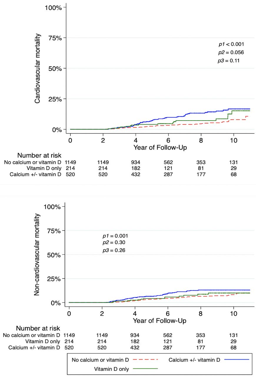
Impact of calcium and vitamin D supplementation on the cumulative incidence of cardiovascular and non-cardiovascular deaths. Survival analyses were performed using the Kaplan-Meier non-parametric method. Patients supplementing with calcium±vitamin D were at higher crude risk of mortality from both cardiovascular and non-cardiovascular causes compared with those who did not supplement. In each graph, p values are calculated using the log-rank test and labelled according to the comparison groups, with p1=calcium±vitamin D versus no supplementation; p2=vitamin D alone versus no supplementation; p3=calcium±vitamin D versus vitamin D alone.
Univariate and multivariable Cox proportional-hazard regression analyses of all-cause, cardiovascular and non-cardiovascular mortality, stratified by status of receiving aortic valve replacement, and secondary outcome of aortic valve replacement
Patients who supplemented with calcium alone (n=115) had similar all-cause mortality (HR=1.24, 95% CI (0.77 to 1.99); p=0.38) but a higher risk of AVR (HR=2.7, 95% CI (1.76 to 4.08); p<0.001) (results not shown). No survival differences were found in those who underwent AVR (table 3). AVR was performed more often in the calcium±vitamin D cohort relative to non-supplementers (513 (49.7%) vs 143 (11.1%), respectively; AR=88 per 1000 person-years, AR increase=71 per 1000 person-years, p<0.001) (table 2). On subgroup analyses that stratified by osteoporosis status to eliminate confounding by indication, differences in survival and AVR persisted between groups (results not shown).
Secondary outcomes
Figure 3 reveals the temporal changes in AS parameters across the study period, with AS progression among all groups evidenced by a significant trend in AVA reduction and MG and PG increase, irrespective of AVR status (online supplemental figure 4). In all groups, greater than one-third of patients developed severe AS by year 5, with no significant differences in the proportion of valve severity at all time points. On multivariable longitudinal mixed-effects linear regression, any supplementation was not associated with progression to severe AS, irrespective of AVR status (table 4). Baseline AVA had no impact on the rate of AS progression, and no differences were observed in mean AVA on nearest echocardiogram immediately prior to AVR between calcium±vitamin D versus vitamin D alone versus no supplementation groups (0.84±0.28 cm2 vs 0.82±0.25 cm2 vs 0.74±0.26 cm2, respectively, p=0.33).

Progression of aortic stenosis as measured by (A) aortic valve (AV) area, (B) mean AV gradient and (C) peak AV gradient, according to the study groups. Longitudinal analyses were performed using the linear mixed-effects models. All groups demonstrate a similar but significant increase in mean and peak gradients and reduction in AV area across the study duration.
Univariate and multivariable linear mixed-effects model to predict progression of aortic valve stenosis according to type of supplementation, stratified by status of receiving aortic valve replacement
Discussion
In this retrospective longitudinal analysis of a large cohort with mild to moderate AS followed over a median duration of 69 months, oral supplementation of calcium with or without vitamin D was significantly associated with all-cause mortality, CV mortality and AVR (figure 4). The risks of all-cause and CV mortality were also higher in patients supplementing with calcium who did not undergo AVR. Strengthened by its large sample size and extended follow-up period, our study suggests that calcium supplementation does not confer any CV benefit, and instead may reflect an elevated overall risk of AVR and mortality especially in those not undergoing AVR.

Association of supplemental calcium and vitamin D with mortality and aortic valve replacement in patients with mild to moderate aortic stenosis. In a retrospective longitudinal cohort analysis of patients followed over a median duration of 69 months, oral supplementation of calcium with or without vitamin D was significantly associated with higher all-cause mortality, cardiovascular mortality and aortic valve replacement.
While treatment of osteoporosis has been associated with attenuated AS progression,17 our understanding of the long-term safety of calcium intake and its implications on valvular disease is incomplete owing to the scarce available literature. Of the few studies that assess the role of supplementation in AS, the extremely small sample size limits generalisability and the follow-up periods are inadequate given the slow progression of the disease.15 16 No study to date investigates the clinical and haemodynamic impact of calcium and vitamin D supplementation on mild to moderate AS. The novelty of our study therein lies in its attempt to account for these limitations.
Previous work demonstrates that AS results from an active disease process analogous to atherosclerosis formation, incited by endothelial dysfunction leading to chronic inflammation and lipid deposition.1 7 Yet, coronary artery disease and valvular dysfunction and calcification are not entirely correlated as AS progression may occur independent of atherosclerotic risk factors.18 It is now understood that AS is not simply a degenerative disease intrinsic to ageing, but rather a complex mechanism associated with multiple risk factors.8 Notably, the role of calcium metabolism is well recognised in AS progression as demonstrated in conditions of dysregulated metabolism such as end-stage renal disease.19 Cinacalcet attenuates the progression of valvular calcification in haemodialysis patients, suggesting a beneficial role for reducing serum calcium.20 Importantly, despite the strong interplay between AS progression and ossification,21 the recent SALTIRE2 trial revealed that targeting molecular triggers of valvular calcification with denosumab or alendronate did not slow AS progression.22
With these data in mind, there are multiple tenable explanations for the lower survival among those supplementing with calcium who did not undergo AVR. First, this cohort comprises an undoubtedly higher risk profile at baseline, and therefore, may be more vulnerable to the effects of calcium supplementation on both valvular dysfunction, as evidenced by the observed lack of mortality difference in those receiving AVR, and convincingly, on alternative non-valvular mechanisms. Prior studies reveal relationships between oral calcium and progressive coronary arterial calcification,23 myocardial infarction24 and stroke.25 Alternatively, the underlying disease states that necessitate calcium supplementation, namely osteoporosis and states of deficiency such as hypoparathyroidism, are potentially identifying patients at greater risk of CV disease and mortality prior to study onset.26 27 While out of the scope of this analysis, it is plausible that these associations are attributed to supplementation leading to increased serum calcium, subsequent deposition of extraosseous calcification, and ultimately, as has been proposed, higher CV mortality through downstream alterations in endothelial function and blood coagulation.28 The acute rise in serum calcium after supplementation may also play a role,28 and perhaps explains the emerging theory that supplemental but not dietary sources are tied to excess CV risk.
These associations could further explain why AVR was performed more often in those supplementing with calcium despite similar rates of AS progression between groups. Our results confirm a significant progression into severe AS among all groups across the study period, with no difference in mean AVA between groups immediately prior to AVR. With class I indications for AVR that include severe AS with symptoms and severe AS while undergoing alternative cardiac surgery, the AVR differential is conceivably due to worsened non-valvular processes, such as atherosclerotic disease and its associated symptoms, in patients already highly comorbid at baseline. Notwithstanding, further mechanistic investigations are warranted to elucidate the effects of calcium intake on vascular and valvular health.
The significance of calcium intake on survival remains unclear with conflicting results in prior trials,29 prospective cohort studies24 and meta-analyses.10 The inconsistent findings suggest that this relationship is subject to a host of factors, including the mode (supplementation vs diet) and dosage of intake, studied population by sex, age or community, and duration of follow-up. Otherwise, supplemental vitamin D alone had no observed impact on survival in our study, consistent with prior randomised trials.30 Importantly, these investigations fail to mention the proportion of patients with concomitant AS and offer no guidance on the safe use of either supplementation in this cohort. The strengths of our analysis, in addition to its large sample size and long follow-up period, include the objective quantification of AS progression and correlation with outcomes of calcium and vitamin D supplementation, often used together, in both sexes.
Limitations
Several study limitations are worth noting. First, this analysis was subject to the inherent biases of a retrospective study, including selection and ascertainment bias. By relying on manual chart review to identify patient compliance with supplement use, similar to prior efforts,15 non-documented use and dietary intake of each supplement was difficult to measure and unaccounted for, which may have impacted the estimated effect of supplementation on the study outcomes. Second, the measured effect of specific supplemental dosages, both at baseline and with any dosage change over time, was not considered. Third, we did not quantify valvular calcification as a measure of AS progression; aortic valve calcification and AVA are strongly associated, and therefore the use of AVA was deemed sufficient. Fourth, the lack of data on cause of death for a significant proportion of patients poses ascertainment bias for the analyses on the associations between supplementation and differentiated mortality, and therefore, our findings should be interpreted with caution. Finally, given the degree of clinical differences between groups at baseline, there was a risk of residual confounding that may have impacted our findings; we attempted to mitigate this with our statistical model.
Conclusions
Oral calcium supplementation with or without vitamin D is associated with lower survival and a greater need for AVR in elderly patients with mild to moderate AS. Our findings suggest that supplemental calcium in this population does not confer any CV benefit, and instead these relationships should be thoughtfully considered in light of growing evidence and concern for CV harm particularly with unnecessary supplementation.
Key messages
What is already known on this subject?
Dysregulated calcium metabolism has long been implicated in aortic valvular calcification. The supplemental use of oral calcium and vitamin D is highly prevalent and continues to rise.
What might this study add?
Supplemental calcium with or without vitamin D is associated with lower survival and greater aortic valve replacement in elderly patients with mild to moderate aortic stenosis.
How might this impact on clinical practice?
Healthcare practitioners may further consider these relationships in elderly patients with mild to moderate aortic stenosis who supplement with oral calcium.
Data availability statement
Data are available upon reasonable request.
Ethics statements
Patient consent for publication
Ethics approval
This study was approved by the Cleveland Clinic Institutional Review Board (#18-1031). Informed consent was waived owing to the retrospective study design with anonymised data.
Acknowledgments
This study was made possible by a generous gift from Jennifer and Robert McNeil.
References
Supplementary materials
Supplementary Data
This web only file has been produced by the BMJ Publishing Group from an electronic file supplied by the author(s) and has not been edited for content.
Footnotes
Twitter @kassisMD, @EssaHariri, @DesaiMilindY, @tavrkapadia
NK and EHH contributed equally.
Contributors NK, EH, AKK and SK conceived and designed the study. NK, EH, AKK, KA, HL, AS, MG, MK and NB collected, analysed and interpreted the data. NK, EH, HL, BG, ZBP, SCH, MYD and SK drafted and critically revised the manuscript. BG, ZBP, SCH, MYD and SK supervised the study. NK, EH and SK are responsible for the overall content and serve as guarantors. All authors read and approved the final manuscript.
Funding This work was supported by unrestricted philanthropic support to the Cleveland Clinic Heart, Vascular, and Thoracic Institute.
Disclaimer The funding source had no role in the design or conduct of the study; the collection, management, analyses, or interpretation of the data; the preparation, review, or approval of the manuscript; or the decision to submit the manuscript for publication.
Competing interests None declared.
Provenance and peer review Not commissioned; internally peer reviewed.
Supplemental material This content has been supplied by the author(s). It has not been vetted by BMJ Publishing Group Limited (BMJ) and may not have been peer-reviewed. Any opinions or recommendations discussed are solely those of the author(s) and are not endorsed by BMJ. BMJ disclaims all liability and responsibility arising from any reliance placed on the content. Where the content includes any translated material, BMJ does not warrant the accuracy and reliability of the translations (including but not limited to local regulations, clinical guidelines, terminology, drug names and drug dosages), and is not responsible for any error and/or omissions arising from translation and adaptation or otherwise.