Article Text

Abstract
Background Functional mitral regurgitation (FMR) is a common finding in patients with heart failure (HF), but its effect on outcome is still uncertain, mainly because in previous studies sample sizes were relatively small and semiquantitative methods for FMR grading were used.
Objective To evaluate the prognostic value of FMR in patients with HF.
Methods and results Patients with HF due to ischaemic and non-ischaemic dilated cardiomyopathy (DCM) were retrospectively recruited. The clinical end point was a composite of all-cause mortality and hospitalisation for worsening HF. FMR was quantitatively determined by measuring vena contracta (VC) or effective regurgitant orifice (ERO) or regurgitant volume (RV). Severe FMR was defined as ERO >0.2 cm2 or RV >30 ml or VC >0.4 cm. Restrictive mitral filling pattern (RMP) was defined as E-wave deceleration time <140 ms. The study population comprised 1256 patients (mean age 67±11; 78% male) with HF due to DCM: 27% had no FMR, 49% mild to moderate FMR and 24% severe FMR. There was a powerful association between severe FMR and prognosis (HR=2.0, 95% CI 1.5 to 2.6; p<0.0001) after adjustment of left ventricular ejection fraction and RMP. The independent association of severe FMR with prognosis was confirmed in patients with ischaemic DCM (HR=2.0, 95% CI 1.4 to 2.7; p<0.0001) and non-ischaemic DCM (HR=1.9, 95% CI 1.3 to 2.9; p=0.002).
Conclusion In a large patient population it was shown that a quantitatively defined FMR was strongly associated with the outcome of patients with HF, independently of LV function.
- Heart failure
- diastolic dysfunction
- cardiomyopathy dilated
- mitral regurgitation
Statistics from Altmetric.com
Patients with heart failure (HF) are frequently affected by functional mitral regurgitation (FMR) owing to the distortion of valve apparatus secondary to global and local remodelling of the left ventricle.1 The effect of volume overload in a primary failing ventricle is deleterious and stimulates further modifications at molecular, cellular, tissue and cardiac chamber level.2 3 Mitral regurgitation increases diastolic wall stress, which is associated with an increase of extracellular matrix turnover, and neurohormone and cytokine activation, which leads to further eccentric hypertrophy, ventricular dilatation and failure. Despite this pathophysiological background, there are some inconsistencies about the impact of FMR on the prognosis of patients with HF. Although FMR is most often considered as a risk factor associated with adverse prognosis, some studies show that FMR is not associated with prognosis after adjustment for left ventricular ejection fraction (LVEF), suggesting that FMR is just a mere consequence of ventricular dysfunction.4–6 This clinical challenge has important consequences. FMR is not simply another variable associated with adverse prognosis but a risk factor which might potentially be treated. However, the decision to treat FMR surgically or percutaneously is based on the assumption that FMR is a key factor in the functional and prognostic impairment of patients with HF independently of LV dysfunction.
One aspect that might partly explain the controversial results of the clinical studies on the prognostic effect of FMR is the methodology used to evaluate the severity of FMR. Most negative studies used a semiquantitative method based on the evaluation of the jet area within the left atrium.4–6 This method is easy and widely used but is negatively influenced by important shortcomings. First, although the jet area is strongly correlated with the regurgitant volume, there is wide scattering, leading to low accuracy in individual patients.7 Furthermore, different centres defined semiquantitatively the severity of FMR using different scales and this makes comparability of different studies challenging and the value of the cut-off point used to define severity, ambiguous. For these reasons the European Association of Echocardiography recently stated that the colour jet area is not recommended for evaluating the severity of mitral regurgitation.8 On the contrary, analysis of the proximal convergent zone and the vena contracta allows quantitative determination of FMR severity and these are the recommended approaches for evaluating FMR.8 These measurements can be applied in routine clinical practice and have contributed prognostic insights into organic and ischaemic mitral regurgitation.9 10 Furthermore, the American Society of Echocardiography recommendations confirm previously issued guidelines advocating the use of quantitative methods involving Doppler echocardiography for determining thresholds of severity in different clinical settings.11
The aims of our study were (1) to evaluate the independent prognostic effect of FMR in a large population of patients with HF due to ischaemic and non-ischaemic dilated cardiomyopathy (DCM); (2) to validate the threshold values of quantitatively defined FMR as proposed by previous clinical studies, to define prognostically severe FMR.
Methods
Patients with chronic HF due to DCM of both ischaemic and non-ischaemic aetiology were retrospectively reviewed. Patients with FMR, with a structurally normal mitral valve, underwent a quantitative evaluation of the degree of regurgitation. Patients were recruited in four Italian centres (Pisa: 539; Milan 374; Verona 292; Brescia 51 patients). Exclusion criteria were the presence of significant aortic valve disease and recent myocardial infarction (<6 months).
The primary composite end point of the study was all-cause mortality or hospitalisation for worsening of HF and the secondary end point was all-cause mortality and hospitalisation for worsening HF considered separately.
End-point information was gathered from clinical charts or by telephone call or interview with the referring doctor or relatives.
Echocardiography
LV diastolic and systolic diameters were measured. LVEF was quantitatively determined by volume determinations, M-mode or visual estimation. Diastolic function was classified by the presence or absence of a restrictive mitral filling pattern (RMP), defined in the presence of E-wave deceleration time <140 ms.
The evaluation of FMR was assessed by measuring vena contracta (VC) or regurgitant volume (RV) or effective regurgitant orifice (ERO) according to the methodology preferred in each centre. The VC as it emerges from the valve leaflets was measured in both parasternal and apical views and then averaged. The RV and ERO were measured by the proximal isovelocity surface area method. The apical four-chamber view was most commonly used for optimal visualisation of the convergent flow. The radius of the proximal isovelocity surface area was measured at mid-systole in three different cardiac cycles and then averaged. ERO was obtained using the standard formula and RV was obtained by the standard proximal isovelocity surface area method or by a simplified method as previously published.12 According to previous studies, FMR was defined as severe when the mitral RV was >30 ml or ERO >0.2 cm2 or VC width was >0.4 cm.10 13 Three groups of patients were then considered: group 1 without any FMR, group 2 with mild to moderate FMR (RV ≤30 ml or ERO ≤0.2 cm2 or VC ≤0.4 cm) and group 3 with severe FMR (RV >30 ml or ERO >0.2 cm2 or VC >0.4 cm). If more than one parameter was measured in a single patient, ERO was considered the reference if the results were discordant.
Statistical analysis
Continuous data are presented as mean ±SD. Comparisons of all measurements were made using the unpaired t test or χ2 test or analysis of variance (ANOVA), as appropriate. Logistic regression analysis was used to analyse the independent association of symptom severity with clinical variables.
Multivariate Cox proportional hazard ratio (HR) was used to demonstrate that the association between FMR and outcome was independent of LV systolic and diastolic function and of other clinical variables that more strictly characterised patients divided according to survival status (considering a p value <0.0001).
HRs for severe FMR, LVEF <30% and presence of RMP were obtained using multivariate models, with follow-up and survival status at different time intervals in order to describe variations over time.
Survival rate was analysed by the Kaplan–Meier method and survival curves were compared by the log-rank test. A p value <0.05 was considered statistically significant. Statview 5.0 (Abacus Concepts, SAS Institute) was used for statistical analysis.
Results
The study population consisted of 1256 patients (mean age 67±11; 78% male) with chronic heart failure due to DCM. The mean EF was 32±8 and 35% of patients had RMP. Twenty-seven per cent of patients did not have FMR; mild to moderate functional FMR was present in 49% and severe FMR in 24% of the overall population. Clinical and echocardiographic characteristics of patients divided according to FMR severity are shown in table 1.
Clinical and functional parameters of patients divided according to severity of functional mitral regurgitation (FMR)
Patients without FMR had a higher LVEF than patients with FMR, but subgroup analysis demonstrated that only patients with severe FMR had a significantly lower LVEF (29±8% p-ANOVA <0.0001) while patients with mild to moderate and those without FMR had a similar level of LV dysfunction (LVEF 33±8 and 34±8, respectively; p=0.4). In contrast, a gradual and significant increase in the prevalence of RMP was seen in patients with different degrees of FMR (20%, 30% and 62% in patients without FMR, with mild to moderate FMR and severe FMR, respectively; p<0.0001).
Symptomatic status
Symptomatic status was available in 1023 patients, of whom 150 (15%) were in functional New York Heart Association (NYHA) class I, 500 (49%) in class II, 316 (31%) in class III and 57 (6%) in class IV. There was a significant difference in the prevalence of FMR according to NYHA class (figure 1). Increasing age, decreasing LVEF and the presence of RMP were all significantly associated with the severity of symptoms (p=0.0006 for age, p<0.0001 for LVEF and RMP).

Prevalence of functional mitral regurgitation (FMR) according New York Heart Association class.
Logistic regression analysis was performed to determine correlates of severe symptomatic status (NYHA III-IV vs I-II). In a multivariate model, severe FMR (OR=1.8, 95% CI 1.1 to 2.8; p=0.01) was significantly associated with symptomatic impairment compared with patients without FMR. In the same model LVEF (OR=0.93, 95% CI 0.9 to 0.95; p<0.0001) and age (OR=1.02, 95% CI 1.009 to 1.04; p=0.002) were also independently associated but RMP showed a borderline association with severity of symptoms (OR=1.4, 95% CI 0.97 to 1.9; p=0.07).
Survival analysis
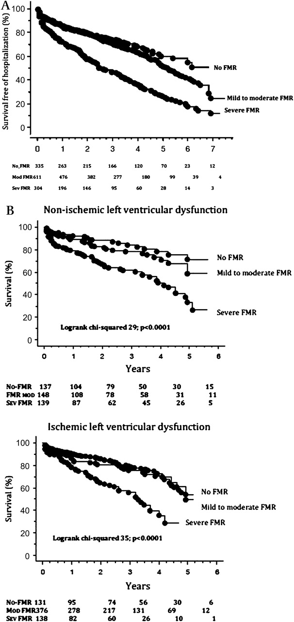
After a mean follow-up of 2.7±2.0 years (median 2.47; range 0.03–7.9 years), the primary end point was reached by 525 patients (300 deaths and 225 hospitalisations for heart failure). Clinical and echocardiographic characteristics of patients divided according to survival status are shown in table 2. The degree of FMR was strongly associated with adverse outcome. As shown in figure 2A, the overall survival free of hospitalisation for worsening heart failure at the end of follow-up was 40% in patients without FMR, 25% for patients with mild to moderate FMR and 7% for those with severe FMR (log-rank Mantel Cox χ2=94; p<0.0001). The hazard risk associated with severe FMR was 2.7 (95% CI 2.1 to 3.5; p<0.0001) and with mild to moderate FMR was 1.3 (95% CI 1.04 to 1.7; p=0.02) compared with patients without FMR. Each single indicator of severe FMR showed a strong association with outcome with similar HR (ERO >0.2 cm2: 1.8 (95% CI 1.3 to 2.6; p=0.0009); RV >30 ml: 2.0 (95% CI 1.3 to 2.6; p=0.001); VC >0.4: 2.5 (95% CI 2.1 to 3.2; p<0.0001)).
Clinical and function parameters of patients divided according to survival free of hospitalisation status

(A) Kaplan–Meier plots showing time to the combined end point of all-cause mortality and heart failure hospitalisation for patients without functional mitral regurgitation (FMR), with mild to moderate FMR and with severe FMR. (B) Kaplan–Meier plots showing time to all-cause mortality alone in patients with ischaemic dilated cardiomyopathy and non-ischaemic cardiomyopathy.
After adjustment for LVEF and RMP, the risk of severe FMR was 2.0 (95% CI 1.5 to 2.6; p<0.0001) and for mild to moderate FMR 1.2 (95% CI 0.96 to 1.6; p=0.09). This was confirmed at different time points of follow-up (figure 3). The result of the analysis did not change when the LVS was substituted for LVEF (severe FMR 6.7, 95% CI 2.1 to 21.4; p=0.001). The presence of severe FMR (severe FMR vs mild to moderate or no FMR) maintained a significant association with outcome (HR=1.5, 95% CI 1.2 to 1.9; p=0.001) even after adjustment for both clinical (NYHA I-II vs III-IV, age <65 years) and functional parameters (LVEF <30% and RMP) (see table 3 for details).

Hazard ratio of severe functional mitral regurgitation (MR) (white dots) was stable at different time intervals. At each point the association between severe FMR and outcome was independent of ejection fraction (EF) (black dots) and restrictive mitral filling (RMP) (triangles).
Cox proportional hazard model (multivariate analysis) considering both primary and secondary end points
The prognostic role of FMR was confirmed when secondary outcomes were used separately (table 2). FMR was independently associated with the risk of all-cause mortality. Figure 2B shows that the mortality rate is similar according to FMR severity for ischaemic and non-ischaemic DCM. For the risk of hospitalisation for worsening heart failure, severe FMR was associated with double the risk of hospitalisation independently of symptom severity at baseline, while LVEF and RMP did not add any significant information to FMR and NYHA (see table 3 for details).
Interestingly, in less symptomatic patients (NYHA I-II) FMR was the only predictor of future hospitalisation for HF (severe FMR, HR=2.9, 95% CI 1.6 to 5.1; p=0.0001; mild to moderate FMR, HR=2.1, 95% CI 1.3 to 3.4; p=0.003) and LVEF, RMP and NYHA class did not have any univariate association with the risk of HF.
Given the possible overestimation of LVEF in the presence of mitral regurgitation, LVEF was artificially decreased by a factor of 4 in patients with mild to moderate FMR and of 8 for severe FMR, as suggested by other authors.14 After adjustment for the modified LVEF, the HR of severe FMR was 1.7 (95% CI 1.3 to 2.2; p=0.0001) and for non-severe FMR, 1.1 (95% CI 0.9 to 1.4; p=0.3).
The presence of severe FMR significantly predicted the primary end point in different subgroups of patients (figure 4).

Hazard ratio and interval confidence of severe functional mitral regurgitation (FMR) in different subgroups of patients. CAD, coronary artery disease; EF, ejection fraction, NYHA, New York Heart Association; RMP, restrictive mitral filling.
Discussion
The main finding of this study is that severe FMR, defined as RV >30 ml or ERO >0.2 cm2 or VC >0.4 cm, is associated with a twofold increased risk of adverse events after adjustment for LVEF and RMP in patients with HF due to DCM. Accordingly, FMR should not be considered just a mere consequence of ventricular remodelling but a major predictor for the outcome of patients with HF, suggesting that in patients with severe FMR all therapeutic options of pharmacological and non-pharmacological treatment should be considered.
The natural history of patients with mitral regurgitation due to flail leaflet is characterised by a high risk of morbidity and mortality.14 This risk is gradually increased with the degree of mitral regurgitation, even in patients with normal ventricular function.9 As a consequence, it would be expected that FMR, which is superimposed on a primary dysfunctional left ventricle, should be an even more serious condition. It has recently been demonstrated that a moderate mitral regurgitation after an acute myocardial infarction affects the degree of ventricular remodelling through different molecular and cellular mechanisms.15 This suggests that FMR is not simply a bystander but has a direct effect on excess remodelling, enhanced risk of atrial fibrillation16 and subsequent clinical events.17
Nevertheless, the clinical implications of FMR in patients with HF is still dubious, because different studies have shown opposite results and there is no consensus about whether FMR might affect prognosis independently of the degree of ventricular dysfunction.4–6 10 13 The reasons for this incongruence are varied and are mostly due to small study series with relatively low overall event rates and insufficient power to discriminate the relative importance of FMR and also to the methodology used to evaluate FMR. The majority of studies that failed to demonstrate the independent prognostic role of FMR used semiquantitative methods based on the jet area within the left atrium. Although this method is easy, rapid and widely used, it should be acknowledged that the accuracy in detecting severe FMR is unacceptable owing to a high overestimation in the case of the central jet.18 Patel and co-authors demonstrated that FMR was not associated with outcome when using either semiquantitative methods or quantitative measurements of ERO and RV.4 However, that study, in our opinion, had an important shortcoming: measurements of ERO and RV were available in 81% of patients with moderately-severe and severe FMR but in only 34% of moderate or mild FMR, leading to an important bias in patient selection. On the contrary, quantification of ERO and RV is possible and feasible in all ranges of FMR and for this reason we believe that the clinical usefulness of these quantitative parameters is still unrecognised.
Our study overcomes the limitations of previous studies showing that in a large patients population the presence of severe FMR, defined quantitatively as ERO>0.2 cm2 or RV>30 ml or VC>0.4 cm, provides relevant clinical information independently of left ventricular function in patients with both ischaemic and non-ischaemic DCM and in patients with severe and not severe HF. The clinical usefulness of quantitative parameters is highlighted by the striking coherence of the results provided by different studies. In this study the survival rate of patients with ischaemic FMR was 30% after 5 years, which is almost identical to the survival rate described by a previous investigation where the same values of ERO and RV were used to define severe ischaemic FMR.10
A finding which corroborates the association of FMR with outcome is the observation that ACE inhibitors, β blockers and resynchronisation therapy concomitantly increase survival and decrease the severity of FMR in patients with HF.19–21 However, this link is difficult to demonstrate because all these treatments have profound anti-remodelling effects. The surgical treatment of FMR, operating directly and solely on the mitral valve, may have solved this question. Despite contrasting results, mitral valve surgery improves symptoms22 but fails to improve long-term outcome compared with medical treatment.23 This is at variance with the hypothesis of FMR as a key factor independently of LVEF, but some limitations of these studies must be acknowledged. First, they are non-randomised trials.23 Second, there is a consistent technical problem which causes a very high percentage of recurrence of FMR, eliminating the potentially beneficial effect in the mid–long term.24 Third, patients were recruited for surgery mainly using diagnosis based on semiquantitative methods, which have important limitations as discussed above and might have induced a bias in patient selection.
The interaction between FMR and LVEF is complicated since they are intrinsically inter-related. LVEF is a marker of ventricular remodelling, which is the cause of FMR owing to LV enlargement and papillary muscle displacement, but FMR can artificially increase LVEF through its volume overload. Consequently, extrapolating the independent effect of each variable on outcome is possible only when large populations are studied and when FMR is directly defined by the use of quantitative parameters. Furthermore, our study has demonstrated the independent effect of FMR even when LVEF was downgraded by a factor of 4 and 8 points in patients with mild to moderate and severe FMR respectively, in order to avoid the potential artificial effect of volume overload on ventricular function. The interaction between FMR and RMP is even more subtle. RMP has been described as a marker of increased left atrial pressure,25 decreased LV diastolic function26 and a powerful marker of adverse prognosis in patients with LV systolic dysfunction.6 However, mitral regurgitation induces predictable changes in mitral inflow and pulmonary vein velocities in exactly as diastolic function does27 and, accordingly, in this study a strong and graded association between the degree of FMR and RMP was seen. This suggests that mitral regurgitation may be a main determinant of RMP beyond left atrial pressure elevation and may contribute to the important prognostic effect of RMP.
Our study is retrospective and has limitations inherent in its design. Important clinical information such as the presence of diabetes or renal function or pharmacological treatment were not reported since this information could not be obtained for all patients. Likewise, information about resynchronisation therapy and mitral surgery was not complete for all patients and, consequently, the potential impact of these therapeutic approaches cannot be evaluated. However, it is unlikely that the lack of this information would have influenced the impact of severe FMR on prognosis because not considering the use of treatments which reduce the burden of mitral insufficiency would, if anything, have led to underestimation of the clinical severity of FMR. Another important limitation which has to be acknowledged is the lack of the use of each parameter in single patients, which would have allowed us to evaluate which quantitative parameters might be more useful in clinical practice. Given that the study was multicentric, a possible lack in standardisation of the end-point collection might be a limitation. The relative prognostic relevance of FMR compared with natriuretic peptide was not assessed.28
Finally, the demonstration of a clear and powerful association between FMR and prognosis might only suggest that treatment of FMR may improve outcome. However, particularly for the percutaneous approach to FMR, the effectiveness of these procedures can be demonstrated only by randomised trials.
In conclusion, FMR is strongly associated with outcome after adjustment for LVEF and RMP in patients with both ischaemic and non-ischaemic DCM.
References
Footnotes
Competing interests None to declare.
Provenance and peer review Not commissioned; externally peer reviewed.