Article Text

Abstract
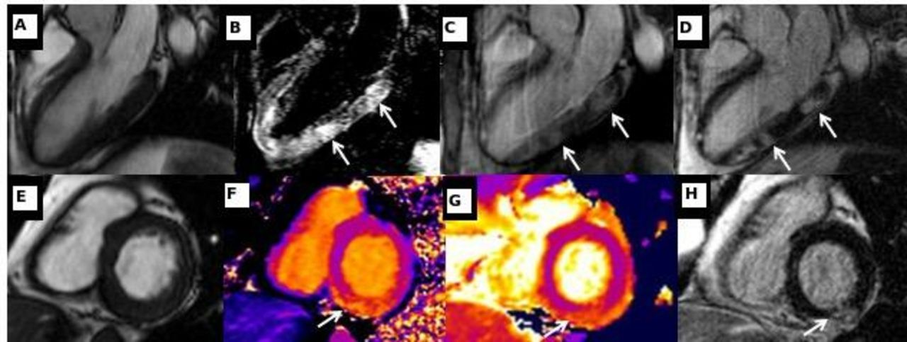
Clinical introduction A 45-year-old man with ulcerative colitis was admitted with bloody diarrhoea and chest pain. Inflammatory markers and high-sensitivity troponin were elevated (C reactive protein 57 mg/L, white cell count 10.65×10⁹/L, neutrophil 6.6×10⁹/L, Troponin-I 663 mmol/L). The ECG showed inferior ST-elevation. Urgent coronary angiography revealed unobstructed coronary arteries. Inpatient cardiovascular magnetic resonance (CMR) was arranged to determine the aetiology of the myocardial infarction with non-obstructive coronary arteries. The imaging protocol at 1.5 T included balanced steady-state free precession cine images, T2-weighted oedema sequences, and early and late gadolinium enhancement (LGE). Native T1 and T2 mapping images provided advanced tissue characterisation (figure 1).
Question What is the most likely diagnosis based on the MRI findings?
Multiple embolic myocardial infarctions in the right coronary artery territory.
Acute autoimmune myocarditis.
Cardiac sarcoidosis.
Stress (Takotsubo) cardiomyopathy.
Multiple embolic myocardial infarctions in the left circumflex coronary artery territory.

(A) Balanced steady-state free precession (bSSFP) left ventricular long-axis, three-chamber view. (B) T2 short-tau inversion recovery. (C) Early gadolinium enhancement demonstrating high signal intensity indicative of hyperaemia with capillary leakage (arrowed). (D) Late gadolinium enhancement with high signal intensity indicative of increased extracellular space (arrowed). (E) bSSFP left ventricular short-axis view. (F) Native myocardial T1 mapping with elevated native T1 mapping values in the inferior wall (arrowed). (G) Native myocardial T2 mapping with elevated native T2 values in the inferior wall, indicative of oedema (arrowed). (H) Late gadolinium enhancement with high signal intensity indicative of increased extracellular space (arrowed).
- cardiac magnetic resonance (cmr) imaging
- acute coronary syndromes
- myocarditis
This is an open access article distributed in accordance with the Creative Commons Attribution Non Commercial (CC BY-NC 4.0) license, which permits others to distribute, remix, adapt, build upon this work non-commercially, and license their derivative works on different terms, provided the original work is properly cited, appropriate credit is given, any changes made indicated, and the use is non-commercial. See: http://creativecommons.org/licenses/by-nc/4.0/.