Article Text

Abstract
Transition from congenital junctional ectopic tachycardia to complete AV block was observed in an 8 month old girl, over a 36 hour period, during initial hospital admission. Two years later she had evidence of a rapidly increasing left ventricular end diastolic diameter, associated with lowest heart rates during sleep of < 30 beats/min. A transvenous permanent pacemaker was therefore implanted. This finding supports the idea that a pathological process in the area of the AV junction, initially presenting as junctional ectopic tachycardia may later extend to sudden complete atrioventricular block.
- junctional ectopic tachycardia
- heart block
- congenital heart disease
- paediatrics
Statistics from Altmetric.com
Junctional ectopic tachycardia (JET) is one of the rarest forms of supraventricular tachycardia in infancy. The mechanisms of termination of the congenital form of JET are unclear from most clinical series.1 ,2 Histological studies have shown His bundle degeneration and Purkinje cell tumours as well as fibroelastosis.3 Progression to complete atrioventricular block has been proposed,3 but has not been described in the clinical setting. We present a girl who presented with JET at the age of 8 months who went into complete AV block during hospital admission 36 hours later.
Case report
An 8 month old girl presented to her local hospital after a period of crying, apnoea, and cyanosis at home. After stimulation she had recovered fully but at the hospital tachycardia was noted at a frequency of 230 beats/min. The ECG at admission showed a regular, narrow QRS complex tachycardia with AV dissociation consistent with the diagnosis JET (fig 1). Echocardiography showed an anatomically normal heart with decreased contractility (shortening fraction 15%) and left ventricular dilatation, suggesting longstanding tachycardia. Intravenous adenosine was ineffective, and digoxin was started (10 μg/kg at 8 hourly intervals). Thirty six hours later she exhibited complete AV dissociation with QRS rate of 100 beats/min (fig 2), and was discharged from hospital. At follow up ventricular function had normalised within three months (shortening fraction 38%). Two years later digoxin was discontinued. Routine Holter recordings at follow up showed no episodes of tachyarrhythmia and she was asymptomatic. At the age of 2 years and 8 months, she had evidence of a rapidly increasing left ventricular end diastolic diameter, associated with lowest heart rates during sleep of < 30 beats/min. A transvenous permanent pacemaker was therefore implanted.
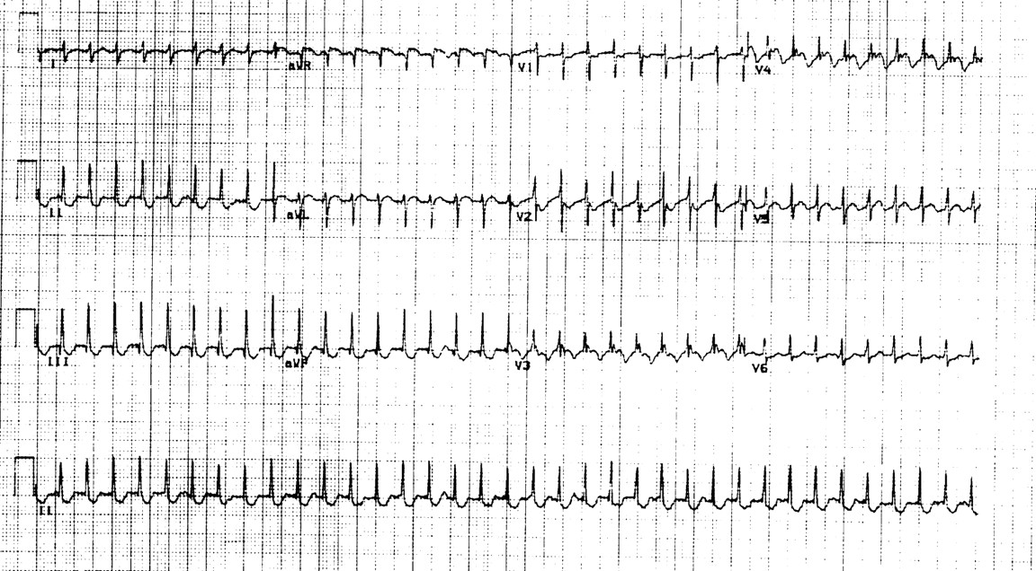
12 lead ECG demonstrating a regular, narrow QRS tachycardia with AV dissociation.

12 lead ECG 36 hours later, demonstrating resolution of tachyarrhythmia with persistence of AV dissociation and narrow QRS complexes.
Discussion
Congenital JET is a rare form of supraventricular tachycardia with an obscure aetiology. An association with late onset AV block has been postulated, and degeneration of the His bundle with fibroelastosis, as well as His-Purkinje cell tumours have been demonstrated by histology in patients with JET who died suddenly.3 The outcome is generally poor. Villain et al showed that congenital JET is associated with a high incidence of congestive heart failure and high mortality, (sometimes despite adequate medical control).1 The results of conventional drug treatment have been unsatisfactory, with JET continuing at slower rates in most patients receiving antiarrhythmic medication. In refractory cases, His bundle ablation and permanent pacemaker implantation have been proposed, but these procedures have also been associated with deaths.1 Late AV block has been postulated as a cause of sudden death in some patients in whom the tachycardia was satisfactorily controlled with medications.1 In most patients in whom medical treatment was successful, normal AV conduction was re-established, with intermittent episodes of JET occurring when the sinus rate decreased. The clinical course in our patient supports an ongoing pathological process in the region of the His bundle presenting as JET and finally resulting in permanent loss of AV conduction.