Article Text

Abstract
Background: Tumour necrosis factor α (TNF α) is implicated in the pathophysiology of heart failure. Plasma TNF α is raised in patients with myocardial dysfunction in proportion to the symptoms.
Objective: To determine whether this genetic variant is over represented in heart transplant recipients.
Patients: 175 heart transplant recipients and a control group of 212 healthy volunteers were studied. The reason for transplantation was severe symptomatic myocardial dysfunction in all cases.
Methods: The TNF α genotype was determined by polymerase chain reaction and gel electrophoresis. The populations were compared for their fit to Hardy–Weinberg equilibrium by calculating the expected frequencies of each genotype and comparing them to the observed values. A χ2 test was used to determine the significance of the difference between the observed and expected values.
Results: No differences were found in the frequency of the TNF2 allele between all heart transplant recipients taken together (54/175, 31%) and healthy volunteers (58/212, 27%). A higher proportion of TNF2 allele carriers was present in cardiac recipients with a pretransplant diagnosis of viral mediated or idiopathic heart failure than in those with ischaemic myocardial dysfunction (26/69 (37.7%) v 28/106 (26.4%), p = 0.03). Patients with a non-ischaemic aetiology had a higher prevalence of TNF2 than healthy volunteers (26/69 (37.7%) v 58/212 (27%), p = 0.05).
Conclusions: The TNF2 allele is overrepresented in patients with end stage non-ischaemic myocardial dysfunction. This may represent a genetic predisposition in a small subset of patients who could respond favourably to anti-TNF α treatment.
- tumour necrosis factor α
- heart failure
- cytokines
- PCR, polymerase chain reaction
- TNF, tumour necrosis factor
Statistics from Altmetric.com
In 1985, patients with septic shock were found to have a circulating myocardial depressant substance.1 This was later found to be tumour necrosis factor α (TNF α).2 TNF α has since been strongly implicated in the pathogenesis of heart failure. Circulating concentrations of TNF α are raised in heart failure patients, and there is a correlation between increasing concentrations and severity, symptoms, cardiac cachexia, and survival.3,4 Animal studies have shown that overexpression5,6 or chronic infusion7,8 of TNF α leads to myocarditis, systolic dysfunction, ventricular dilatation and hypertrophy, myocardial fibrosis, myocyte apoptosis, and increased mortality, all of which can be reversed by TNF α binding proteins.9 Early human clinical trials of TNF α inhibition suggest that an improvement in symptoms and exercise capacity,10 as well as an increase in ejection fraction,10,11 can be achieved.
Levels of TNF α after lymphocyte12 or monocyte13 stimulation vary between healthy individuals. This is partly the result of a guanine to adenosine substitution polymorphism located at position −308 in the promoter region of the gene.14 The “TNF2” allele is a more powerful transcriptional activator than the common allele, with a six- to sevenfold increase in the inducible level of TNF α gene transcription.15 Cell stimulation studies have shown that the TNF2 allele is associated with greater TNF α production in vitro.16 The allele is also in strong linkage disequilibrium with other genetic determinants of TNF α production, for example HLA-DR3.12
We hypothesised that the TNF2 genetic variant would be overrepresented in patients with end stage heart failure who required a heart transplant.
METHODS
Patients
We studied recipients of human cardiac allografts transplanted between 1987 and 1996. The control group consisted of 212 unrelated healthy volunteers.17
Sample collection
Patients gave informed consent to the collection and storage of blood, isolation of DNA, and determination of the TNF2 polymorphism. Ethical approval was obtained from the South Manchester medical research ethics committee. Blood samples were mixed with EDTA and stored at −80°C before further processing. DNA or stored leucocytes from deceased patients were acquired from the tissue typing laboratory, Manchester Royal Infirmary.
Primer sequences
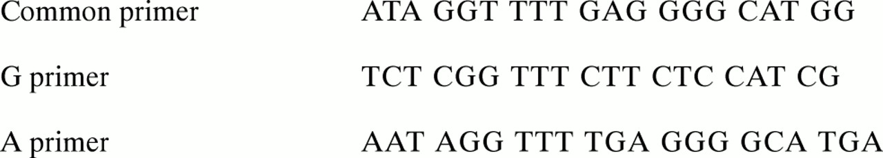
Specific oligonucleotide primers were designed for the TNFα–308 polymorphism, based on the published sequence (fig 1). Lyophilised oligonucleotide probes were manufactured by Genosys, UK. The primers were prepared to a working concentration of 50 mM by the addition of measured quantities of PCR grade water.

Primer sequences for the TNF α–308 polymorphism.
Polymerase chain reaction
Each polymerase chain reaction (PCR) mixture comprised (in relative quantities) 10× reaction buffer (× 0.22), 2 mM dNTP mix (× 0.22), 25 mM MgCl2 (× 0.13), 60% (wt/vol) sucrose (× 0.42), thermoprime DNA polymerase (× 0.011). A 50 μl aliquot of each of the common and specific primers (G or A) was mixed with 150 μl of PCR grade water. Equal volumes of the reaction and primer mixtures were placed in separate thin walled PCR tubes. One tenth this volume of test DNA was added before PCR.
A Techne Genius (Techne Cambridge, Duxford, UK) controlled the thermal cycles of 95°C for one minute followed by 95°C for 15 seconds, 65°C for 50 seconds, and 72°C for 40 seconds (10 cycles), and 95°C for 20 seconds, 61°C for 50 seconds, and 72°C for 50 seconds (20 cycles). A final holding temperature of 4°C was used.
Electrophoresis of PCR products
The genotype was identified by running the PCR products through 2% agarose gel containing 5 mg/100 ml ethidium bromide. The PCR product size was 184 base pairs. A and G homozygotes (high and low producers, respectively) have one product band only in their respective lanes; GA heterozygotes (high producer) have a band in each lane.
Statistical analysis
The data were tested for their fit to the Hardy–Weinberg equilibrium by calculating the expected frequencies of each genotype and comparing them to the observed values. A χ2 test was used to determine the significance of the difference between the observed and expected values.
RESULTS
No differences were found in the frequency of the TNF2 allele between all heart transplant recipients taken together—regardless of disease aetiology (54/175, 31%)—and healthy volunteers (58/212, 27%) (table 1). Carriers of the TNF2 allele were overrepresented in heart transplant recipients with a pretransplant diagnosis of viral mediated or idiopathic myocardial dysfunction (that is, non-ischaemic), in comparison with those with ischaemic myocardial dysfunction (26/69 (37.7%) v 28/106 (26.4%), p = 0.03) and healthy unrelated volunteers (26/69 (37.7%) v 58/212 (27%), p = 0.05) (table 2). The high producing TNF allele was therefore equally prevalent between patients with pretransplant ischaemic heart disease and the control cohort (26.4% v 27%, NS).
Frequency of the TNFα–308 polymorphisms in the transplant recipient, transplant donor, and healthy unrelated populations
Frequency of the TNF2 polymorphism in transplant recipients and a healthy unrelated population
DISCUSSION
TNF α has been strongly implicated in the pathogenesis of heart failure.3–11 In this study we showed that the TNF2 allele, which is associated with a six- to sevenfold increase in the inducible level of TNF α gene transcription,15 was more prevalent in transplant recipients with a preoperative diagnosis of non-ischaemic myocardial dysfunction. This finding suggests that a subset of individuals could have a genetic predisposition to end stage heart failure requiring transplantation. These patients may develop left ventricular dysfunction through a TNF α dependent pathway triggered, for example, by a viral infection. Our results are supported by a Japanese study of idiopathic dilated cardiomyopathy,18 in which the frequency of TNF2 in affected patients was significantly higher than in a healthy control group. In a non-transplant population, however, the polymorphism was not associated with the presence of heart failure,19 but this might reflect the select nature of our transplant cohort.
The development of virus induced autoimmune myocarditis is associated with local production and secretion of TNF α.20 Administration of TNF α can promote coxsackievirus B3 myocarditis in genetically resistant B10A mouse models.21 In a mouse model of encephalomyocarditis virus infection, exogenous recombinant TNF α increased the virus concentration in the myocardium, produced myocardial necrosis and cellular infiltration, and exacerbated the myocarditis. TNF α antibodies improved survival and the myocardial lesions.22 Murine models of cardiac myosin induced myocarditis closely resemble the chronic stages of virus induced myocarditis in humans. Antibodies against TNF α also had a beneficial effect in this condition.23 Raised serum TNF α was found in 46% of patients presenting with viral myocarditis, compared with 35% with dilated cardiomyopathy.24
Possible mechanisms of TNF α mediated heart failure include reduction in intracellular calcium,25 desensitisation of cardiac myocytes to calcium, interference with nitric oxide synthase function, activation of metalloproteinases, direct negative inotropic effects,26 cardiomyocyte apoptosis,27 or increased production of superoxide anions.28 Cardiac myocytes appear to be the main source of TNF α.5 Alternatively, raised TNF α may be secondary to heart failure and not a causative factor. TNF α concentrations may reflect a common pathophysiological background resulting from peripheral hypoperfusion and ischaemia29 or increased right atrial pressure resulting in splanchnic congestion. This may lead to increased bowel permeability with consequent bacterial translocation, endotoxin release, and increased systemic TNF α production.29 Also, increasing myocardial wall stress could elicit TNF α expression.
The findings of this study could have practical implications. If a subgroup of patients do have TNF α mediated heart failure then they may respond to anti-TNF α therapy. Treatment with TNF α binding protein prevents and reverses the negative inotropic effects in vitro.15 An adenovirus encoding the 55 kDa TNFα receptor–IgG fusion protein improved left ventricular size and myocardial inflammation in TNFα overexpressor mice.30 Clinical trials of TNFα inhibition in humans using pentoxifylline11 or TNF α receptor (p75) fusion protein10 suggest an improvement of symptoms and exercise capacity, an increase in ejection fraction, and a reduction in ventricular size and mass.10 TNF α, however, is an auto/para/juxtacrine agent with biological activity determined by the target cell type as well as by the intracellular milieu. Results obtained with systemic administration of a cytokine inhibitor must be interpreted with this in mind.
Conclusions
We have found that the TNF2 allele, which confers increased TNF α production, is over represented in patients with end stage non-ischaemic myocardial dysfunction. This may represent a genetic predisposition in a small subset of patients who could respond favourably to anti-TNF α treatment.
Acknowledgments
CGD was supported by a grant from the National Heart Research Fund. The study was funded by the Wythenshawe Cardiology Research Fund and the New Heart, New Start appeal. Many thanks to Dr Steve Sheldon, St Mary's Tissue Typing Laboratory, for supply of some DNA and leucocyte samples.