Article Text

Abstract
Objective: To assess the clinical utility and cost effectiveness of a personal ultrasound imager (PUI) during consultation rounds for cardiac evaluation of patients with suspected cardiac disease.
Methods: 107 unselected patients from non-cardiac departments (55% men) were enrolled in the study. After the physical examination the consultant cardiologist performed an echocardiographic study with a PUI. The final report was given instantly to the referring physician. All patients subsequently underwent a study with a standard echocardiographic device (SED). For each patient the consultant cardiologist noted whether the findings of the PUI were adequate for final diagnosis. The total cost when full echocardiography was used was compared with the cost when the PUI was used. The time interval from request to diagnosis was also compared.
Results: In 84 (78.5%) patients no further examination with an SED was regarded as necessary. Twenty three patients (21.5%) required a further detailed examination with the SED because of the need for haemodynamic information. There was an excellent agreement for the detection of abnormalities between the two devices (96%). The total cost was €132 per patient with the SED and €75 per patient with the PUI. According to this study, the use of the PUI can lead to a 33.4% reduction of total cost. The mean time from request to diagnosis at the authors’ institution was four days for the SED and instantly for the PUI, for additional potential cost savings.
Conclusions: Immediate echocardiographic assessment during consultation rounds can lead to significant cost savings and can shorten the time to diagnosis.
- cost effectiveness
- personal ultrasound imager
- hand held ultrasound device
- clinical usefulness
- PUI, personal ultrasound imager
- SED, standard echocardiographic device
Statistics from Altmetric.com
During consultation rounds in non-cardiology departments the consulting cardiologist is confronted with specific clinical questions (such as presence of pericardial effusion, left ventricular function, source of embolism, or inferior vena cava collapse). It has been proved that echocardiography is superior to physical examination in diagnosis of cardiac disorders, especially in the early stages of disease.1–3 However, transporting a standard echocardiographic device (SED) during ward rounds is unpractical and therefore of limited usefulness. Recently, small hand held ultrasound imagers have been developed. Being ultraportable and having a high accuracy and low cost, they can have a big impact on daily clinical practice.
The aim of the present study was to evaluate the clinical utility and cost effectiveness of a small personal ultrasound imager (PUI) (SonoHeart System, SonoSite, Inc, Bothell, Washington, USA) during consultation rounds for evaluation of unselected patients with suspected cardiac disease. The results from a high end SED were used for performance comparison and verification.
PATIENTS AND METHODS
Study population
We studied 107 consecutive unselected patients with suspected cardiac disease (55% men) with a mean (SD) age of 53 (17) years, for whom a consultation by the cardiologist was requested.
Study design
The main inclusion criterion for this study was the request from a physician in a non-cardiac department for cardiac evaluation of a patient. In addition to the physical examination, an echocardiographic study was performed with the PUI by the consulting cardiologist at the patient’s bedside. The final cardiac report was given instantly to the referring physician for a management decision. The need for an echocardiographic study with the SED was noted by the consultant cardiologist after the clinical examination of the patient and the PUI study. As part of the study, all of the patients also underwent echocardiography by means of an SED (Sonos 5500, Hewlett Packard, Andover, Massachusetts, US or System V, Vingmed, Horten, Norway). These results were reported by a second investigator blinded to the results of the PUI examination.
Routine logistical procedures for echocardiographic examinations in the echolaboratory were not changed in this study. Echocardiographic data were obtained in standard cardiac views and basic linear measurements of structures and cavities were taken.4,5
The average cost of the normal procedure when a patient is referred to full echocardiography was calculated and compared with the cost when the PUI was used.
Furthermore, the time intervals between the request for an echocardiographic examination and the final cardiac report for both the PUI and the SED were compared.
The study was approved by our institutional medical ethical committee and written informed consent for the study was obtained from all patients.
The PUI
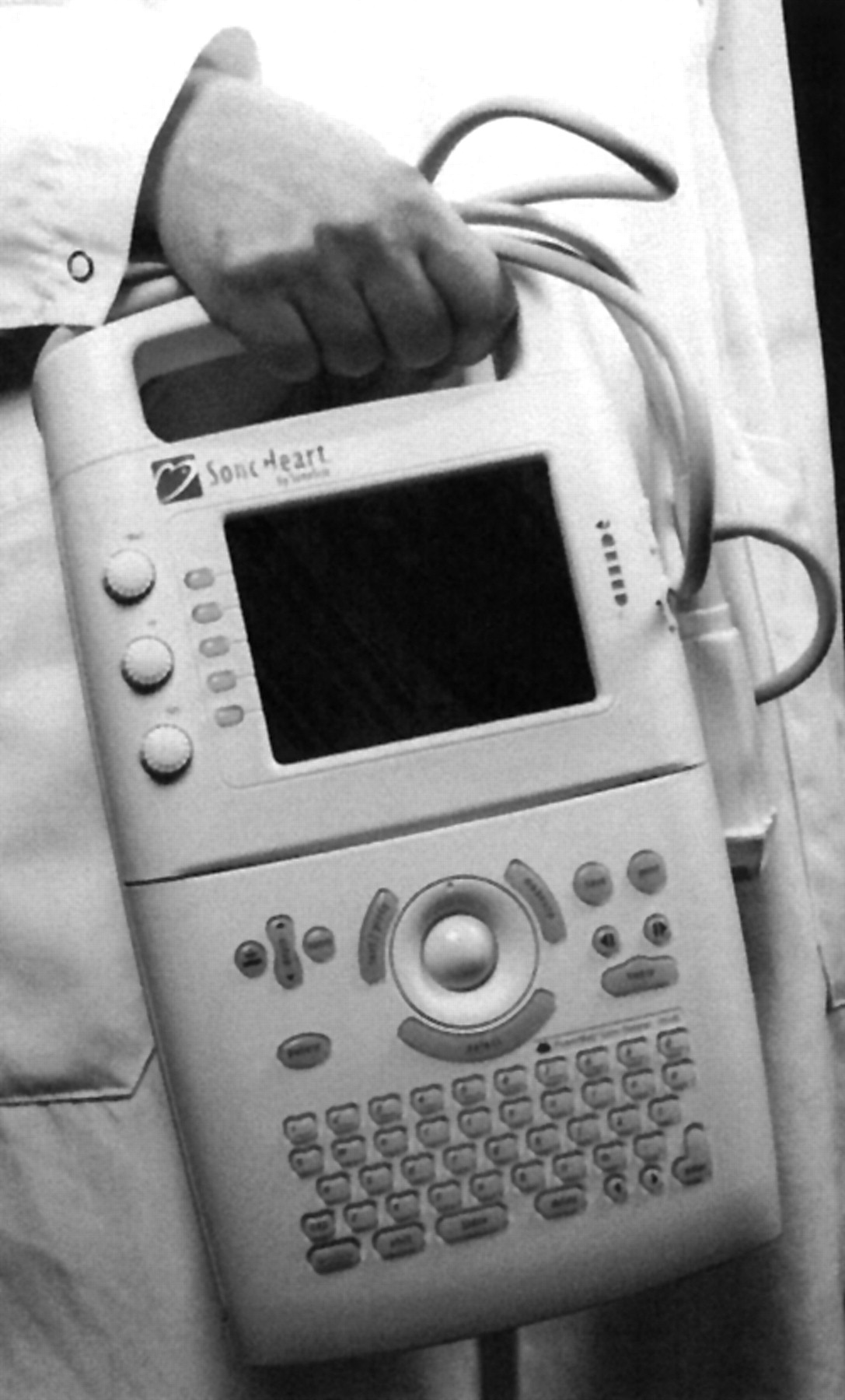
The SonoHeart system (fig 1) is a small hand held ultrasound device equipped with a 2–4 MHz phased array broadband transducer and operating on a rechargeable lithium ion battery or alternating current power. Two dimensional control settings similar to those on an SED and colour power Doppler flow mapping are integrated in the unit. Distance measurements are possible with inclusive callipers. SonoHeart has a storage memory of 50 images and can be connected to a videorecorder, a printer, or an external monitor.

The SonoHeart device, a hand held ultrasound imager (weight 2.4 kg) which was used in this study.
Statistical analysis
Descriptive statistics are reported as mean (SD) or frequency percentages. The agreement for detection of abnormalities was assessed from 2 × 2 tables using weighted κ statistics. Values of κ of 0.4, between 0.4–0.75, and > 0.75 were considered to indicate poor, fair to good, and excellent agreement, respectively, based on Fleiss’s classification.6
In addition, specificity, sensitivity, and positive and negative predictive values of the PUI in detecting abnormalities were calculated.
RESULTS
Cardiac visualisation by PUI
In all of the patients, visualisation adequate to answer the request was achieved.
Agreement
Table 1 lists the most common referral questions for which a cardiac evaluation was requested. The yardstick SED examination detected 71 clinically significant findings (table 2). The agreement in identifying abnormalities between the PUI and the SED was 96% (κ = 0.93; table 3).
Reasons for a cardiac consultation request for the 107 patients from non-cardiac departments
Abnormal findings detected with the standard echocardiographic device in 107 patients referred for cardiac evaluation during consultation rounds
Agreement in detection of patients with abnormalities between the SonoHeart and a standard echocardiographic device, showing the number of patients
The PUI provided to the cardiologist sufficient information on 84 (78.5%) patients seen during consultation rounds. In 23 or 107 patients (21.5%) a further detailed examination with the SED was considered necessary despite the echocardiographic examination with the PUI. In 18 of these 23 patients the Doppler study was required to evaluate the severity of regurgitant or stenotic lesions (16 patients) and for diagnosis of pulmonary hypertension (two patients). In two of the 23 patients an SED examination was requested to verify severe wall motion abnormalities, and in one patient for further investigation of a dilated ascending aorta. In two of the 23 patients there was a false positive diagnosis of endocarditis. In both cases the referral physician had a clinical suspicion of endocarditis. Because with the PUI the aortic valve had an echodense appearance, the clinical suspicion was enhanced and the cardiologist requested further analysis with an SED. The SED did not add any further information and the diagnosis of endocarditis was finally rejected on the basis of the transoesophageal echocardiographic study that followed.
Two major abnormalities were missed with the PUI: a moderate mitral regurgitation in a patient with a referral question of left ventricular function, and pulmonary hypertension in a patient with a referral question of cor pulmonale. However, the second patient was referred to SED examination by the cardiologist.
Calculation of cost effectiveness of the PUI
The average cost of an SED study was estimated by calculating the cardiologist’s consultation fee (€72) and the cost charged for a full echocardiographic study (€60). The final cost was €132 per patient.
Echocardiographic examination by the PUI is considered to be part of the physical examination and is therefore not charged. However, we incorporated in the final cost the capital investment of such a device, which is about €15 000. The five year equipment depreciation of this amount is €3 000 per year, which results in €3 per patient on a basis of 1000 patients seen during consultation rounds per year. Thus, the cost of a consultation visit with the use of the PUI was calculated to be €75 per patient.
Applying these data to our study results, the total cost for the 107 echocardiographic examinations performed was €14 124 for the standard procedure using the SED. However, with the PUI the total cost was €9 405, since only 23 patients were considered to need further investigation with an SED. Thus, with the use of PUI a cost reduction of 33.4% was achieved.
In addition, the mean time interval between a request for an echocardiogram by the consultant cardiologist and the final echocardiography report was reduced substantially. At our institution the average time was four days when standard echocardiography was requested, whereas the PUI provided results instantly. Figure 2 shows the logistical flowcharts of an echocardiographic study request for inpatients after a consultation visit by the cardiologist.

Logistical flow charts used at the Erasmus Medical Centre Rotterdam, the Netherlands, for a routine echocardiographic study requested for patients from non-cardiac departments after consultation with the cardiologist.
DISCUSSION
This study describes the potential utility of a small hand held ultrasound device during consultation rounds in the evaluation of patients in non-cardiac departments with suspected cardiac disease. In 84 patients (78.5%) the PUI provided the physician with efficient instant information indicating that further examination with SED could have been avoided. In patients in whom a complete echocardiographic study was considered necessary, it was due mostly to the need for haemodynamic assessment by Doppler. With the addition of this feature in the next generation PUIs the need for SED for patients seen during consultation rounds may be reduced even further.
Prior studies using limited imaging protocols have provided evidence that a limited imaging study is feasible for both the diagnosis and evaluation of most of the important cardiac pathologies (hypertrophic cardiomyopathy,7 left ventricular hypertrophy,8–11 mitral valve prolapse,12 and abdominal aortic aneurysm).13,14 Such a limited echocardiographic strategy can be effectively implemented with a small hand held ultrasound device.
Today, small hand held ultrasound devices aim to couple the physical examination and echocardiography at the point of care. By being ultraportable and easy to use they are practical for carrying while undertaking consultation rounds. Our group showed in a recent previous study the efficacy and high accuracy of this small imaging device in assessing pathomorphology and the function of the heart, thus enhancing and extending the physical examination and allowing goal oriented examination.4,5 The current study further confirmed these results.
Cost effectiveness of PUI during consultation rounds
Like all technological breakthroughs, the PUI has to be evaluated in financial terms, as well as for its clinical effectiveness, to gain wide acceptance. The capital investment in such a device is economic (about one 12th of the cost of an SED) and the maintenance costs are low.
In our hospital 1125 inpatients from non-cardiac departments were referred for an echocardiographic examination during 2001. Thus, for the year 2001, the total cost for the 1125 consultation visits that required an echocardiographic examination with the SED was €148 500. According to our study the cost can be reduced to €98 901 (66.6% of the initial amount) with the use of the PUI.
Eighty five per cent of the patients in our study were preoperative patients. In fact, in our hospital the majority of inpatients referred for cardiac consultation are preoperative patients. The usual request from anaesthesiologists and surgeons is to evaluate the systolic left ventricular function or a murmur, which can reliably be answered by an echocardiographic examination. Thus, the standard approach for these patients is to request an echocardiographic study further to the physical examination. The instant answer to a request can prevent potential delay in a patient who is planned for surgery and can therefore lead to cost savings. But this is only a hypothesis that has to be investigated.
Recently, Kimura and colleagues15 reported that the consequence of the presence of an abnormal initial limited echocardiographic examination in the emergency department was that patients had a significant length of hospital stay (that is more than two days). Furthermore, their study showed that, in the setting of the emergency department, a limited echocardiographic examination has better diagnostic accuracy than a physical examination in identifying cardiac abnormalities.
The present study was performed by cardiologists with experience in echocardiography. Immediate decision making diagnosis based on the echocardiographic examination with a PUI during consultation rounds requires level II or III training in echocardiography.16 Kimura and colleagues15 proved that it is feasible to train health care providers to obtain a parasternal long axis view and to interpret significant abnormalities. However, training and licensing non-cardiologists to use these devices will become an important issue in the future.
Study limitations
In the present study we did not specifically address the impact of the use of the PUI on hospitalisation stay. This may form the basis for future studies.
The PUI that was used for this study had no Doppler modalities to obtain haemodynamic data. Spectral Doppler and colour Doppler are now integrated in the new generation of PUIs.
Conclusion
During consultation rounds, a PUI can help to make an instant diagnosis at the bedside, leading to a shortened time to diagnosis with efficacy equal to that of an SED and with lower cost.