Article Text

Abstract
Background: Anderson–Fabry disease is an X-linked glycosphingolipid storage disorder caused by deficient activity of the lysosomal enzyme α-galactosidase A. This leads to a progressive accumulation of globotriaosylceramide (Gb3) in the lysosomes of cells throughout the body that ultimately results in premature death from renal, cardiac or cerebrovascular complications. Until recently, there was no effective therapy available for this disease. The present study was designed to assess the safety and efficacy of enzyme replacement therapy with agalsidase alfa on the cardiac manifestations of Anderson–Fabry disease.
Method: The effects of therapy with agalsidase alfa on cardiac structure and function were assessed in a randomised, double-blind, placebo-controlled study of 15 adult male patients with Anderson–Fabry disease. The following parameters were measured at baseline and 6 months: left ventricular mass, QRS duration and levels of Gb3 in cardiac tissue, urine sediment and plasma. After 6 months of the randomised trial patients were enrolled in a 2-year open-label extension study.
Results: Left ventricular mass, as measured by MRI, was significantly reduced following 6 months of treatment with agalsidase alfa compared with placebo (p = 0.041). A mean 20% reduction in myocardial Gb3 content as assessed by serial transvenous endomyocardial biopsies was demonstrated over the 6 months of enzyme replacement compared to a mean 10% increase in patients receiving placebo (p = 0.42)
Conclusion: Enzyme replacement therapy with agalsidase alfa resulted in regression of the hypertrophic cardiomyopathy associated with Anderson–Fabry disease.
Statistics from Altmetric.com
Anderson–Fabry disease is an X-linked glycosphingolipid storage disorder caused by deficient activity of the lysosomal enzyme α-galactosidase A.1 This leads to the progressive accumulation of globotriaosylceramide (Gb3) in the lysosomes of cells in most body tissues. Particularly affected are cells of the cardiovascular system, nervous system and kidneys. Quality of life is severely compromised, and premature death usually occurs in the fourth or fifth decade from renal, cardiovascular or cerebral complications. Both males and females are affected, although the clinical signs and symptoms of the disease are generally more severe in males. The true incidence of Anderson–Fabry disease is unknown with estimates in males ranging from 1:3100 in Italy,2 1:117 000 in Australia,3 and 1:476 000 in The Netherlands.4 Previous studies have shown that between 3% and 9% of male patients with otherwise unexplained hypertrophic cardiomyopathy have low serum levels of α-galactosidase A, indicating that they may have undiagnosed Anderson–Fabry disease.5–7
In addition to left ventricular hypertrophy, the cardiovascular manifestations of Fabry disease include symptomatic valvular abnormalities, arrhythmias and conduction disturbances.8 In some patients cardiac abnormalities may be the predominant manifestations of Anderson–Fabry disease.9 10
Previously, there has been no effective treatment for Anderson–Fabry disease, other than palliative care. Recently, replacement therapy with α-galactosidase A has been shown to reduce plasma, urinary sediment and tissue levels of Gb3.11–13 Six months of treatment with agalsidase alfa has been shown to reduce neuropathic pain, stabilise renal function and shorten QRS duration compared with patients given placebo; and an observational study of the effects of agalsidase beta on cardiac structure has shown a progressive decrease in the interventricular septum thickening and in left ventricular (LV) mass.11–14
The present study set out to assess the efficacy of agalsidase alfa on left ventricular mass and myocardial Gb3 content in Anderson–Fabry disease.
METHODS
Patients
Fifteen hemizygous male patients, over 18 years of age, with Anderson–Fabry disease were included in the study. Entry criteria included a confirmed diagnosis of Anderson–Fabry disease by leucocyte α-galactosidase A activity and genotyping, evidence of increased left ventricular mass by M-mode and two-dimensional echocardiography, and absence of permanent cardiac pacemaker or contraindication for cardiac biopsy. The study protocol received local research ethics committee approval. All patients gave their written informed consent before participating.
Study design and treatment regimen
The study was randomised and double blind for 6 months followed by an open-label extension for 2 years. Randomisation to the active treatment group (n = 7) or the placebo group (n = 8) occurred after completion of baseline evaluations and confirmation of eligibility criteria.
Agalsidase alfa (Replagal, Shire Human Genetics, UK) was administered at a dose of 0.2 mg/kg every 2 weeks for 6 months by intravenous infusion over 40 min. Agalsidase alfa is produced in a genetically engineered continuous human cell line (Shire HGT, Cambridge, MA, USA). Agalsidase alfa in the cell culture supernatant is harvested and the enzyme purified by a series of conventional chromatographic steps. The purified agalsidase alfa is formulated and placed in vials containing sodium phosphate as a buffering agent, polysorbate 20 as a stabilising agent, and sodium chloride as an isotonic agent. The drug is diluted in 100 ml physiological saline for administration. The enzyme is 99.5% pure, with a specific activity of 3.4×10⁶ nmol/hour/mg protein. Placebo infusions (excluding the α-galactosidase A) were identical to the enzyme infusions in composition, appearance and method of administration.
Following the randomised study, patients were enrolled in an open-label follow-up study. Ten patients completing the open label study each received 0.2 mg/kg agalsidase alfa every 2 weeks for an additional period of 2 years. Subsequent to the trial, 11 of the original 15 patients continued to receive uninterrupted infusions of 0.2 mg/kg agalsidase alfa every 2 weeks at home.
Efficacy measures
The primary efficacy endpoint was myocardial Gb3 content; reduction of the left ventricular mass by MRI assessment was a secondary efficacy endpoint.
Cardiac Gb3 content
Endomyocardial biopsies were taken from the right ventricular side of the interventricular septum via femoral vein catheterisation at baseline and at weeks 13 and 24 of the study. Four biopsy samples were taken on each occasion. One patient was excluded from follow-up biopsies after developing an arrhythmia during the baseline biopsy procedure. Myocardial samples were analysed for Gb3 content using a validated high performance liquid chromatography (HPLC) assay (for further details on methods, see supplementary online-only files).
Assessment of left ventricular mass by MRI
Magnetic resonance imaging of the heart was performed at baseline and after 13 and 24 weeks using a Siemens Magnetom 63SP (1.5 Tesla) scanner with standard phased-array body coil. All sequences were ECG gated. Three plane LV scouts, 6–8 multislice short axis LV cines and axial ascending (and descending) aortic phase contrast flow quantification were performed and the following parameters derived: PLVW (mm). septum (mm), LVSV (ml), LVEDV (ml), LVESV (ml), LVEF (%), LVED mass (g), LV mass index (g/m2).
One patient suffered from claustrophobia and was excused from his week 24 MRI. During the open-label phase MRI was performed every 6 months.
Echocardiography
Two-dimensional and M-mode echocardiograms were performed at baseline and at 24 weeks, using a Sonatron CFM800C coupled with a 2.35 or 3.25 MHz transducer.
During the open-label phase of the trial patients underwent annual echocardiographic monitoring. Left ventricular (LV) mass was calculated using M-mode recordings using Devereux formula.15
Electrocardiography
Twelve-lead electrocardiograms were performed every 2 weeks for the first 6 months for safety monitoring. QRS duration was compared at baseline and 6 months. Electrocardiograms were recorded as 50 mm/s paper tracings.
Gb3 in urinary sediment and plasma
Urine and blood samples were collected at baseline and during weeks 9, 17, 23 and 24 (randomised) and 37, 51, 65, 79, 105 and 131 (open label) for measurement of Gb3 in urinary sediment and plasma, respectively (see supplementary online-only files).
Safety
All adverse events were recorded, irrespective of whether they were assessed by the investigator to be related to the study medication. Events were treated appropriately and patients were monitored at suitable intervals until the events resolved.
Statistical methods
A sample size of 12 patients per group was calculated to have 90% power in detecting a difference in the mean change from baseline cardiac Gb3 content of 3.3 nmol/mg cardiac tissue. This assumed a mean change from baseline of 0 nmol/mg for the placebo group and 3.3 nmol/mg for the active treatment group, and a common SD of 2.2 nmol/mg using a two-sample t test with a 0.05 two-sided significance level. However, the rarity of Anderson–Fabry disease and the inclusion criteria, requiring that left ventricular hypertrophy be present at baseline, resulted in enrolment being terminated after 15 patients had entered the study.
For the randomised phase, data were analysed by treatment group with respect to demographics, baseline characteristics, and safety and efficacy variables. Statistical analysis was carried out using SAS System, version 6.12 and 8.0. Analysis of covariance (ANCOVA) was used to compare baseline with week 24 data for both the intention-to-treat and per-protocol populations. Statistical tests were two-sided, and a value of p⩽0.05 was considered significant. The Shapiro–Wilks test for normality (p = 0.15) indicated that the normality assumption for the ANCOVA was not violated. Heteroscedasticity of the response variable was evaluated informally using graphical methods, such as box plots and scatter plots, which indicated no major assumption violations. The assumption of parallel slopes for the treatment regression lines was tested using the F test of equality of slopes (p = 0.46).
The per protocol set excluded one patient from the myocardial Gb3 analysis as a result of the development of an arrhythmia during his baseline cardiac biopsy procedure and one placebo from MRI LV mass analysis because of development of claustrophobia.
RESULTS
All 15 patients completed the initial 6-month randomised study period. Age distribution, ethnic origin, weight, blood pressure, and left ventricular mass at baseline were comparable in the two study groups as were left ventricular ejection fraction and glomerular filtration rate (supplementary table 1; see supplementary online-only table). Mean (SE) left ventricular mass index at baseline for the treatment and placebo groups were 176 (10) g/m2 and 186 (18) g/m2, respectively (p = 0.7), compared with a normal left ventricular mass, as assessed by echocardiography, of 134 g/m2. All patients fulfilled the inclusion criteria of increased LV mass by M-mode and 2D echocardiography. However, using MRI only 10 of the 15 patients enrolled in the study had a left ventricular mass above 225 g, which is the upper 95% confidence limit for left ventricular mass by MRI in normal.
There was no change in blood pressure during the randomised trial. Mean baseline GFR showed a statistically significant increase within the normal range (106–131 ml/min p = 0.007) in the drug group whereas the placebo groups did not show any statistically significant change throughout the 6-month study. During the study there was no significant change in body weight of the drug group (0.7 (0.7) kg, p = 0.345) whereas the placebo group gained 1.3 (0.5) kg (p = 0.047). Supplementary table 2 (see supplementary online-only table) lists the clinical characteristics of the individual patients, together with their mutations/genotypes and the leucocyte α-galactosidase A measurements.
Myocardial Gb3 content
At baseline, the myocardial Gb3 levels in the groups receiving agalsidase alfa and placebo were 0.71 (0.18) nmol/μg protein and 0.58 (0.08) nmol/μg respectively (p = 0.47). There was no correlation between LV mass and myocardial GB3 content (R2<0.1). During the 6 months of the study, there was a mean increase in myocardial Gb3 content of approximately 10% in the placebo group (by 0.05 (0.08) nmol/μg protein; n = 8) in contrast to mean decrease of approximately 20% (–0.13 (0.16) nmol/μg protein; n = 6) in the group receiving agalsidase alfa (fig 1). Four of the six agalsidase alfa treated patients (66.7%) compared to only one of eight placebo patients (12.5%) showed a decrease in myocardial Gb3 content over the relatively short 6-month study period (p = NS). Myocardial Gb3 contents for individual patients at baseline, 13 weeks and 24 weeks are given in supplementary table 3 (see supplementary online-only table).

Left ventricular mass
A mean decrease in LV mass of 11.5 g was measured by MRI in the group receiving agalsidase alfa, in contrast to a mean increase of 21.8 g in the placebo group (p = 0.041; per-protocol ANCOVA, n = 14) (fig 2; supplementary table 4, available online-only). This equated to mean increase in LV mass index for the placebo group of 12 g/m2 compared to a decrease in the treated group of 6.4 g/m2 (p = 0.02 vs placebo).

Echocardiographic measurements of the effect of agalsidase alfa on LVH produced qualitatively similar results. Patients receiving placebo showed a 6.3% mean increase in LV mass (21.5 (20.4) g) whereas there was a mean 6.2% decrease (–20.4 (27.2) g) in patients given agalsidase alfa (p>0.05; intention-to-treat ANCOVA).
MRI measurements in the 10 patients, who continued to receive agalsidase alfa in the open-label extension phase of the trial demonstrated individual heterogeneity in response (fig 3). After 2 years of active treatment mean LV mass was not significantly changed (p = 0.18). There were statistically significant reductions in LV posterior wall and septal thickness and at 1.5 and 2.0 years compared to baseline (mean change LV posterior wall thickness at 2 years −1.9 (0.73) mm p = 0.01, and mean change septal thickness at 2 years −3 (0.9) mm, p = 0.01) indicating cardiac remodelling while on agalsidase alfa. At 2 years 7/10 patients demonstrated maximal LV wall thickness above the normal range compared to 9/10 at baseline.
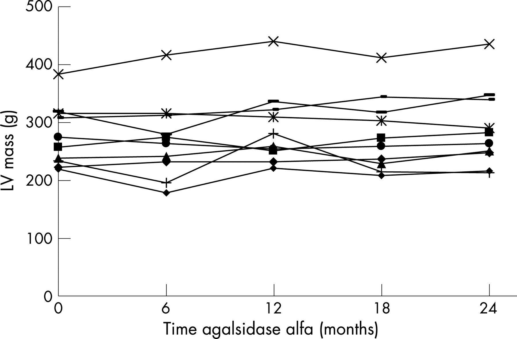
Subsequent to the trial 11 patients have continued to receive uninterrupted therapy with agalsidase alfa and have been monitored by annual echocardiography. After 4 years seven patients demonstrated a reduction in LV mass while four show increases compared to their pre-treatment baseline.
Left ventricular function
Echocardiographic measurements of LV function at baseline and after 6 months of treatment with agalsidase alfa are given in supplementary table 5 (see supplementary online-only table). Ejection fraction was normal or hypernormal (>65%) at baseline and remained normal or hypernormal after 6 months in all patients. There was no statistically significant change in fractional shortening after 6 months in either group.
QRS duration
In the enzyme treated group there was a mean reduction in QRS duration of 12.9 (11.8) ms over the 6 months of the study, compared with an increase (+4 ms) in the placebo group (p = 0.8; intention-to-treat ANCOVA). However, this large mean reduction in QRS duration in the treated group was driven largely by one patient who showed resolution of complete right bundle branch block.
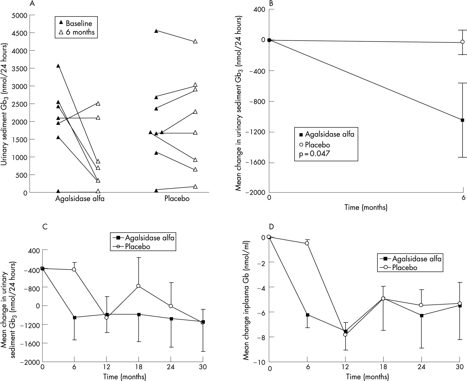
Plasma and urinary sediment Gb3
There was with a mean decrease of 45% (–6.22 (1.05) nmol/ml) in the group given agalsidase alfa compared with no change (–0.55 (0.35) nmol/ml) in plasma Gb3 in the placebo group between baseline and 6 months (p<0.001 vs placebo). Following initiation of active treatment in the open-label phase, the original placebo group demonstrated a similar statistically significant decrease in plasma Gb3 (−7.3 (1.1) nmol/ml; p<0.001 vs baseline). Reductions in plasma Gb3 were maintained throughout the rest of the open-label extension study.
Similarly, the reduction in mean urinary sediment Gb3 from baseline was significantly greater in patients receiving agalsidase alfa treatment (–52%; 1052 (457) nmol/24 h) compared to those given placebo (−5.9%; −25 nmol/24 h) (p = 0.047 vs placebo) (fig 4). After crossover to active therapy the original placebo group demonstrated a 52% mean decrease in urine Gb3 content at week 51 (p = 0.081 vs placebo). At 18 months an increase in the urinary Gb3 was observed in two members of the original placebo group. This coincided with detection of neutralising IgG antibodies and resolved over the following 12 months.
Safety
Infusions were in general well tolerated. Only one patient experienced an infusion-related reaction during the 131 weeks of the study and was pre-treated with intravenous hydrocortisone and oral antihistamines. IgG antibodies to agalsidase alfa were detected in three patients at one or more time points using an enzyme-linked immunoabsorbent, assay. No patients developed IgE, IgA or IgM antibodies. There were no serious adverse events related to the agalsidase alfa infusions. Ten patients received their final 12–18 study infusions in their homes with no patient experiencing a serious adverse event or terminated an infusion due to a serious adverse event.
DISCUSSION
Cardiac involvement is responsible for a significant proportion of the morbidity and mortality associated with Anderson–Fabry disease.16 The pathological changes that occur in the heart of patients with Anderson–Fabry disease include accumulation of Gb3 within myocytes, coronary endothelium, intimal and medial cells and valve tissue. This glycosphingolipid storage results in LV hypertrophy, conduction abnormalities, valve thickening, heart failure, anginal chest pain and less frequently to acute myocardial infarctions.
The majority of the patients (10/15) enrolled in the present study had LV hypertrophy at baseline, as assessed by MRI. In other cardiac conditions, LV hypertrophy has been shown to be an independent risk factor for cardiovascular events and premature mortality.17 Therefore, the significant reduction in LV mass observed after 6 months of treatment with agalsidase alfa is likely to be clinically relevant. Indeed, in this progressive disease, agalsidase alfa had a positive therapeutic effect (regression or stabilisation of LV hypertrophy) in the majority of patients. Lack of statistical significance of the change in LV mass measured by echocardiography may be due to the higher variability of measurements by this technique.
The observed reduction in myocardial Gb3 content, while not reaching statistical significance, is consistent with the observed mean decrease in LV mass in agalsidase alfa treated patients. This failure to reach statistical significance for the primary efficacy endpoint of cardiac Gb3 may be the result of the study being underpowered, (only 15 of the planned 24 patients were enrolled), or to its relatively short duration. Although the degree and mechanism of enzyme penetration into organs is unclear, the observed reduction in myocardial Gb3 content suggests effective exposure of cardiac tissues to agalsidase alfa.
The observed decrease in plasma and urine sediment Gb3 levels after 6 months of agalsidase alfa treatment reflects increased metabolism of accumulated Gb3 and likely a reduction in its pathogenic tissue storage. Significant reductions in plasma Gb3 were maintained throughout the 2-year open-label extension study. In two patients who developed neutralising IgG antibodies, urinary but not plasma GB3 levels were transiently elevated at the time of antibody detection but fell in the following 12 months, suggesting natural tolerance. LV mass was also noted to increase in these two patients during the time of antibody detection.
In summary, we have described a short-term placebo-controlled trial of enzyme replacement therapy with agalsidase alfa in patients with Anderson–Fabry disease. We have shown regression of the cardiac hypertrophy in association with a reduction in intramyocardial Gb3 content. The subsequent open-label extension trial and two observation periods demonstrated ongoing response or stablisation in the majority of patients and that long-term enzyme replacement in patients with Anderson–Fabry disease is safe and well tolerated. Inter-individual variation in response warrants further investigation in larger studies.
Acknowledgments
The authors acknowledge that Transkaryotic Therapies (TKT) Inc, Cambridge, MA, USA, which funded the study, was involved in the study design and analysis, including statistical analysis. However, the study was double-blind and placebo-controlled for submission to regulatory agencies and all data were independently analysed by Royal Free investigators. The data remained blinded until the database was locked and the statistical analyses performed. The data are stored with the Royal Free investigators and will eventually be made available for public scrutiny. We also thank Oxford Pharmagenesis for assistance with early drafts of the manuscript; however we confirm that only the authors named are responsible for the manuscript.
REFERENCES
Supplementary materials
web only appendix 94/2/153
Files in this Data Supplement:
Footnotes
Competing interests: A Mehta, D Hughes and P Elliott have received travel grants, educational grants, and/or honoraria for speaking engagements from Shire Human Genetics.
Linked Articles
- Editorial