Article Text

Abstract
Objective In patients with bicuspid aortic valve (BAV), coronary anatomy is variable. High take-off coronary arteries have been described, but data are scarce, especially when associated with complex congenital heart disease (CHD). The purpose of this study was to describe coronary patterns in these patients.
Methods In 84 postmortem heart specimens with BAV and associated CHD, position and height of the coronary ostia were studied and related to BAV morphology.
Results High take-off right (RCA) and left coronary arteries (LCA) were observed in 23% and 37% of hearts, respectively, most frequently in hearts with hypoplastic left ventricle (HLV) and outflow tract anomalies. In HLV, high take-off was observed in 18/40 (45%) more frequently of LCA (n=14) than RCA (n=6). In hearts with aortic hypoplasia, 8/13 (62%) had high take-off LCA and 6/13 (46%) high take-off RCA. High take-off was seen 19 times in 22 specimens with perimembranous ventricular septal defect (RCA 8, LCA 11). High take-off was associated with type 1A BAV (raphe between right and left coronary leaflets), more outspoken for the RCA. Separate ostia of left anterior descending coronary artery and left circumflex coronary artery were seen in four hearts (5%), not related to specific BAV morphology.
Conclusion High take-off coronary arteries, especially the LCA, occur more frequently in BAV with associated CHD than reported in normal hearts and isolated BAV. Outflow tract defects and HLV are associated with type 1A BAV and high take-off coronary arteries. Although it is unclear whether these findings in infants with detrimental outcome can be related to surviving adults, clinical awareness of variations in coronary anatomy is warranted.
- Word count: 3239
Statistics from Altmetric.com
Introduction
Bicuspid aortic valve (BAV) is the most common congenital heart defect (prevalence 0.5%–2%). BAVs with different morphology subtypes exist and BAV morphology correlates to outcome with respect to valve function and aortic dilation.1 2 In the majority of BAVs the right and left coronary leaflets are conjoined, in a minority the right and non-coronary leaflets and in only a small group the left and non-coronary leaflets are conjoined.1 3
Variations in coronary anatomy have been described in patients with BAV, including increased incidence of separate ostia of the left anterior descending coronary artery (LAD) and left circumflex coronary artery (LCX), and of a left dominant coronary system.4 5 Also differences in coronary ostial positions as related to the aortic sinus have been noted.6 7 In normal hearts, the coronary arteries arise mostly from the aortic sinuses below the sinotubular (ST) junction. Coronary ostia can be located a few millimetres above the ST junction in 5%–22% of normal hearts, although literature is scarce.8 9 Coronary ostial take-off well above the ST junction, referred to as high coronary take-off, is rare, occurring in <1% of the normal population.10 11 Data concerning coronary height in patients with BAV are scarce and not consistent, mostly confined to case reports.6 7 Lerer et al reported the left coronary artery (LCA) in patients with isolated BAV above the aortic ST junction in 29% of cases, whereas the right coronary artery (RCA) is located too high in 11%.12 This study did not include hearts with additional congenital anomalies. In patients with Turner syndrome, only 1 out of 50 (2%) patients had a high take-off RCA.13 This patient also had a BAV, whereas all other patients had a tricuspid aortic valve.
The significance of recognising high take-off coronary arteries lies in potential challenges encountered during catheter cannulation or surgery. When unrecognised, there have been cases of inadvertent transection and coronary cross-clamping during open-heart procedures.14 15 High take-off coronaries have also been associated with intramural coronary course11 and may impair coronary blood flow due to formation of an acute angle with the ostial ridge and angulation may increase with increasing aortic dimensions.16
Currently available studies have largely been performed in patients with BAV as isolated anomaly. The influence of associated complex congenital heart disease (CHD) on coronary anatomy is unknown. Developmental issues may be of relevance, as many processes during heart development are influenced by signalling events from cell types that also influence coronary development.17 Coronary arteries derive from angiogenic sprouts in the sinus venosus region, initially not connected to the aorta.18 19 Ingrowth in the aorta will occur during later development, orchestrated by epicardial-derived cells, whereas endothelial cells will contribute to the proximal coronary orifices.19–21 Disturbance in this process may lead to a too high position of the coronary arteries as related to the aortic sinus (ie, high take-off) and perhaps also to erroneous ingrowth of a coronary artery, usually the left, in the pulmonary artery,22 although the developmental mechanism of the latter is still enigmatic.
The aim of the current study is to describe coronary patterns in postmortem hearts with BAV associated with complex CHD, as related to BAV morphology.
Methods
Study population
This study was undertaken in accordance with the local ethics committee and Dutch regulations for proper use of human tissue for medical research purposes.
Eighty-four postmortem heart-lung specimens with BAV associated with other CHD from the Leiden collection of malformed hearts (Department of Anatomy and Embryology, Leiden University Medical Center, Leiden, The Netherlands) were studied macroscopically. This collection includes hearts preserved in ethanol and glycerin, dating from the 1950s to the current era.
Anatomical studies
Two experienced observers investigated the hearts. Cardiac morphology was assessed using sequential segmental analysis,23 performed as completely as possible. All cardiovascular anomalies as well as detailed morphology of the aortic valve and position and height of coronary ostia were noted. In some specimens, cardiovascular anomalies could not be assessed due to incompleteness of the specimens.
Hypoplastic left ventricle (HLV) was considered present when the lumen of the LV was smaller than the right ventricle (RV), but the apex was formed by the LV, or when the apex was formed by the RV, including cases with hypoplastic left heart syndrome (HLHS). HLHS was considered present when there was severe underdevelopment of the left heart with significant hypoplasia of the LV including atresia, stenosis or hypoplasia of the aortic or mitral valve, or both valves, and hypoplasia of the ascending aorta and aortic arch.24 The ascending aorta was considered hypoplastic if the aortic orifice diameter was smaller than expected as compared with the pulmonary orifice diameter. Tubular hypoplasia of the aortic arch was defined as a narrowed (often elongated) aortic arch segment. Aortic coarctation was defined as a localised obstructive ridge within the arch consisting of a constriction of the aortic wall and/or a shelf protruding into the aortic lumen.25
BAV classification and terminology
BAV morphology was defined by orientation of the leaflets with respect to each other, determined by which of the leaflets (right coronary, left coronary, non-coronary) were conjoined and presence and position of a raphe. BAV morphology was classified as type 1, if the right and left coronary leaflets were conjoined; type 2, if the right and non-coronary leaflets were conjoined and type 3, if the left and non-coronary leaflet were conjoined. ‘A’ was added for valves in which a raphe could be recognised or ‘B’ for strictly bicuspid valves (no raphe) (figure 1). A raphe was defined as a ridge located in the conjoined area of two leaflets, presumably representing a malformed commissure.

Schematic overview of different BAV morphologies. Drawings are oriented in anatomical view of the aortic valve, that is, as seen from above. Upper panel: BAVs with a raphe; type 1 (‘fusion’ right and left coronary leaflets), type 2 (‘fusion’ right and non-coronary leaflets) and type 3 (‘fusion’ left and non-coronary leaflets). Lower panel: strictly bicuspid valves (without a raphe), defined as a subgroup B. Leaflet size and symmetry as well as the position of the commissures may vary from the schematic overview depicted here. BAV, bicuspid aortic valve; LC, left coronary leaflet; LCA, left coronary artery; NC, non-coronary leaflet; RC, right coronary leaflet; RCA, right coronary artery. Modified after Schaefer et al.1
Coronary ostia
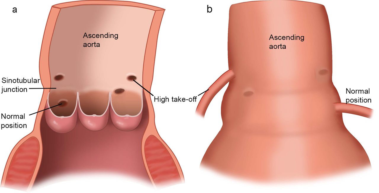
In each specimen, the number and position of the coronary arterial orifices were recorded. The position of each orifice was determined in relation to the ST junction and described as above, below or at the junctional level (figure 2). A coronary orifice was classified as being located above the ST junction when there was no communication between the coronary ostium and the ST junction.

(a) Schematic drawing of the aortic root with the aortic sinuses. The coronary ostia can be below, on or above the sinotubular (ST) junction. Drawing shows one normal coronary ostial position, one coronary ostium on the ST junction and two coronary ostia above the ST junction. Modified after Loukas et al.11 (b) Aortic root from another perspective showing one high take-off coronary artery with sharper angulation and one normal position of the coronary artery.
Statistical analysis
Continuous variables are presented as mean±SD or as median and IQR. Categorical variables are presented as numbers and percentages. Cross-tabulations were made for binary categorical data, on which χ2-square goodness-of-fit-tests were performed to test for independence. For comparing numerical data in more than two categories, logistic regression was used. Statistical analysis was performed using SPSS software (V. 20.0, SPSS, Chicago, Illinois, USA). A p Value <0.05, by a two-sided test, was considered statistically significant.
Results
Specimen characteristics
A total of 84 hearts was studied: 48 male and 35 female. Age at demise ranged from prenatal to24 years. Table 1 provides an overview of patient characteristics including cardiovascular abnormalities.
Patient characteristics and associated cardiovascular abnormalities
The most frequent anomalies were HLV (49%) and aortic coarctation (43%). Detailed morphological information of each of the 84 cases examined is provided in online supplementary table S1.
Aortic valve morphology
In 45 out of 84 hearts (54%), the raphe in the aortic valve was located between the left and right coronary leaflets (type 1A), whereas 9/84 (11%) had a type 2A morphology. In 5/84 (6%) hearts, the raphe was located between the left and non-coronary leaflet (type 3A). In the BAVs without raphe, 16/84 (19%) had type 1B BAV, 5/84 (6%) type 2B and 4/84 (5%) type 3B, together comprising 25 cases (30%, figure 1).
Coronary anatomy
In 84 specimens, 54 (64%) coronary variations or anomalies were seen, of which 50 high take-off coronaries and separate ostia of the LAD and LCX in 4/84 (5%) specimens (figure 3). The latter was not restricted to a specific valve morphology (two had type 1A BAV, one had type 1B and one had type 3A BAV) and not associated with high take-off coronaries (high take-off LCA in 2/4 cases, no high take-off RCA).

Representative examples of postmortem heart specimens with variable valvular and coronary anatomy. (a, b) Type 3A BAV with separate ostia of the LAD and LCX (case 5577). The box (a) points out the origin of the LCA with a zoom in of the separate ostia (b). (c) Type 1B BAV (case 5932) with both coronary ostia located above the ST junction. (d) Type 2A BAV (case 3642) with the LCA located above the ST junction and the RCA on the ST junction (*). (e, f) Specimens with a hypoplastic LV (e) and type 1A BAV (f) (case 6012). The raphe is not visible in this picture. Ao, aorta; BAV, bicuspid aortic valve; LAA, left atrial appendage; LAD, left anterior descending coronary artery; LCA, left coronary artery; LCX, left circumflex artery; LV, left ventricle; Pu, pulmonary artery; RAA, right atrial appendage; RCA, right coronary artery; RV, right ventricle; ST, sinotubular; VS, ventricular septum.
Overall, high coronary take-off was observed more often in the LCA than RCA (table 2).
BAV morphology and height of coronaries in different anomalies
The RCA arose from above the ST junction in 19/83 (23%) hearts (in one heart the RCA could not be evaluated). The LCA arose from above the ST junction in 31/84 (37%) hearts. Although the LCA more often showed high take-off than the RCA, this could not be correlated to a specific BAV morphology. Interestingly, in type A (ie, with raphe) BAVs ostial height of the RCA was significantly more often above the ST junction than in type B (no raphe) BAVs (29% vs 8%, p<0.05, table 3).
Coronary height in different BAV subtypes
This was not observed for the LCA (table 3). Examples of high take-off coronary arteries are shown in figure 3C,D.
No significant difference was found in height of the coronary ostia when comparing type 1 with type 2 or type 3 BAVs (table 3).
Subgroup analyses
As the included specimens comprise a heterogeneous group of associated congenital anomalies, subgroup analyses of valve morphology and coronary anatomy were performed, separating specimens with LV hypoplasia and outflow tract malformations from specimens with inflow tract anomalies and septal defects. Given their developmental origin, hearts with perimembranous ventricular septal defect (VSDs) were clustered among the outflow tract defects26 (table 2).
LV hypoplasia. In 40/82 hearts (49%, table 1), LV hypoplasia was present, in two hearts the LV could not be evaluated. BAV morphology in this group was predominantly type 1A (24/40, 60%). Figure 3E,F shows HLV and a type 1A BAV. In 19/40 (48%) hearts, HLV was associated with aortic coarctation and in 9/40 (23%) with aortic hypoplasia (table 4).
Arch anomalies and height of coronary arteries in LV hypoplasia
In 12/40 (30%) hearts, HLV was not associated with aortic arch anomalies. High coronary take-off was observed in 18/40 (45%) specimens with HLV, more frequently of the LCA (n=14) than the RCA (n=6, table 4). Almost all specimens with HLV and one or more high take-off coronaries(16/18, 89%) had an abnormal aortic arch. All specimens with high take-off RCA had type 1A BAV (tables 2 and 4). The majority of specimens with high take-off LCA also had type 1A BAV, but the percentage was lower (57%) (table 2).
Outflow tract defects
In a total of 77 specimens in which the aortic arch could be evaluated, 13 (17%) showed aortic arch hypoplasia (9 in association with HLV) (tables 1 and 2). BAV morphology in this group was also predominantly type 1A. Again, a high take-off LCA was observed most frequently: 6/13 (46%) had high take-off RCA and 8/13 (62%) high take-off LCA. Specimens with aortic coarctation (33/77 (43%), 19 of which associated with HLV) showed high take-off RCA in 4/33 (12%) and high take-off LCA in 8/33 (24%) cases (table 2). High take-off was observed in none of the two specimens with transposition of the great arteries and three times in six hearts with double outlet right ventricle. Most of the hearts with an outflow tract defect and high take-off coronary arteries also had type 1A BAV (table 2). Although a high take-off LCA was more frequent, especially high take-off RCA was correlated with type 1A BAV (table 2).
Also in the group with perimembranous VSD subtype 1A was dominant. In these specimens (n=22), 19 times a high take-off coronary artery was observed (8 RCA, 11 LCA). Most of the hearts with high take-off coronaries in this group had type 1A BAV (table 2). Again, there was a correlation of high take-off RCA with type 1A BAV: 6/8 (75%) of the RCA with high take-off had type 1A BAV (table 2).
Inflow and septal defects
In the group with muscular VSD (n=20), 11 high take-off coronaries were seen (3 RCA, 8 LCA). Twelve out of these 20 specimens had type 1A BAV, in these six high take-off coronary arteries were seen (table 2). In 11 hearts with ASD, seven high take-off coronaries were seen, 2/2 (100%) high take-off RCA had type 1A BAV and 2/5 (40%) high take-off LCA had type 1A BAV (table 2). In mitral hypoplasia and atresia (related to HLV), the majority showed type 1A BAVs (13/25 (52%)) (table 2).
In general, in both subgroups with inflow and outflow tract defects, high take-off RCA was strongly related to type 1A BAV, whereas a high take-off LCA was more divided over different BAV subtypes (table 2).
Discussion
To our knowledge, this is the first study that describes morphology of BAV and coronary anatomy in a large postmortem group of specimens of BAV associated with complex CHD.
Key findings are as follows: (1) high take-off coronary arteries occur frequently in BAV associated with complex CHD; (2) high take-off LCA occurs more frequently than high take-off RCA; this cannot be correlated to BAV morphology; (3) high take-off RCA is strongly related to type 1A BAV; (4) the subgroups of LV hypoplasia and outflow tract defects are associated with type 1A BAV and high take-off of both coronary arteries.
Coronary ostia and position in complex BAV
Separate ostia of the LAD and LCX were observed in 5% of specimens, considerably more than in the normal population but slightly less than our previous study in patients with isolated BAV (11%). In contrast to that study, where separate ostia were linked to type B BAVs,5 in the current study there was no clear correlation to BAV morphology.
In normal hearts, high take-off coronary arteries from well above the ST junction are rare (prevalence <1%10 11). In isolated BAV, this prevalence was described as high as 11% (RCA) to 29% (LCA).12 In the current study, we observed an even higher prevalence (37% for LCA and 23% for RCA), possibly due to a more complex study population with associated CHD. Similar to studies in isolated BAV, a predisposition for high take-off LCA was noted as compared with the RCA, possibly because left and right coronary ostia are patterned by different mechanisms. Animal studies indicate a left-right difference in coronary arterial development, as the transcription factor Tbx1, expressed in the outflow tract, promotes development of the left, but not right proximal coronary artery and ostium.21
Correct positioning of coronary artery stems depends on proper outflow tract morphogenesis.27 During development, the coronary arterial network develops from angiogenic sprouts from the sinus venosus18 19 and will during later development connect to the aorta, influenced by epicardial derived cells.28 Given the heterogenous composition of the aortic vascular wall,29 it is likely that signalling from other cell populations, including neural crest and second heart field, also play a role. Inhibition of cardiac neural crest and second heart field development results in outflow tract septation/rotation defects, associated with abnormally placed coronary stems on the aorta.27
Although a high take-off LCA was observed more frequently than a high take-off RCA, this was not correlated to BAV morphology. In contrast, RCA ostial height was significantly more often above the ST junction in type A (with raphe) than type B (no raphe) BAVs. A striking finding was that high take-off RCA was in almost all cases associated with type 1A BAV morphology, again indicating a potential right-left difference in coronary development. Whether or not this relates to a differential left-right contribution of epicardial (derived) cells,30 necessary for coronary ingrowth, is as yet undetermined.
Coronary anatomy in HLHS and outflow tract anomalies
HLHS is caused by left-sided obstruction and has also been linked to BAV,31 32 although no correlation has been made to BAV subtype. Aortic arch anomalies are associated with HLHS. In the current study, HLV was observed in 49% of hearts, although most specimens did not meet the criteria of HLHS. Aortic arch anomalies, including aortic arch hypoplasia and coarctation, were observed in most of these cases. In the current study, the majority of HLV hearts were associated with high take-off of both coronary arteries and had subtype 1A BAV morphology. Clinical consequence of this is unclear. In patients with HLHS, the arterial duct is the main source of systemic blood flow and provides retrograde filling to the coronary arteries.33 High take-off coronary arteries have been associated with acute angulation,16 potentially forming an impediment to this retrograde coronary flow. Whether this is a mechanism contributing to fetal demise in cases with HLV and the consequences after Norwood procedures is as yet unknown.
BAV morphology–developmental considerations
Different aortic valve morphologies are considered to have different developmental backgrounds. BAVs with left-right coronary leaflet ‘fusion’ patterns (type 1A BAV) most likely result from abnormal septation of the proximal outflow tract, due to distorted behaviour of neural crest cells,34 whereas the right non-coronary leaflet fusion pattern is considered an abnormality in epithelial-to-mesenchymal transformation.34 Indeed, in the current study, the majority of hearts with outflow defects showed type 1A BAV.
Study limitations
This is a descriptive study of a heterogeneous group of postmortem specimens, comprising different forms of CHD. Many variations of CHD are encountered in clinical practice. This interindividual variation makes a one-to-one correlation of current findings with clinical practice challenging. As this was a postmortem group with therefore per definition a detrimental outcome, prevalence rates of cardiovascular abnormalities cannot be applied to the general CHD or BAV population. On the other hand, this is the largest group of postmortem specimens with BAV associated with complex CHD described to date and different forms of CHD could roughly be clustered in order to compare groups based on a potential common developmental background.
Conclusion and clinical implications
In this study in postmortem specimens with BAV and associated complex CHD, prevalence of high take-off coronary arteries was high, especially in the group with outflow tract malformations. A predisposition to type 1A morphology was found, underlining a potential developmental background related to disturbed contribution of cardiac neural crest, as previously suggested based on animal studies.34 The clinical significance of high take-off coronary arteries is still debated. Potential harmful effects cannot be excluded as they have been linked to sudden cardiac death and ischaemia due to impaired coronary blood flow.14 15 Diagnosing these coronary anomalies is clinically relevant as a high take-off coronary artery can be injured during aortic clamping in cardiac bypass surgery. They can also cause technical difficulties during coronary angiography.11 Identifying separate ostia of LAD and LCX is of clinical importance in using coronary artery perfusion as a means of myocardial protection during surgical aortic valve replacement, and in performing coronary angiography.
In conclusion, in patients with BAV with associated complex CHD—especially patients with HLV and/or outflow tract defects and type 1A BAV—clinicians should be aware of variations in coronary anatomy like high take-off coronary arteries and separate ostia of LCX and LAD, potentially increasing risk of complications during procedures and affecting outcome. It is unclear whether the current findings in infants with detrimental outcome are representative of the population of surviving adults.
Key messages
What is already known on this subject?
Variations and anomalies in coronary anatomy have been described in patients with bicuspid aortic valve (BAV), including an increased incidence of separate ostia of the left anterior descending coronary artery and the left circumflex coronary artery, an increased incidence of a left dominant coronary artery system, but also differences in coronary ostial positions as related to the aortic sinus have been noted. High take-off coronary arteries (above sinotubular junction) have been described, but data are scarce, especially when associated with complex congenital heart disease (CHD).
What might this study add?
In the current study, we found high take-off coronary arteries, especially the left coronary artery, to occur more frequently in BAV associated with complex CHD than reported in structurally normal hearts and hearts with isolated BAV. We also found that outflow tract defects and hypoplastic left ventricle are associated with type 1A BAV and high take-off of both coronary arteries in the selected group of postmortem specimens.
How might this impact on clinical practice?
High take-off coronary arteries have been linked to sudden cardiac death and ischaemia due to impaired coronary blood flow. In addition, diagnosing these coronary anomalies is clinically relevant as a high take-off coronary artery can easily be injured during the clamping of the aorta in cardiac bypass surgery. They can also cause technical difficulties during coronary angiography. Whether the current findings in infants with detrimental outcome are representative of the population of surviving adults is unclear.
References
Footnotes
Contributors WMCK is the main author of this article and planned and executed most of the research, analysis and reporting. MMB has contributed to the study conception, data collection and interpretation and has critically reviewed the article with valuable suggestions. RB has contributed to the study conception and data collection and has thoroughly reviewed the article and made some helpful suggestions. ACGdG has contributed to the study conception and reviewed the article thoroughly with valuable suggestions. MCD has thoroughly reviewed the article and has made some valuable remarks. MJS has contributed to the study conception and reviewing it thoroughly with valuable remarks. MRMJ has contributed to conception of the article, data analysis and interpretation, drafting the article and critically reviewing it several times. Authors responsible for overall content: WMCK, MRMJ. All authors approved of the final version of the article to be published.
Funding The research of MCDR on bicuspid aortic valve disease is funded by a grant of the Dutch Heart Foundation (grant NHS2013T093).
Competing interests None declared.
Provenance and peer review Not commissioned; externally peer reviewed.