Article Text

Statistics from Altmetric.com
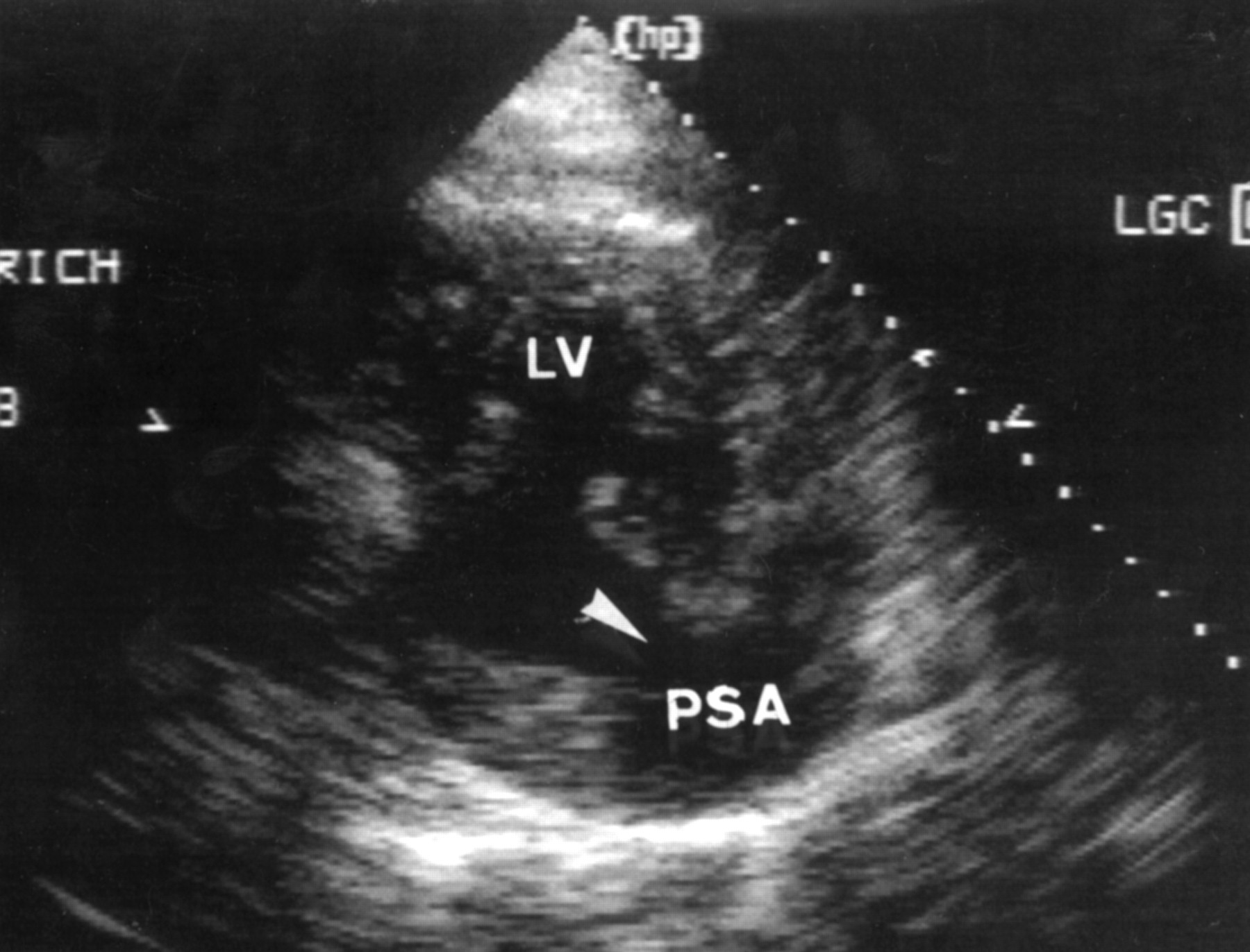
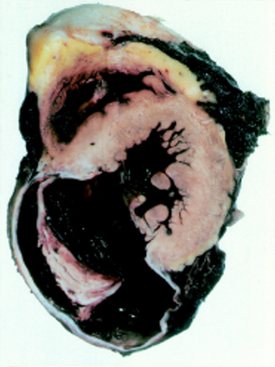
A 66 year old man presented to the emergency room of a community hospital with a 12 hour history of severe retrosternal pain. The ECG showed ST-T segment elevation in the inferior leads. Assuming acute inferior myocardial infarction, systemic thrombolytic treatment with rt-PA was given. Following thrombolysis left sided hemiparesis was noted. A computed tomography (CT) scan of the brain showed no signs of cerebral haemorrhage. To exclude aortic dissection CT imaging was extended to the thoracic region. Surprisingly, these images (top left) revealed posterior ventricular rupture. At that time the patient was haemodynamically stable. He was transferred to our hospital for emergency repair; unfortunately, he deteriorated rapidly and surgery could not be performed. A ruptured posterior ventricular aneurysm and pericardial tamponade was demonstrated by echocardiography (lower left; LV, left ventricle; PSA, pseudoaneurysm). The patient died in electromechanical dissociation. Postmortem examination showed a pseudoaneurysm of the posterior wall with a large parietal thrombus and rupture into the pericardial space (lower right). The brain section showed no haemorrhage or infarction.

