Article Text

Statistics from Altmetric.com
A normal mammal cardiovascular system consists postnatally of a double—pulmonary and systemic—circuit, connected in series, powered by a double pump—the “right” and “left” heart.
Many complex cardiac malformations are characterised by the existence of only one functional ventricle. This “single” ventricle then has to maintain both the systemic and the pulmonary blood circulation, which are not connected in series but in parallel (fig 1A, B). Such a circuit has two major disadvantages: arterial desaturation, both at rest and increasing during exercise, and a chronic volume overload to the single ventricle. Chronic volume overload will in time impair ventricular function, causing from the third decade on a gradual attrition due to congestive heart failure, with few survivors beyond the fourth decade.
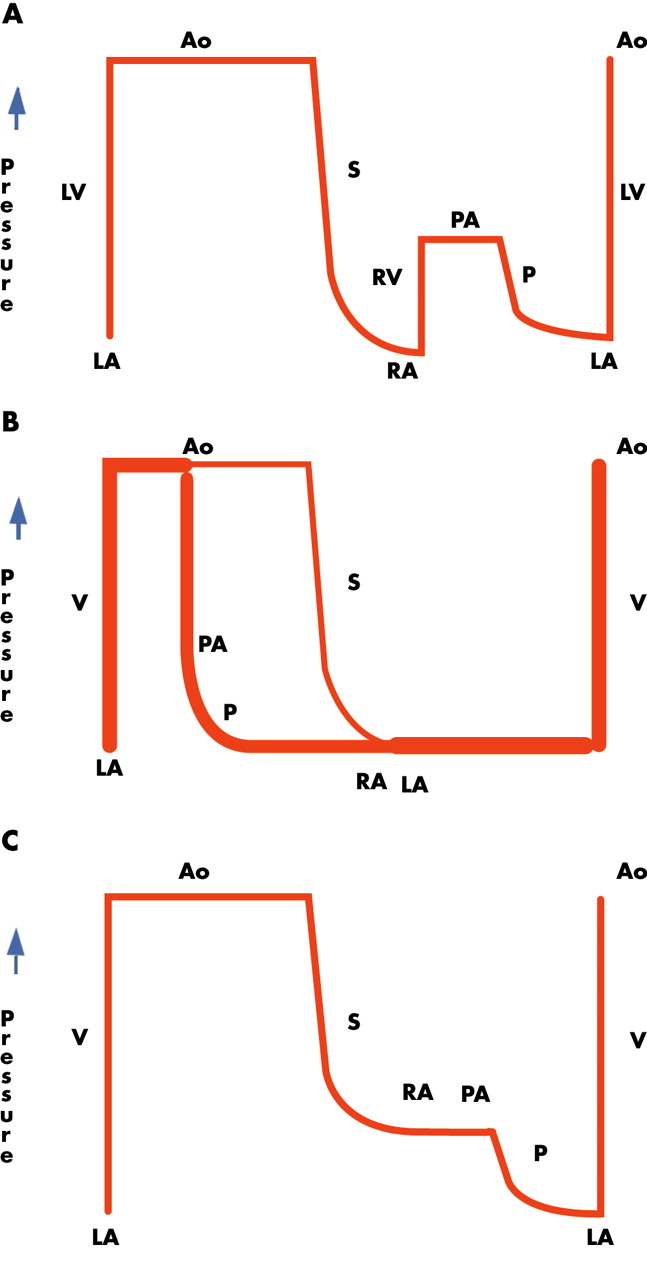
(A) The normal cardiovascular circulation. The pulmonary circulation (P) is connected in series with the systemic circulation (S). The right ventricle maintains the right atrial pressure lower than the left atrial pressure, and provides enough energy to the blood to pass the pulmonary resistance. (B) The patient with a univentricular heart. The systemic and pulmonary circuits are connected in parallel, with a considerable volume overload to the single ventricle (V). The width of the line reflects the degree of volume load. There is complete admixture of systemic and pulmonary venous blood, causing arterial oxygen desaturation. (C) The Fontan circulation. The systemic and pulmonary circulations are connected in series. The right atrium (RA) or systemic veins are connected to the pulmonary artery (PA). The volume overload to the single ventricle is now less than expected for body surface area. In the absence of fenestration, there is no more admixture of systemic and pulmonary venous blood, but the systemic venous pressure is notably elevated. Ao; aorta; LA, left atrium; LV, left ventricle, RV, right ventricle.
In 1971 Francis Fontan1 from Bordeaux, France, reported on a new approach to the operative treatment of these malformations, separating the systemic and pulmonary circulations. In a “Fontan circulation” the systemic venous return is connected to the pulmonary arteries without the interposition of an adequate ventricle, and all shunts on the venous, atrial, ventricular and arterial level are interrupted (fig 1C). In such a Fontan circuit the postcapillary energy is no longer “wasted” into the systemic veins, but collected and used to push the blood through the lungs. Advantages of a Fontan circuit include (near) normalisation of the arterial saturation, and abolishment of the chronic volume overload; the cost for such a circulation includes chronic “hypertension” and congestion of the systemic veins, and decreased cardiac output both at rest and during exercise.2 Typically for this circuit, cardiac output is no longer determined by the heart, but rather by transpulmonary flow (itself mainly determined by pulmonary vascular resistance).
INDICATIONS FOR A FONTAN CIRCUIT
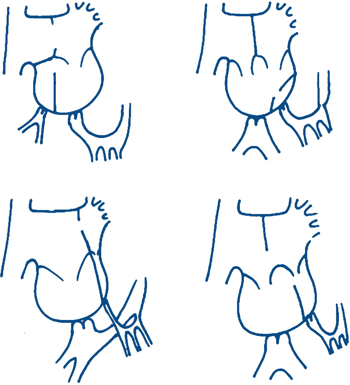
A univentricular Fontan repair can be considered in cardiac malformations with a single functional ventricle, usually because of the absence of an adequate atrioventricular valve or pumping chamber—for example, tricuspid atresia, pulmonary atresia with intact ventricular septum, double inlet ventricle, hypoplastic left heart syndrome (fig 2). In other very complex malformations with high surgical risk morbidity, or need for “high maintenance” (frequent conduit replacement), a Fontan strategy may offer a lower surgical risk and lower incidence of reinterventions for a similar clinical and functional long term result.

Schematic representation of cardiac malformations that are suitable for Fontan repair. Top left: tricuspid atresia. Top right: double inlet left ventricle. Bottom left: hypoplastic left heart syndrome. Bottom right: unbalanced atrioventricular septum defect.
SELECTION OF PATIENTS
In 1978 Choussat and Fontan3 described their recommendations for a successful Fontan operation, defined as having a good cardiac output at an acceptable systemic venous pressure. These rules have been refined by many centres, but all reflect that after repair left atrial pressure must be low (determined by good ventricular function), and that the transpulmonary gradient must be low (determined by the pulmonary vasculature). Cardiac requirements nowadays are: unobstructed ventricular inflow (no atrioventricular valve stenosis, no regurgitation), a reasonable ventricular function, and unobstructed outflow (no subaortic stenosis, no arterial hypertension, and no coarctation). Pulmonary requirements include a non-restrictive connection from systemic veins to the pulmonary arteries (Fontan connection), good sized pulmonary arteries without distortion (at repair and later during growth), a well developed distal vascular bed, (near) normal pulmonary vascular resistance < 2.5 U/m2, and unobstructed pulmonary venous return. As soon as possible following birth, the pre-Fontan management must aim to reach these goals; some deviations are acceptable, however, with increased operative mortality and increased late morbidity and late mortality. The haemodynamic evaluation just before the Fontan surgery must try to predict how lungs and heart will interact after Fontan completion. This can be very difficult because at the time of Fontan surgery ventricular preload will significantly decrease (and according to Frank-Starling also contractility), and with current treatment strategies pulmonary flow will increase, influencing the transpulmonary gradient. All clinicians involved in the management of these patients have learned that these predictions are, at best, just that—predictions.
IS EVERY FONTAN CIRCUIT ALIKE?
Since its original description, the Fontan circuit has known numerous modifications. Early on surgeons used valves (cavo-atrial, atrioventricular, or atriopulmonary), and created various connections between the right atrium and the pulmonary artery (anterior atriopulmonary connection, with or without inclusion of a small hypoplastic right ventricle, posterior atriopulmonary connection), and with different materials (valved conduits, homografts, patches, direct anastomosis). The very high incidence of late reoperations, reaching 40% in some series, reflects the poor design of the first Fontan circuits and the less than ideal surgical techniques used in the early series. Most of the older circuits are no longer created and are considered obsolete; however, many patients still survive on such circuits. When assessing a patient with a “Fontan circuit”, the clinician needs to know exactly which connection has been made with what material.
During the last decade the total cavopulmonary connection has emerged as being superior (fig 3).4 The caval veins are connected to the pulmonary artery, bypassing not only the right ventricle but also the right atrium. The superior caval vein is connected to the pulmonary artery (bidirectional Glenn shunt). There are two variants to connect the inferior caval vein: the lateral tunnel, and the extra cardiac conduit. Introduced in the mid ’80s, the lateral tunnel provides a tubular path between the inferior caval vein and the pulmonary artery, consisting of a prosthetic baffle and a portion of the lateral atrial wall. This circuit has growth potential and can therefore be created in children as young as 1 year; it leaves a minimal amount of atrial tissue exposed to high pressure, which in time may cause atrial arrhythmia. The extracardiac conduit was introduced in 1990, and consists of a tube graft between the transsected inferior caval vein and the pulmonary artery. This circuit leaves the entire atrium at low pressure, has none to minimal atrial suture lines, and can be performed without aortic cross clamping or even cardiopulmonary bypass; however it has no growth potential and therefore will be offered to patients large enough to accept a graft adequate for an adult’s inferior caval vein flow.
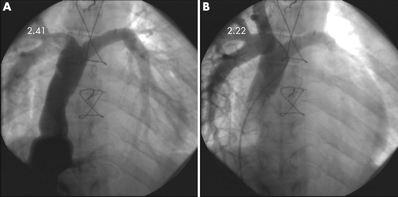
Angiogram of total cavopulmonary connection. (A) Injection in inferior caval vein which is connected with a 22 mm Goretex graft to the pulmonary artery; note mild preferential flow to left pulmonary artery. (B) Injection in right superior caval vein which is connected to the right pulmonary artery; note mild preferential flow to right pulmonary artery.
HOW TO ACHIEVE A FONTAN CIRCUIT CURRENTLY
At birth, it is impossible to create a Fontan circulation. The pulmonary vascular resistance is still raised for several weeks, and the vessels—caval veins and pulmonary arteries—are usually too small, making any cavopulmonary shunt impossible during that period. Even when resistance has fallen, a staged approach is preferred connecting the superior and inferior caval veins at separate occasions. Such a staged approach allows the body to adapt progressively to the very different haemodynamic conditions, and reduces the overall operative morbidity and mortality. A staged approach also allows a better patient selection and intermediate preparatory interventions.
Initially in the neonatal period, management must aim—if not provided by nature—to achieve unrestricted flow from the heart to the aorta (if required, coarctectomy, Damus-Kaye-Stansel, Norwood repair), a well balanced limited flow to the lungs (if required, pulmonary artery band, shunt (modified Blalock-Taussig, central), stent in duct), and unrestricted return of blood to the ventricle (if required, Rashkind balloon septostomy). The infant is then allowed to grow for several months. During this time, the heart is submitted to a chronic volume overload which is good for the development of the pulmonary vasculature, but if excessive is detrimental for ventricular function. The infant will have mild desaturation, inversely related to mild cardiac failure.
At the age of 4–12 months, most centres will introduce a cavopulmonary connection or bidirectional Glenn shunt: the superior caval vein is connected to the pulmonary artery (bilateral if present). If no other blood flow is allowed to the lungs, the volume load to the heart is significantly decreased to slightly less than normal for body surface area (BSA). The patient at this stage will remain slightly cyanotic, as the desaturated blood from the inferior caval vein is still allowed to flow to the aorta.
At 1–5 years of age, depending on centre preference, growth of vascular structures, and cyanosis at rest and during exercise, the Fontan circuit is completed by connecting the inferior caval vein to the pulmonary artery. As mentioned before, two techniques are currently used: the lateral tunnel and the extracardiac conduit. Frequently a small fenestration is created between the tunnel conduit and the pulmonary atrium, either routinely or only in “high risk” patients.5 Such fenestration will allow a residual right-to-left shunt, thereby limiting caval pressure and congestion, and increasing preload of the systemic ventricle and cardiac output, at the expense of cyanosis. Such fenestration has been shown to reduce operative mortality and morbidity (pleural drainage); the fenestration can be closed later (weeks to months) after adaptation of the body to the new haemodynamic condition.
HOW DO OLD (AND CURRENTLY ADULT) CIRCUITS COMPARE WITH CURRENT CIRCUITS?
There are several reasons why adults with a Fontan circuit do not reflect where the current cohort of patients will be in several decades.
The longest survivors of a Fontan operation, currently adults, were frequently less than ideal candidates for this type of surgery, with many significant residua and sequelae related to the cardiac malformation and palliative procedures. A shunt procedure undertaken from the 1960s to the 1980s was evaluated by its long term relief of cyanosis. The fact that this shunt could induce mild pulmonary vascular disease, ventricular dysfunction, or pulmonary artery distortion was not the first preoccupation of the surgeon, as it is now. The success of a shunt is currently evaluated by an acceptable relief of cyanosis without a significant volume overload, by its induction of pulmonary growth without an impairing effect on the pulmonary vasculature, even not during growth after Fontan repair, and by allowing the patient to reach an age at which a Fontan circuit can be safely created.
Moreover, many centres have attempted to determine which combination of criteria could be ignored without compromising a “successful” outcome.6 Fontan circuits have previously been performed on patients who would probably not now be considered for such repair. Those unsuitable candidates who survived the operation obviously represent the poorer end of the clinical spectrum of results after Fontan correction, and will distort all reports on long term results.
The change in management has contributed to a higher proportion of patients with a univentricular heart that fulfil most criteria for a successful Fontan repair. Excessive ventricular dilation, spherical reconfiguration, cardiac overgrowth, ventricular dysfunction, and mild pulmonary vascular disease or distortion are now rarely seen.
Operative mortality has significantly decreased to less than 5%, with better circuits having “a life time” potential. We expect the current cohort of patients to perform better than the early series, also at a later age.
COMPLETE FONTAN VERSUS BIDIRECTIONAL GLENN WITH ADDITIONAL FLOW
When creating the Glenn shunt, the option exists to maintain the original pulmonary blood flow source (pulmonary stenosis or band, shunt contralateral to the superior caval vein). If this flow is limited, the extra pressure load on the superior caval vein is usually well tolerated, with the benefit of extra growth of the pulmonary arteries and higher arterial saturations during this stage of palliation. It was hoped that such a circuit might be a satisfactory final palliation. However, most children outgrow the limited pulmonary blood flow, with increasing cyanosis especially during exercise. Most of these patients will eventually be converted to a “full” Fontan circulation.
COMPLICATIONS OF THE FONTAN CIRCULATION
Complications after Fontan repair are common and related to the increased venous pressure and congestion, and chronic low cardiac output. Complications include early and late mortality, mild to moderate exercise intolerance, residual cardiomegaly, ventricular dysfunction, rhythm and conduction disturbances, hepatomegaly, lymphatic dysfunction with protein losing enteropathy, systemic venous thrombi, ascites, and peripheral oedema.
Late mortality
When assessing long term results from a centre, it should be clear that in all series a profound bias occurs, firstly when patients are accepted for a Fontan repair, and secondly by the survival following the operation. Both are unavoidable effects when studying long term survivors. Many patients have survived for several decades. Late death is directly related to the number of risk factors for a Fontan operation.7 Poor candidates for a Fontan operation, if they survive due to excellent surgery and outstanding postoperative care, remain at higher risk for late death. Good Fontan physiology allows good survival for several decades, but unfavourable haemodynamics and risk factors are associated with an increased early and late attrition.
Functional status and exercise tolerance
Despite the abnormality of the circuit, clinicians are frequently impressed by the ability of most patients with a Fontan circulation to lead a nearly normal life, including mild to moderate sport activities. More than 90% of all hospital survivors are in New York Heart Association (NYHA) functional class I or II. Most patients progress through the education system just like the standard population and can pursue a wide variety of professional careers.8 However, with time there is a progressive decline of functional status in some subgroups. A Fontan circulation is palliative in nature, with good results in patients with an ideal haemodynamic profile, but with significant ongoing morbidity and mortality if some criteria are not met.
Ventricular function
All studies have reported the ventricle of a functionally univentricular heart to be dilated, hypertrophic and hypocontractile, certainly after completion of a Fontan type of circuit. Ventricular dysfunction can be caused by the congenital malformation itself, previous surgical interventions, or the very abnormal working conditions of the ventricle at the various stages of palliation, both before and after Fontan.
During the first months after birth, the ventricle will always be volume overloaded, either by a band or by an aorta–pulmonary shunt. This volume overload will lead to dilation and spherical reconfiguration, cardiac overgrowth, and eccentric hypertrophy. After unloading at the time of a Fontan operation, some regression to normalisation will occur, but will be frequently incomplete. Before the introduction of the cavopulmonary shunt in the 1990s, a large volume overload persisted for several years. Therefore, patients born before the ’90s (currently adults) will frequently have residual dilation, cardiac overgrowth, and hypertrophy even into adult age. With current treatment strategies only a small shunt is allowed to persist for several months, thereby avoiding such a situation.
The preload to the ventricle is, at the time of a Fontan operation, reduced to levels well below normal for BSA (50–70%), and even more when expressed in relation to ventricular size (25–70%). The ventricle thus evolves from being volume overloaded and overstretched, to overgrown and (severely) underloaded. It should therefore not be a surprise that the deprived ventricle in a Fontan circuit shows systolic and diastolic dysfunction. The ventricle may now enter a vicious cycle whereby the low preload results in remodelling, reduced compliance, poor ventricular filling, and eventually continuously declining cardiac output. This phenomenon of progressive “disuse hypofunction” occurs at a chronic preload of less than 70% of the “due” preload.9 Our knowledge of low flow conditions, chronic congestion, and limited ventricular preload is, however, still very limited.
The congenital malformation itself may predispose to ventricular dysfunction. A left ventricle with its unique ellipsoid shape and complex fibre orientation is “tuned” to maintain systemic pressure for a life time. In contrast, a morphologic right ventricle, and even more an undetermined primitive ventricle—both frequently encountered in functionally univentricular hearts—may fail after some years of systemic loading. Furthermore, a tricuspid valve or a common atrioventricular valve poorly tolerate the initial volume overload, and also have problems with the chronic systemic afterload.
The treatment of ventricular dysfunction in the setting of a Fontan circuit is very frustrating for a cardiologist. Lack of reaction to classic treatment strategies has given the ventricle in a Fontan circuit a very bad reputation. Several studies, both acute and chronic, have shown little impact on ventricular function of medication such as inotropes, afterload reducing agents, vasodilators, and β blockers, as these have no impact on the reduced preload which is the dominant limiting factor.
Arrhythmia
Patients with a Fontan circulation are predisposed to develop atrial dysrhythmias. Many old circuits have atrial wall incorporated into the circuit causing atrial dilation and hypertrophy; furthermore, most patients have had an atriotomy, and possible injury to the sinus node or its arterial supply or innervation. Clinically, tachycardia is mostly prevalent in patients with a suboptimal haemodynamic result, especially if the right atrium is dilated and the pulmonary vascular resistance is increased. The prognosis therefore remains worrying, even if the patient can be reconverted to sinus rhythm. Arrhythmias occur with increasing incidence after Fontan repair in up to 40% of the patients 10 years after surgery.
The most common atrial tachycardia is intra-atrial re-entry tachycardia or atypical atrial flutter. Such tachycardia can quickly lead to severe haemodynamic deterioration. Survival depends on the ventricular contractility and the vascular resistance, which can be critically altered by most antiarrhythmic drugs. The safest option is therefore immediate direct current (DC) shock reconversion. During the subsequent workup, the clinician should obtain a complete haemodynamic evaluation in every patient with a new tachycardia, as this may be the late but first clinical manifestation of pathway obstruction and/or thrombi. Full anticoagulation should be considered in every Fontan patient with atrial arrhythmias in view of the significant risk of a right atrial thrombus.
Long term treatment of atrial arrhythmia can involve medication and ablation; progression of the arrhythmia and functional decline is frequently observed. The best long term investment for these patients is conversion of the old Fontan circuit to an extracardiac cavopulmonary connection, together with a right atrial maze and a reduction plasty (combined with a DDD epicardial pacemaker if indicated).10
It is speculated that the incidence of late atrial tachycardia will decrease with the use of cavopulmonary connections. Only time will tell whether this theoretical advantage will come true, but results after similar follow up are promising.
Ventricular arrhythmias are rare in patients with a Fontan circulation. If present, they are probably related to very poor Fontan haemodynamics or severe ventricular dysfunction.
Hypoxaemia caused by residual–new shunts
Patients with a Fontan circulation are slightly desaturated with values in the low 90s, even when a residual atrial septum defect had been refuted. The coronary sinus in all recent circuit modifications drains to the left atrium, causing a modest arterial desaturation of ±2%. Several anatomic substrates may result in a significant residual/progressive right-to-left shunt and desaturation: a residual atrial septum defect or fenestration, a coronary sinus to left atrium shunt through a defect or through cardiac veins (in older circuits), intrapulmonary arteriovenous fistulae, and abnormal systemic venous drainage to the pulmonary venous atrium (for example, a left superior caval vein to the left atrium). These connections may be closed percutaneously as they cause symptomatic cyanosis.
Residual left-to-right shunt
Left-to-right shunting may be present because of aorta–pulmonary collaterals, incomplete ligation of the main pulmonary artery, or failed occlusion of previous shunts. These shunts usually cause an unimportant volume load for the systemic ventricle, but also put a useless and sometimes detrimental volume/pressure overload on the pulmonary flow. Any systemic vessel that upon injection gives a pulmonary capillary blush with opacification of the pulmonary veins should be occluded percutaneously.
Pulmonary circulation
A low pulmonary vascular resistance is mandatory for a good functioning Fontan circuit. Apart from a few exceptions, pulmonary vascular resistance will control the cardiac output. The circuit runs on “autopilot”, with little interference possible by the clinician. This has frustrated many cardiologists as they have experienced that “their organ” no longer controls cardiac output. Cardiac output can be increased by improving flow to and into the lungs (streamlined non-obstructive Fontan connection, stenting of hypoplastic or stenotic pulmonary artery), or by bypassing the lungs with a fenestration. The effect of pulmonary vasodilators is usually very modest.
Pre-Fontan management must aim at perfect growth of pulmonary vasculature. We have now started to realise that an initial mild pulmonary overflow is beneficial for adequate growth of pulmonary vasculature. The optimal balance between a small short lived shunt and a large longstanding shunt, leading inversely to either more or less cardiac preservation or pulmonary growth, still needs to be refined.
Pulmonary artery distortion is a recognised late complication of most systemic to pulmonary artery shunts. Clinicians should realise that at the time of Fontan repair these shunts are sometimes not divided, but just occluded with a clip. Therefore, the pulmonary artery is still fixed to a systemic artery, which can cause a distortion of the pulmonary artery late after Fontan repair, especially during growth. Careful clinicians will choose to actively document adequate growth of all connections and structures during and certainly at the end of somatic growth.
The effect of high altitude and decreased oxygen tension varies widely among individuals. Patients with borderline haemodynamics have been reported to deteriorate acutely after moving to moderate altitude above 2000 m. Patients with good haemodynamics and no or little reactivity of the pulmonary vasculature do well at moderate altitude. It remains questionable whether patients whose cardiac output is sensitive to the pulmonary vascular resistance should be living at increased altitude.
Lymphatic dysfunction: protein losing enteropathy, plastic bronchitis
A Fontan circulation operates at—or sometimes just beyond—the functional limits of the lymphatic system. The superior caval vein pressure is elevated and its runoff decreased, impeding drainage of the thoracic duct. Leakage in the interstitium causes lymph oedema or pulmonary oedema, a very lethal complication in the early postoperative period. Leakage into the thorax or pericardium will lead to chylothorax or chylopericardium, a complication which usually only occurs in the perioperative period, but rarely thereafter because of adhesions. Leakage into the gut leads to protein losing enteropathy (PLE), the most frequent lymphatic problem in long term follow up. Leakage of chyle into the bronchus leads to plastic bronchitis, most frequently diagnosed at necropsy.
PLE is caused by intestinal lymphangiectasia with leakage of lymphocytes, chylomicrons, and serum protein such as albumin and immunoglobulin into the gut; PLE results in oedema, ascites, immunodeficiency, and hypocalcaemia. PLE is a relatively rare complication: in an international multicentre study,11 involving 35 centres and 3029 patients with Fontan repair between 1975 and 1995, PLE occurred in 114 patients (3.8%, range across the 35 centres 0–25%). The diagnosis of PLE usually yields a very poor prognosis: after diagnosis, the five year survival rate was 59%, with less than 20% of patients surviving beyond 10 years.
Problems in the Fontan circuit (gradient at connection, poor design), problems of the pulmonary vasculature (increased pulmonary vascular resistance, hypoplasia, distortion, loss) or cardiac problems (atrioventricular–valve regurgitation, myocardial dysfunction) can frequently be identified. An autoimmune or inflammatory cause, sometimes triggered by an infection, has also been isolated in some patients.
Several treatment strategies have been identified. A diet high in calories, high protein content, and medium chain triglyceride fat supplements with low salt content is usually recommended. Diuretics can more or less limit peripheral oedema in most patients. Protein infusions (albumin, globulin) can be required on a weekly or monthly basis. In some patients specific anti-infection measures are necessary (chronic antibiotics, vaccines). Clinical resolution of PLE with only these measures is rare (< 1%).
Attempts to augment cardiac function or output with inotropic drugs or systemic vasodilators are usually unsuccessful. Corticosteroids, high molecular weight heparin, and octrioide acetate may occasionally improve PLE. Some patients are “cured” for several weeks, months, or years with a single episode of medication; some patients will have an ongoing need for medication, while many patients will show no reaction at all. Side effects in the long term are important.
Hypoxaemia induced pulmonary vasoconstriction can lead to PLE; a long stay at high altitude or even a long flight can trigger PLE in some patients. Most patients with a Fontan circuit will, however, tolerate such altitude conditions without a problem. Cardiac pacing in patients with excessive bradycardia or atrioventricular dissociation can critically improve cardiac function and may cure PLE. Resection of the most affected part of the gut resulting in clinical improvement has been reported.
Several surgical options have been reported: relief of obstruction, conversion to streamlined cavopulmonary connection, atrioventricular–valve repair/replacement. However, many of these procedures yield a high mortality, especially if there is significant atrioventricular–valve regurgitation and ventricular dysfunction. Late takedown has been performed in some patients, usually with high mortality. Of all the surgical procedures, heart transplantation is the most predictive, but obviously with its own long term problems. Heart transplantation will improve the haemodynamic condition by increasing cardiac output and decreasing venous congestion; chronic anti-rejection therapy may also influence PLE. If the patient survives the transplantation (increased risk from cachexia, immunologic deregulation), PLE resolves in most patients; rarely it persists or relapses after some years.
Several haemodynamic problems can be solved with catheter techniques: balloon dilation, stent implantation of a circuit stenosis or residual vessel hypoplasia; residual left to right shunts (native, surgical, arterial venous collaterals) can be embolised; a fenestration12 with stent can safely be created through the percutaneous (or transhepatic) route, even in circuits with an extracardiac conduit. Such fenestration will decrease venous congestion, improve runoff and increase cardiac output, at the expense of arterial saturation, however. PLE nearly always improves with a fenestration, but the range between “acceptable” PLE and “unacceptable” cyanosis may be narrow; this range frequently further narrows over months, probably because of secondary changes in the pulmonary vasculature. Many such patients will therefore require heart transplantation in the medium term follow up.
Plastic bronchitis is a very rare but potentially lethal complication occurring weeks to months after the Fontan correction: it leads to obstruction of major airways by solid fibrinomucoid material. Persistent segmental atelectasis, large airway obstruction, or expectoration of tenacious fibrinomucoid material should prompt early consideration of diagnostic and therapeutic bronchoscopic lavage. Treatment is very difficult and similar to that for PLE.
Reproduction: pregnancy
Most females after Fontan repair have normal menstrual patterns, making it apparent that pregnancy is possible. Pregnancy and delivery clearly put an increased load on the cardiovascular system. Normal pregnancy is associated with an increase in cardiac output of 30–40% by 24 weeks of gestation, an increase in the circulating blood volume by 30–40%, but a decrease in systemic vascular resistance. Fluid retention and a hypercoaguable state are frequently observed. These changes result in an increased systemic venous pressure and may trigger complications of “right heart” failure such as atrial arrhythmias, oedema, and ascites. Any right-to-left shunt through a residual atrial septal defect will increase; the resultant decrease in arterial saturation, however, is usually well tolerated. There is an increased risk for venous thrombosis and pulmonary embolus. Nevertheless, successful pregnancy with delivery of normal children is possible. Women with a good haemodynamic result apparently tolerate pregnancy and normal delivery well.13 Therefore, if a woman is doing well both clinically and haemodynamically, and strongly desires to become pregnant, it seems currently unreasonable to discourage her. However, she and her husband must be made aware that the risk to her and the child is increased.
The Fontan circulation: key points
-
Good Fontan physiology enables survival for several decades, but unfavourable haemodynamics and risk factors are associated with poor exercise tolerance and an increased early and late attrition
-
Following birth, the ventricle tries to adapt to different and very abnormal circulations, evolving from overloaded and overstretched while shunted, to underloaded and overgrown after a Fontan type of circulation
-
The absence of a clinical problem does not equal a satisfactory haemodynamic result; possible problems must actively be searched and resolved before they become clinically relevant
-
Knowledge of low flow conditions, chronic congestion, and limited ventricular preload is still very limited
-
The Fontan circulation can be a source of frustration for the cardiologist as, apart from a few exceptions, the heart in a Fontan circuit no longer controls cardiac output. Conventional anti-failure treatment will yield no clinical result
-
Management of atrial tachycardia: the faster the rhythm, the poorer the patient’s condition, the more scars on the chest, the less the clinician understands, the faster he should opt for urgent DC reconversion.
The risk for the fetus of having congenital heart disease is currently unknown, as women with cardiac malformations amenable to Fontan surgery have rarely had offspring. For most malformations the risk will probably vary between 5–10%.
Coagulopathies
Thrombosis is more likely to occur in patients with low cardiac output and more than usual atrial and systemic venous dilatation and blood stasis. Fontan patients have an increased incidence of coagulation factor abnormalities—as in any patient with hepatic congestion: protein C, protein S, and antithrombin III deficiency.14 Thrombosis may complicate an infection, especially when associated with dehydration.
A systemic venous thrombus may embolise to the pulmonary artery. Massive embolisation may result in shock, arrhythmia, or death; it is the most common cause of sudden out-of-hospital death in patients with a Fontan circuit. Chronic multiple pulmonary microemboli may lead to pulmonary vascular obstructive disease, a late complication which is particularly lethal in a Fontan circulation.
Some clinicians recommend anticoagulating every patient with a Fontan circuit; however, there are clearly subgroups of patients with a very low risk. Full anticoagulation should be prescribed in patients with previous thrombi, poor cardiac output (frequently associated with spontaneous contrast on echo), congestion, dilation of venous or atrial structures, and arrhythmia. If coagulation abnormalities are present, oral contraceptives should not be prescribed. It is not clear if a small dose of acetylsalicylic acid or clopidogrel is beneficial for prevention of venous thrombi in these patients. This discussion over whether or not to anticoagulate all patients will dissipate once the new generation of antithrombotic drugs becomes available.
The failing Fontan
A Fontan circulation may become “failing” and unbearable because of persistent congestion with oedema, low cardiac output with very limited exercise tolerance, relapsing tachycardia, protein losing enteropathy, or plastic bronchitis. As explained before, treatment with inotropes, vasodilators, and diuretics will show little result and will frustrate any “conventional” cardiologist. Frequently a more aggressive approach will be needed aimed at optimising the circuit (stenting of stenosis, embolising collaterals, conversion to cavopulmonary connection, right atrial maze); if this is insufficient, fenestration and heart transplantation should be considered.
CONCLUSIONS
The long term results of the Fontan operation performed in patients with a low risk profile show good overall results: arterial hypoxaemia is eliminated, volume overload to the single ventricle is abolished, the expense of chronic systemic venous hypertension is usually acceptable, and most patients enjoy a good quality of life. However, sequelae persist in less than ideal patients, which are frequently progressive and associated with a declining functional result and even premature death. This should remind every clinician that a Fontan operation remains an imperfect solution for a complex cardiovascular problem.
REFERENCES
Linked Articles
- Miscellanea
- Miscellanea