Article Text

Statistics from Altmetric.com
- Echocardiography
- three dimensional
- pulmonary arterial hypertension
- pulmonary hypertension
- Pulmonary arterial hypertension (PAH)
Pulmonary arterial hypertension (PAH) is a severe disease characterised by a progressive increase of pulmonary pressure and resistance leading to right heart failure. PAH is commonly diagnosed at a late stage of the disease and is associated with progressive clinical deterioration and premature death. According to the most recent consensus conference, pulmonary hypertension (PH) is categorised into five main groups: group 1, PAH; group 2, PH associated with left sided heart diseases; group 3, PH associated with lung disease and/or hypoxaemia; group 4, PH due to chronic thrombotic and/or embolic disease; and group 5, miscellaneous—summarising a variety of rare and not well characterised disorders characterised by non-specific signs and symptoms.1 Screening for PH is usually made by transthoracic echocardiography on the basis of the velocity of the regurgitant tricuspid jet, and final confirmation is done by right heart catheterisation.2
Without treatment, the prognosis for patients with significant PH is poor. The reported median life expectancy of idiopathic PAH is 2.8 years from the diagnosisw1; however, recent meta-analysis of trials in the field of PAH have provided indications of a beneficial influence of PAH treatments on survival.3 Current PAH specific medications, including prostanoids, endothelin receptor antagonists (ERA), and phosphodiesterase 5 inhibitors (PDE5i), have sought to address the pulmonary vascular endothelial dysfunction and vasoconstriction associated with this condition. Several novel drugs that have already produced encouraging results in animal models aim to target more directly the structural vascular changes (remodelling) and are currently under clinical investigation (eg, soluble guanylyl cyclase activators/stimulators, tyrosine kinase inhibitors, 5-HT2B receptor antagonists).
This article reviews the clinical classification, which has been recently updated. Moreover, established approaches to evaluate PH and the currently recommended treatments for PAH are summarised.
Definition of PH
The haemodynamic definition of PH has not been evidence based. Under normal circumstances, resting mean pulmonary artery pressure (mPAP) was shown to be 14.0±3.3 mm Hg, which was independent of gender and ethnicity.4
Thus, during the 4th World Symposium on PH held in Dana Point 2008, new thresholds for mPAP were introduced. An mPAP <21 mm Hg was defined as normal, from 21–25 mm Hg was categorised as borderline, and an mPAP >25 mm Hg was designated as manifest PH. Correspondingly, echocardiographic systolic tricuspid regurgitant velocity thresholds <2.5 m/s is defined as normal, 2.5–2.8 m/s as borderline, and >2.8 m/s is highly indicative for a manifest PH (table 1).w2 mPAP during exercise is dependent on exercise level and age. During mild exercise, PAP was 19.4±4.8 mm Hg in subjects <50 years compared to 29.4±8.4 mm Hg in subjects >50 years. While the mPAP at rest is virtually independent of age and rarely exceeds 20 mm Hg, exercise mPAP is age related and frequently exceeds 30 mm Hg, especially in elderly individuals, which makes it difficult to define normal mPAP values during exercise.4 Therefore, exercise values of mPAP were not included in the new definition of PH.1
New thresholds of pulmonary hypertension determined at the 4th World Symposium held in Dana Point 2008
Updated classification of PH
The World Health Organization has hosted conferences on PH introducing a classification scheme in 1973 and proposing the Evian classification in 1988.w3 This classification was based on similar pathophysiological mechanism, clinical presentation and therapeutic options, and was modified further in Venice in 2003, where the term idiopathic pulmonary arterial hypertension (IPAH) replaced the term primary pulmonary hypertension.w4 The other prominent change made at the Venice meeting was to move pulmonary veno-occlusive disease (PVOD) and pulmonary capillary haemangiomatosis (PCH) from separate categories into a single subcategory of PAH. In 2008 the 4th World Symposium on PH provided the opportunity to slightly modify the previous clinical classification (figure 1).1

Updated clinical classification of pulmonary hypertension according to the proposals of the 4th World Symposium on Pulmonary Hypertension held in Dana Point 2008.
PAH (group 1)
Idiopathic and heritable PAH
Due to the fact that PH occurs in different clinical conditions, IPAH corresponds to sporadic disease without family history of PAH and typical risk factors. If PH is detected in a familial context, mutations of the bone morphogenetic protein receptor type 2 (BMPR2) gene can be documented in approximately 70% of cases.5 However, given the fact that in 30% of families with PAH no BMPR2 mutation has been identified, it was decided to abandon the term ‘familial PAH’ in the new classification and to replace it with the term ‘heritable PAH’. Heritable forms of PAH include germline mutations (BMPR2, activin-receptor like kinase 1, endoglin) and familial cases without identified germline mutations.1 However, this new category does not imply a recommendation for general genetic testing of patients with IPAH or in familial cases of PAH. Instead, genetic testing should be performed as part of an extensive programme that includes genetic counselling and discussion of risk, benefits, and limitations of such testing.6
Drug and toxin induced PAH
PAH is also a rare side effect of certain weight reducing agents, such as fenfluramine and dexfenfluramine. However, the incidence of drug induced PAH is decreasing as these agents are no longer available. Recently, a class effect of selective serotonin reuptake inhibitors was claimed after use in pregnant women. In a case–control study, after 20 weeks of gestation newborns showed an increased risk for developing PAH.
Additionally, the occurrence of PAH after methamphetamine abuse (inhaled, smoked, or intravenous), which has already caused a significant increase of drug related new PAH cases in the south west of the USA, could be documented.
IPAH and many forms of the formerly called secondary PAH share histopathological commonalities, natural history and response to treatment, and on this basis these conditions are considered as being ‘associated pulmonary arterial hypertension’. These forms include the following entities.
PAH associated with connective tissue disease
PAH is a well recognised complication of connective tissue diseases such as systemic sclerosis and systemic lupus erythematosus (SLE). It can, but must not necessarily, occur in conjunction with different degrees of interstitial lung diseases. The prevalence of PAH in patients with systemic sclerosis has been reported to be up to 16%. Patients with PAH associated with systemic sclerosis have a particularly poor prognosis compared to those with systemic sclerosis without PAH.6 w5
PAH associated with congenital heart disease
Congenital heart disease is relatively common, affecting around 1% of the population. Within this population 15% eventually develop PAH.7 As determined by the level of pulmonary vascular resistance, the most severe form of PAH is Eisenmenger's syndrome, which is associated with the reversal of an initial left to right shunt causing cyanosis and limited exercise capacity.w6 During the Dana Point meeting, the pathologic and pathophysiologic classification of congenital heart disease with systemic-to-pulmonary shunts were redefined to provide more detailed description of each condition.1
PAH associated with HIV infection
PAH is a rare (estimated prevalence in patients with HIV is 0.5%) but relatively well documented complication of HIV infection.8 With the advent of highly active antiretroviral therapy and notably improved survival, PAH and other non-infectious manifestations of HIV infection are increasingly responsible for HIV associated morbidity and poor prognosis. In patients with HIV, the HIV-1 envelope glycoprotein GP120 may stimulate the production of endothelin by macrophages.9 HIV associated PAH shows a clinical picture similar to IPAH and seems to be independent of the degree of immunosuppression. The prevalence of HIV associated PAH was evaluated more recently and showed a stable prevalence of 0.46%.
Portopulmonary hypertension
Portal hypertension might lead to development of PAH which is known as portopulmonary hypertension (POPH). Prospective studies have shown that 2–6% of patients with portal hypertension have PH. Risk factors for development of POPH are identified as female sex and autoimmune hepatitis. The prognosis is related to the presence and severity of cirrhosis and to cardiac function.10 w7
PAH associated with schistosomiasis
Since 2008 PAH associated with schistosomiasis has been added to group 1. In the previous classification this form of PH had been categorised in group 4. It was thought that embolic obstruction of pulmonary arteries by schistosoma eggs was responsible for the development of PAH. However, more recent publications point to similarities of PAH associated with schistosomiasis and IPAH showing the same histological findings—for example, plexiform lesions. From the latest data it is assumed that more than 200 million people are infected with any of the three species of schistosoma and that 4–8% of patients develop PH in association with hepatosplenic disease.11 12
Chronic haemolytic anaemia
There is growing evidence that chronic hereditary haemolytic anaemias, including sickle cell disease (SCD), thalassaemia, hereditary spherocytosis, stomacytosis, and microangiopathic haemolytic anaemia, are today associated with PAH. It is estimated that PAH occurs most frequently in patients with SCD. However, the mechanism leading to PAH in patients with SCD is not fully elucidated. Chronic haemolysis might be followed by high rates of nitric oxide (NO) consumption, thereby not activating smooth muscle guanosine monophosphate, a potent vasodilator/antiproliferative mediator.13 However, there is some discussion regarding the relevance of moderate elevations of pulmonary artery (PA) pressures in view of the hyperdynamic circulation of patients particularly suffering from SCD, where pulmonary vascular resistance typically is in the normal range; therefore, a proposed benefit from treatment with PAH specific drugs is yet to be shown.
The most recent classification for PH placed PVOD and PCH with PH in a dedicated subgroup of group 1 (so called group 1). In PVOD, histological changes similar to those found in the small pulmonary arteries (ie, intimal fibrosis and medial hypertrophy) of IPAH patients are present in addition in small post-capillary venules. Regarding diagnosis, untreated PVOD is often difficult to distinguish clinically from IPAH. Indications for PVOD can be derived from high resolution computed tomography (CT) scans of the lungs, showing interlobular septal thickening, ground glass opacities, and enlarged mediastinal lymph nodes. PVOD/PCH remains a difficult disorder to treat, as often initiation of PAH specific treatments, particularly intravenous prostanoids, can result in pulmonary oedema formation with severe aggravation of hypoxaemia. Thus, early listing for lung transplantation remains the only option for management of PVOD patients.
PH owing to left heart disease (group 2)
All forms of PH owing to left heart disease are included in group 2. Either left ventricular disease or valvular disease may lead to an increased left ventricular end-diastolic pressure resulting in high pressure in the pulmonary artery. In most affected patients the pulmonary vascular resistance is normal (<250 dyn·s·cm−5) and the transpulmonary gradient is <12 mm Hg. The most recent classification acknowledges the increasing number of patients with left sided heart dysfunction with preserved systolic function. Thus this distinct aetiology has been added to left heart systolic dysfunction and left heart valvular disease.1 Currently no drug is approved for the treatment of this subcategory of PH, and adequate treatment of the underlying cardiac disease remains the recommended standard.
PH owing to lung disease and/or hypoxia (group 3)
Group 3 includes PH associated with lung disease and/or hypoxaemia. This includes chronic obstructive pulmonary disease (COPD) and interstitial lung diseases, as well as PH associated with prolonged stay in high altitudes (‘chronic mountain sickness’). The prevalence of PH in this group remains largely unknown; however, recent data indicate for COPD associated PH an incidence of approximately 25% with manifest PH (defined by mPAP>25 mm Hg) and 1–2% with severe PH (ie, mPAP >40 mm Hg).14 Currently numerous trials are investigating the therapeutic benefit of treating PH, both in patients with interstitial lung disease as well as COPD.
Chronic thromboembolic pulmonary hypertension (group 4)
In the new classification, chronic thromboembolic pulmonary hypertension (CTEPH) remains in group 4. The former Venice classification divided CTEPH into two subgroups—proximal and distal CTEPH. However, since there is no consensus among experts on how to distinguish between proximal and distal CTEPH, only a single category of CTEPH is proposed in the new definition of group 4. Recent prospective data have suggested that the incidence of CTEPH after initial pulmonary embolism may be higher than previously estimated. The incidence of symptomatic CTEPH within 2 years of a first time pulmonary embolism was 3.8%. In addition to the residual clot in the proximal pulmonary arteries, another major problem in CTEPH is the development of varying degrees of small vessel arteriopathy. These small vessel changes can contribute to the development and progression of PH in CTEPH. With few differences, the general pathology of these distal vessels in CTEPH appears to mimic that seen in PAH.15 w8
PH with unclear aetiologies (group 5)
Group 5 comprises several forms of PH for which the aetiology is unknown or multifactorial. Chronic myeloproliferative disorders including polycythaemia vera, essential thrombocythaemia, and chronic myeloid leukaemia are listed; systemic disorders leading to PH (sarcoidosis, Langerhan's cell histiocytosis, lymphangioleiomyomatosis, neurofibromatosis), and metabolic disorders (type I glycogen storage disease, Gaucher disease, hyperthyroidism, hypothyroidism) are also summarised in this group. Lastly, some rare cases of PH have been observed in a number of miscellaneous conditions (tumour, end stage renal failure, mediastinal fibrosis).1
Diagnostics of PH
Clinical presentation
The diagnostic workup of PH requires a series of investigations to confirm the diagnosis and to clarify the clinical class and subtype of PH. However, PAH is difficult to diagnose. The clinical cardinal symptom of PH is dyspnoea (either on exertion or even at rest) and early fatigue; as the disease advances, patients can develop chest pain due to right ventricular dysfunction, palpitations, oedema, and ascites, and can experience syncope.w1 A high level of suspicion is needed to establish the diagnosis. A typical delay of 2–3 years between onset of symptoms and making a diagnosis is still frequently observed.
Electrocardiogram
In PH the electrocardiogram (ECG) may demonstrate signs of right ventricular hypertrophy, such as right axis deviation, T inversion in the anterior leads, persistent S wave, and right bundle branch block (figure 2). However, the ECG's sensitivity (55–73%) and specificity (70%) are inadequate to be an effective screening tool for the detection of PAH. Some features of the ECG in patients with IPAH may have prognostic value: a P wave amplitude in lead II of ≥0.25 mV has shown to predict a 2.8-times higher risk of death over a 6 year follow-up period.7
Chest x-ray
The chest x-ray appears to be inferior to the ECG in detecting PH. Nevertheless, peripheral lung vascular markings, and hilar pulmonary artery prominence with a hilar-to-thoracic ratio >44%, might be specific for the diagnosis of PH (figure 3). Even it is not sensitive, it may still uncover underlying lung disease. Furthermore, changes in heart diameter may reflect beneficial (reduction in diameter) or detrimental (increased diameter) courses of the disease, particularly in response to medical treatments.

Electrocardiogram demonstrating the changes of right ventricular hypertrophy—for example, incomplete right bundle branch block, inversion of the T wave in anterior leads, persistent S wave in V6.

Chest x-ray showing decreased peripheral lung vascular markings, and hilar pulmonary artery prominence with a hilar-to-thoracic ratio >44%, which is specific but not sensitive for the diagnosis of pulmonary hypertension.
Echocardiography
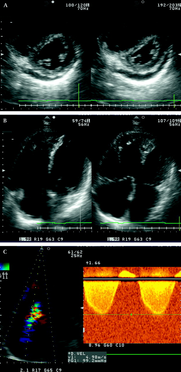
If PH is suspected based on the history, symptoms, and physical examination, transthoracic echocardiography should be performed (figure 4). It provides a quantitative assessment on pulmonary artery pressure (PAP). After Dana Point 2008 new thresholds of the velocity of the tricuspid regurgitation (TR) jet were defined as described above. Utilising the modified Bernoulli equation (4v2) and adding the right atrial pressure, the systolic pulmonary artery pressure (sPAP) can be estimated non-invasively. sPAP >40 mm Hg generally warrants further evaluation in patients with unexplained dyspnoea.w9 Doppler echocardiography can also accurately estimate the right ventricular outflow tract (RVOT) acceleration time, which correlates linearly with sPAP. An acceleration time <100 ms reflects an abnormal PAP. Moreover, the peak early diastolic and the end-diastolic velocities of pulmonary regurgitation flow correlate with the mean PAP and pulmonary artery end-diastolic pressure.

Echocardiographic findings in a patient with pulmonary hypertension include right atrial and ventricular enlargement, flattening or D shape of the interventricular septum, and underfilled left heart chambers. (A) Parasternal short axis view. (B) Apical four chamber view. (C) Doppler analysis of the systolic tricuspid regurgitant velocity.
Echocardiography not only provides Doppler analysis but also helps to exclude valvular, primary myocardial and congenital causes of elevated right sided pressure. Furthermore, morphological aspects of the right ventricle (RV) can be assessed. Most of the patients present with enlarged right sided chambers, RV hypertrophy, and reduced global RV systolic function due to chronic overload by increased pulmonary resistances. A reduced RV systolic function can be measured using either the Tei index or tricuspid annulus plane systolic excursion (TAPSE).16 The Tei index is defined as the isovolumic contraction time (ICT) and isovolumic relaxation time (IRT) divided by the ejection time (ET). The normal value of the index is 0.28±0.04. An increased RV Tei index is associated with left ventricle (LV) abnormalities or increased PAP and correlates with a worse outcome. When the sPAP >40 mm Hg is combined with an RV Tei index >0.36, the accuracy of echocardiography for predicting PAH might increase.17
Anatomic M mode is used to measure TAPSE and has also been reported to predict survival rates in patients with PAH.16 In patients with a TAPSE >18 mm the survival estimates at 2 years were 88% in contrast to 50% in the subjects with a TAPSE <18 mm.
The morphological characteristics of a chronic RV pressure overload include a systolic flattening of the interventricular septum (IVS), with increased thickness and an abnormal IVS/posterior LV wall ratio (>1). The LV appears D shaped with reduced diastolic and systolic volumes, but preserved global systolic function. Pericardial effusion and mitral valve prolapse have also been described in PAH patients.
However, an unequivocal non-invasive diagnosis of PAH with this method is not possible. It does not allow direct and reliable measurements of mean PAP and is dependent on a rather variable assumption of the right atrial pressure. Several studies evaluating the reliability of this method have found an unacceptable variation of sPAP in patients with proven PAH. Thus, Doppler alone should not be used in deciding when to treat patients on the basis of the magnitude of the PAP, and certainly it should not be used alone as a method for monitoring the efficacy of a treatment.
Imaging techniques in the diagnosis of PAH
Every patient with suspected PAH should undergo a ventilation–perfusion (V/Q) scan. Because of its high sensitivity it is widely used for detection of acute and chronic thromboembolic PH. However, complementary tests such as CT and/or pulmonary angiography with right heart catheterisation is mandatory for final diagnosis, staging and qualification for potential surgical treatment (in the case of CTEPH).
High resolution CT might be particularly useful in patients with PAH and suspected interstitial lung disease and/or connective tissue disease. Moreover, CTEPH can be assumed when a mosaic pattern is revealed in the presence of PAH, indicating areas of regional hyperperfusion versus hypoperfusion. Furthermore, high resolution CT can provide additional information regarding the presence or absence of PVOD and PCH (see above).
Magnetic resonance (MR) imaging techniques are being explored to assess RV mass, volumes, and function. Even though there are no general standards in the diagnosis of CTEPH, MR angiography might replace the requirement of classical pulmonary angiography. Markers of PH include changes of septal curvature, RV ejection fraction, non-invasively measured cardiac index, and delayed hyperenhancement.18
Additional tools in the diagnostic setting of PAH
Several tools including pulmonary function test, 6 min walking test, and laboratory tests retain their importance in the diagnosis of PAH.
A pulmonary function test is a required component and can be helpful in patients with unexplained dyspnoea. While significant airway restriction and/or obstruction can be excluded, lung mechanics are commonly abnormal in PAH: in IPAH, forced vital capacity (FVC), forced expiratory volume in 1 s (FEV1), and total lung capacity (TLC) are often slightly reduced. Of note, the diffusing capacity of the lung for carbon monoxide (DLCO) is also reduced in proportion to the PAH severity19; this is not so much due to thickening of the diffusion barrier (such as in interstitial lung diseases) but more because of an overall reduction of gas exchange surface area (due to vascular pruning in PAH).
The 6 min walking test is sensitive to restrictions in cardiac and pulmonary performance, thereby predicting morbidity and mortality in patients with PAH. However, this test is affected by many factors including age, body height, and general fitness. Although commonly used as the primary efficacy end point in clinical trials in PAH, in clinical practice it should be considered complementary to cardiopulmonary exercise testing and other parameters for the judgement of the individual clinical course of the patients.
Laboratory tests should include antinuclear antibodies (ANA) and other markers of autoimmune diseases, screening for HIV and viral hepatitis, and markers of coagulation disorders (eg, protein S and C, lupus anticoagulants, von Willebrand factor). Moreover, the cardiac biomarker brain natriuretic peptide (BNP, NT-proBNP) is a valuable tool in the determination of RV overload and for controlling treatment effects and prognosis prediction.
Right heart catheterisation and vasoreactivity testing
The inability to assess reliably the transpulmonary blood flow and pulmonary venous pressure represents the most relevant limitations of echocardiography in the context of PH assessment. Thus, right heart catheterisation remains the gold standard for diagnosis and allows concomitant vasoreactivity testing.18
Haemodynamics determine prognosis in PAH, but more importantly the measurement of the pulmonary capillary wedge pressure (PCWP) at rest and after application of vasodilators is essential for the differential diagnosis of PH. Moreover, an invaluable component is the transpulmonary gradient (mPAP–PCWP), which is significantly elevated in patients with PAH, but usually not in those with PH due to left heart disease.
According to the new guidelines the diagnosis of PH requires an elevated mPAP >25 mm Hg, and a vascular resistance of more than 3 Wood units, in the presence of a normal PCWP (<15 mm Hg). An elevated peripheral vascular resistance (PVR) represents a robust parameter reflecting an increased transpulmonary gradient and decreased cardiac output. It is only elevated if the vascular obstruction occurs mainly inside the precapillary pulmonary resistance vessels.
A limited number of patients with IPAH may benefit from calcium channel blockers (CCBs). Therefore, the current recommendation for the treatment of PAH proposes that the acute response of the pulmonary circulation to a short acting pulmonary vasodilator should be used as the basis for selecting patients for high dose CCB treatment. According to a recently published analysis, a positive vasoreactive response was defined as a drop in mPAP of >10 mm Hg, a reduction below an absolute value of 40 mm Hg, and concomitant normalisation of cardiac output during administration of a short acting vasodilator such as inhaled NO.20 However, the vast majority of PAH patients (>95%) are non-responders (according to the above mentioned criteria), in whom the use of CCBs should be generally discouraged due to potential detrimental side effects such as negative inotropy and chronotropy.
There are some discrepancies and difficulties in the interpretation of the PCWP after administration of vasodilators. In patients with congestive heart failure and PH, the inhalation of NO results in a reduction of PVR without changes in PAP and cardiac output as a consequence of increased PCWP. In contrast, after application of sildenafil a significant reduction of PAP and an increase of cardiac index could be observed whereas the PVR remains unaffected due to reduced PCWP.2
Updated treatment strategies for PH
At the 4th World Symposium on PH in Dana Point, several important changes in the treatment strategy for patients with PAH were discussed. There are currently approved treatments only for those conditions in group I (PAH); however, a consensus statement was also achieved regarding the treatment of the other PH entities (figure 5).21

Treatment algorithm of pulmonary arterial hypertension (PAH) following the recommendation of the 4th World Symposium on PAH held in Dana Point 2008. APAH, diseases associated with pulmonary arterial hypertension; CCB, calcium channel blockers; ERA, endothelin receptor antagonist; FC, functional class; HLuTx, heart and lung transplantation; IPAH, idiopathic pulmonary arterial hypertension; IV, intravenous; LuTx, lung transplantation; NYHA, New York Heart Association; PDE5I, phosphodiesterase-5-inhibitors; SC, subcutaneous; A–C, level of evidence.
Non-pharmacological treatment
Physical activity, as applied during controlled rehabilitation programmes, may have a positive impact on exercise capacity and quality of life of PAH patients.w10 However, potentially hazardous symptoms such as severe dyspnoea, syncope, and chest pain should be clearly avoided.
Pharmacological treatment
In PAH patients, a specific treatment with ERA or PDE5i is now recommended for patients in functional class II. This is partly based on the interpretation of the results from the EARLY trial, which investigated the effects of treatment with the ERA bosentan in PAH patients in functional class II. After 6 months PVR was reduced significantly in bosentan treated patients (compared to placebo), whereas the 6 min walk distance was unchanged. Moreover, there was a significant delay in time to clinical worsening in the bosentan treated group.
In the ARIES-1 and 2 trials ambrisentan improved exercise capacity after 12 weeks in PAH patients both in functional class II and III, an observation also seen for sildenafil in the SUPER-1 trial. In Europe, bosentan, ambrisentan and sildenafil are approved for the treatment of patients in functional class II.
The treatment recommendations suggest either PDE5i, ERA or inhaled iloprost with the highest degree of evidence for patients in functional class III. Parenteral prostanoids (eg, intravenous epoprostenol, intravenous iloprost, intravenous or subcutaneous treprostinil) should be primarily given to PAH patients in functional class IV.
Combination therapy (ERA and/or PDE5i and/or prostanoids) is proposed if the clinical response to monotherapy is not adequate. An adequate response includes maintenance of/improvement towards functional class I and II, no signs of right heart failure (eg, normal cardiac output, absence of syncope or oedema), and a 6-min walking distance >400 m.
For other forms of PH such as PH due to left heart disease or chronic lung disease, it remains valid that the underlying disease should be treated as efficiently as possible. In patients with chronic lung disease even a mild PH (mPAP 20–30 mm Hg) might be detrimental due to worsening of the physical activity and gas exchange, which are predictors of a worse prognosis. In the absence of controlled clinical trial data, a general recommendation for administration of PAH specific treatments does not exist here. However, a targeted PAH treatment may be beneficial in selected patients with an ‘out of proportion PH’, but these treatments should exclusively be initiated in experienced centres.
Pulmonary endarterectomy (PEA) remains the treatment of choice for CTEPH. If patients are technically inoperable and/or if comorbidities preclude surgical treatment, specific PAH treatment may be considered, but these patients should at present be included in clinical trials.
Since there is currently no cure for PH/PAH, further development and progress in medical treatment are highly desirable. A number of promising novel compounds are currently under investigation. These include soluble guanylate cyclase (sGC) stimulators/activators, tyrosine kinase inhibitors, and serotonin antagonists.
sGC stimulators
Recently, classes of drugs have been developed that can activate sGC independently of NO. The sGC stimulators (eg, BAY63-2521) can stimulate the sGC directly and enhance the sensitivity of the reduced enzyme to low levels of NO. In patients with IPAH a significant elevation of sGC expression could be documented and in several animal models of PAH a positive effect of BAY63-2125 (riociguat) could be observed.22 In a phase II trial the effects of riociguat on pulmonary haemodynamics could be demonstrated.23
Tyrosine kinase inhibitors
Given the PAH induced morphological changes of the vascular wall (‘vascular remodelling’) the disease has been characterised as chronic proliferative disease. These morphological changes are particularly induced by peptidergic growth factors such as platelet derived growth factor (PDGF) that elicit their signals via activation of receptor tyrosine kinases. Hence, there is both experimental and clinical evidence for a therapeutic efficacy of the tyrosine kinase inhibitors, such as imatinib, which provides the basis for ‘reverse remodeling’ strategies and indeed represent a promising novel approach for the treatment of PAH.24 25
Serotonin receptor antagonists
Several observations suggest a role for serotonin (5-HT) in the pathogenesis of PAH. In particular, an increased 5-HT2B receptor expression has been evaluated. Terguride is a partial dopamine-D2 receptor antagonist, an α-2 receptor antagonist, and a 5-HT2A and 5-HT2B receptor antagonist, which showed antiproliferative, antithrombotic, and antifibrotic effects. In monocrotaline induced PAH, terguride results in a dose dependent reduction of PAP and right ventricular hypertrophy. In a phase II trial (TERPAH) the effect of terguride in PAH patients is currently under investigation.
You can get CPD/CME credits for Education in Heart
Education in Heart articles are accredited by both the UK Royal College of Physicians (London) and the European Board for Accreditation in Cardiology—you need to answer the accompanying multiple choice questions (MCQs). To access the questions, click on BMJ Learning: Take this module on BMJ Learning from the content box at the top right and bottom left of the online article. For more information please go to: http://heart.bmj.com/misc/education.dtl
RCP credits: Log your activity in your CPD diary online (http://www.rcplondon.ac.uk/members/CPDdiary/index.asp)—pass mark is 80%.
EBAC credits: Print out and retain the BMJ Learning certificate once you have completed the MCQs—pass mark is 60%. EBAC/ EACCME Credits can now be converted to AMA PRA Category 1 CME Credits and are recognised by all National Accreditation Authorities in Europe (http://www.ebac-cme.org/newsite/?hit=men02).
Please note: The MCQs are hosted on BMJ Learning—the best available learning website for medical professionals from the BMJ Group. If prompted, subscribers must sign into Heart with their journal's username and password. All users must also complete a one-time registration on BMJ Learning and subsequently log in (with a BMJ Learning username and password) on every visit.
Acknowledgments
The authors are thankful for the support given by the Excellence Cluster Cardio-Pulmonary System (ECCPS, Coordinator Professor W Seeger, University of Giessen Lung Center, Klinikstrasse 36, D-35392 Giessen).
References
- ↵
Updated classification of pulmonary hypertension after the 4th World Symposium held in Dana Point 2008.
- ↵
Important paper dealing with distinctive features in the diagnosis of pulmonary hypertension.
- ↵
Epidemiologic registry indicating the improved outcome of PAH patients on optimised treatment.
- ↵
- ↵
- ↵
- ↵
- ↵
- ↵
- ↵
- ↵
- ↵
- ↵
- ↵
- ↵
Excellent review on the state-of-the-art management and treatment of patients with chronic thromboembolic pulmonary hypertension.
- ↵
Important paper on TAPSE indicating its predictive value in PAH patients.
- ↵
- ↵
Expert consensus on the optimal treatment of pulmonary hypertension.
- ↵
- ↵
- ↵
Nice review on pulmonary hypertension associated with left heart disease or chronic lung disease.
- ↵
- ↵
- ↵
- ↵
Supplementary materials
Web Only Data 96/7/552
Files in this Data Supplement:
Footnotes
Competing interests In compliance with EBAC/EACCME guidelines, all authors participating in Education in Heart have disclosed potential conflicts of interest that might cause a bias in the article. HA Ghofrani has received honoraria and research funds from Actelion, Bayer Schering, ErgoNex Pharma, GlaxoSmithKline, Novartis, and Pfizer. F Grimminger has received honoraria and research funds from Actelion, Bayer Schering, Novartis, and Pfizer. C Hamm received speaker fees from Actelion and GlaxoSmith Kline. H Moellmann and H Nef received speakers fee from Actelion.
Provenance and peer review Not commissioned; not externally peer reviewed.