Article Text

Statistics from Altmetric.com
The sudden death of an athlete during a sports event dramatically highlights the need for cardiologists and sports medicine specialists to work together in preventing premature death during exercise across the age spectrum from the teenage student to the professional athlete. In this issue of Heart, we present the first of a series of review articles published collaboratively with the British Journal of Sports Medicine (BJSM). The state-of-the-art review by Harmon and colleagues (see page 1227) summarizes studies describing the incidence of sudden cardiac death (SCD) in athletes and critically examines the methodology and reliability of these estimates. The bottom line is that although defining the exact incidence of SCD in athletes is challenging, a reasonable estimate, based on synthesis of the data, is 1:50,000 in college athletes and between 1:50,000 and 1 in 80,000 in high school athletes. The incidence of SCD may be higher is some subgroups including men, African-Americans and basketball players. Future articles in this series will highlight other issues related to exercise and cardiovascular disease.
Exercise is also the focus of an original research paper by Dr Utomi and colleagues (see page 1264) who compared left ventricular (LV) geometry in endurance or resistant trained elite male athletes to sedentary men. Based on detailed echocardiographic measurements, LV geometry was normal (perhaps surprisingly) in most athletes, regardless of the type of exercise training. Eccentric hypertrophy (increased LV volumes with a proportionate increase in wall thickness) was present in 30% of endurance trained athletes, matching the perception that exercise may be associated with “physiologic” LV hypertrophy. In contrast, concentric hypertrophy (an increase in LV mass due to increased wall thickness) was not seen in any of the resistance trained athletes figure 1.
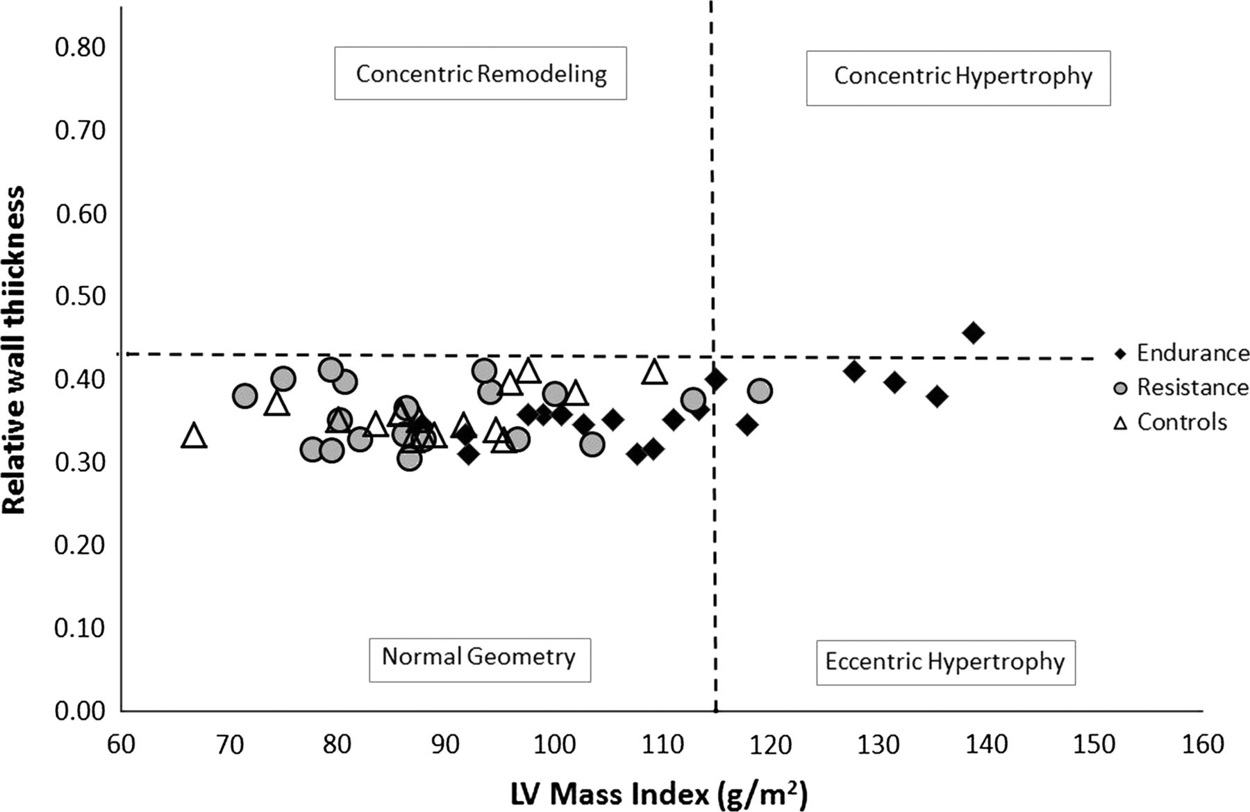
Comparison of the individual distribution of relative wall thickness (RWT) and LV mass index in all athletes. Normal geometry (LV mass ≤115 g/m2 with RWT ≤0.42), concentric remodelling (LV mass ≤115 g/m2 with RWT >0.42), concentric hypertrophy (LV mass ≤115 g/m2 with RWT <0.42) or eccentric hypertrophy (LV mass ≤115 g/m2 with RWT ≤0.42).
In the accompanying editorial, (see page 1225) Professors Haykowsky and Tomczak suggest that the Morganroth hypothesis – that theory the type of exercise determines the pattern of LV hypertrophy –is obsolete. The fact that only 30% of endurance trained athletes had LV hypertrophy also suggests that research is needed on the factors that might account for an increase LV size in some, but not all, athletes. Those who wish to read more about left ventricular function and mural architecture will enjoy the Education in Heart article (see page 1289) on this topic.
There is a wealth of data on the epidemiology, clinical presentation and outcomes of patients with heart failure in developed countries but little information about this diagnosis in sub-Saharan Africa. Compared to the typical demographics in developed countries, Dr Makubi and colleagues (see page 1235) found a younger average age in 427 consecutive heart failure patients admitted to the national referral hospital in Tanzania, In addition, causes of heart failure were different with hypertension in 45%, cardiomyopathy in 28%, and ischemic heart disease in only 9%. Rheumatic heart disease accounted for only 12% of heart failure cases in this series. Multivariate predictors of mortality are shown in figure 2.

Multivariate hazard ratios (HR) for all-cause mortality. BMI, body mass index; CV, cardiovascular; MAP, mean arterial pressure; NYHA, New York Heart Association functional classification.
In the accompanying editorial (see page 1223), Dr. Bukhman notes that while great strides have been made in the diagnosis and treatment of heart failure at national and regional centers in sub-Saharan Africa, 63% of the population remains rural and may not have access to this care. He suggests: “There is a great need for strategies to simplify, decentralize and integrate diagnosis (including echocardiography) and treatment of heart failure at district hospital level in the interest of equity.” We hope to see more studies in Heart on extending effective care to geographically dispersed populations with limited health care resources from many areas around the world.
Ensuring that patients who present with an acute coronary syndrome (ACS) receive appropriate secondary preventative care remains a challenge in every country. In a study from Australia and New Zealand, Dr. Redfern and colleagues (see page 1281) found that only 27% of 2,299 ACS survivors received optimal preventative care at hospital discharge. Patients less likely to receive optimal care included those with unstable angina (rather than myocardial infarction), those who were not treated with percutaneous coronary intervention, patients over age 70 years and those admitted to a private hospital. Professor Alexander Clark proposes (see page 1221) that rather than responding to this data simply as an example of systems-wide failure, we need to “harness failure better”. He states: “Far from being associated with fear and incompetence, modern organisations increasingly see failure to achieve intended outcomes not only as being important to capture with data but also as being integral to subsequent learning and improvement.” He suggests that these data “present a compelling case of the need for providers to do a smaller number of simple things much better: systems are needed to ensure that it is difficult for individual clinicians not to prescribe key medicines for each patient and refer all eligible patients to cardiac rehabilitation.” A goal we all should strive for in every health care setting.
Try the Image Challenge case (see page 1271) to see if you can make the diagnosis from the ECG and chest radiograph in this patient presenting with chest pain.
Linked Articles
- Editorial
- Cardiac risk factors and prevention
- Editorial
- Image challenge
- Heart failure and cardiomyopathies
- Review
- Editorial
- Special populations
- Education in Heart